

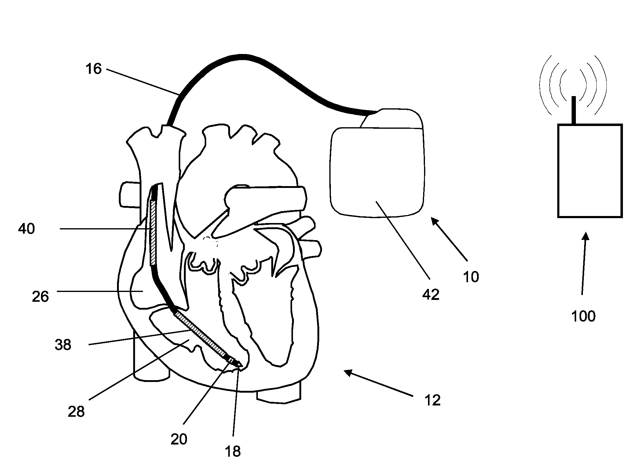
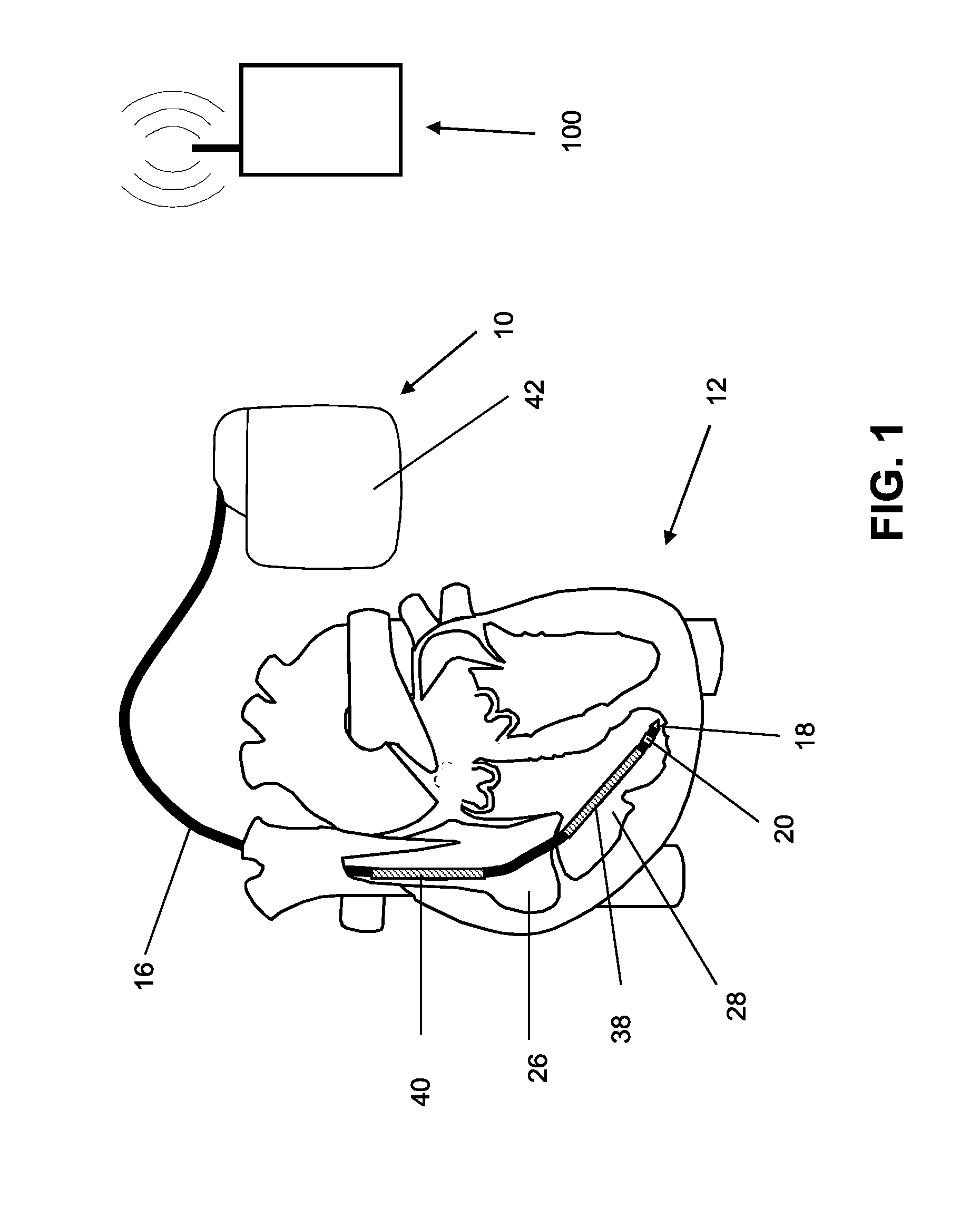
Implantable medical device
a medical device and implantable technology, applied in the field of implantable medical devices, can solve the problems of cardiac insufficiency, inability to further analyze the ballistocardiogram, and inability to establish any relationship with the stroke volume, so as to improve the signal quality and suppress the noise components
- Summary
- Abstract
- Description
- Claims
- Application Information
AI Technical Summary
Benefits of technology
Problems solved by technology
Method used
Image

Examples
example 2
[0062]An implantable medical device designed as a monitoring implant is provided, and has no dedicated treatment function, thought it has sensors such as a 3-axis acceleration sensor and a sensor for ECG detection. The acceleration sensor's output is used to determine the ballistocardiogram (BCG) using the methods described above, as well as determining patient activity. A measure for the stroke volume is determined from the ballistocardiogram, for example from the amplitude of the J-wave, and is transmitted together with the other sensor values to an external device (patient device) and to a home monitoring service center via a telemetry connection. In the home monitoring service center, the stroke volume is included in the assessment of the state of health of the patient. In addition, the effects of treatment (such as medication) can be monitored, and the treatment can be adjusted if necessary.
Combination with Other Sensor Variables
[0063]The calculation and evaluation of the balli...
PUM
 Abstract
Description
Claims
Application Information