
Method, apparatus and computer program product for displaying normalized medical images

a technology of medical images and computer programs, applied in the field of medical images, can solve the problems of compressed images that cannot be manipulated in a consistent manner, the viewer that is to process dicom images is quite complex, and the code library used in the dicom standard is generally not very complex, so as to achieve efficient transmission and processing

Active Publication Date: 2012-10-04
CHANGE HEALTHCARE HLDG LLC
View PDF3 Cites 38 Cited by
  • Summary
  • Abstract
  • Description
  • Claims
  • Application Information

AI Technical Summary

Benefits of technology

[0008]The method, apparatus and computer program product of embodiments of the present invention are configured to process and display normalized DICOM image data such that normalized images may be more efficiently transmitted and processed, thereby facilitating the review of normalized images that were generated from DICOM images on a wider variety of client devices. Thus, the methods, apparatus and computer program product of embodiments of the present invention permit client devices that are mobile and / or that are not dedicated to processing DICOM images to support the display of normalized images that were generated from DICOM images. Thus, the methods, apparatus and computer program product of embodiments of the present invention may provide more widespread access to normalized images that were created based upon DICOM images with less sophisticated client devices.

Problems solved by technology

The DICOM standard generally requires the use of relatively complex code libraries.
As such, a viewer that is to process DICOM images may be quite complex.
As a result, the compressed images may not be manipulated in a consistent manner.
For example, a server configured to process DICOM images may not consistently and in a computationally inexpensive manner supply rectangular sub-areas within an image to a viewer, as some of the compression techniques allowed by the DICOM standard do not provide effective techniques for extracting such sub-areas.
Indeed, in some instances, the entire image would have to be decompressed, clipped and re-compressed in order to supply a rectangular sub-area, thereby resulting in a computationally expensive process.
However, the DICOM standard does not allow for an explicit definition of the logical structure of a study.
As a result, DICOM viewers are generally relatively slow to reveal the complete study structure to the user as the viewer is required to process each and every image in a study in order to determine the respective identifiers and, in turn, the study structure.
Such a specialized proprietary library generally is of a considerable size and complexity, thereby limiting its portability.
As a result of the amount of data that must be transferred to the DICOM viewers in terms of image data and the associated meta-information as well as the complexity of the image processing required at such DICOM viewers, the viewing and processing of DICOM images is generally limited to dedicated viewers with limited, if any, portability.

Method used


Image

  • Method, apparatus and computer program product for displaying normalized medical images
  • Method, apparatus and computer program product for displaying normalized medical images
  • Method, apparatus and computer program product for displaying normalized medical images

Examples


Embodiment Construction

[0020]The present invention now will be described more fully hereinafter with reference to the accompanying drawings, in which some embodiments of the invention are shown. This invention may, however, be embodied in many different forms and should not be construed as limited to the embodiments set forth herein; rather, these embodiments are provided so that this disclosure will be thorough and complete, and will fully convey the scope of the invention to those skilled in the art. Further, the apparatus and method of example embodiments of the present invention will be primarily described in conjunction with medical-imaging applications. It should be understood, however, that the apparatus and method can be utilized in conjunction with a variety of other applications, both in the medical industry and outside of the medical industry. Like numbers refer to like elements throughout.

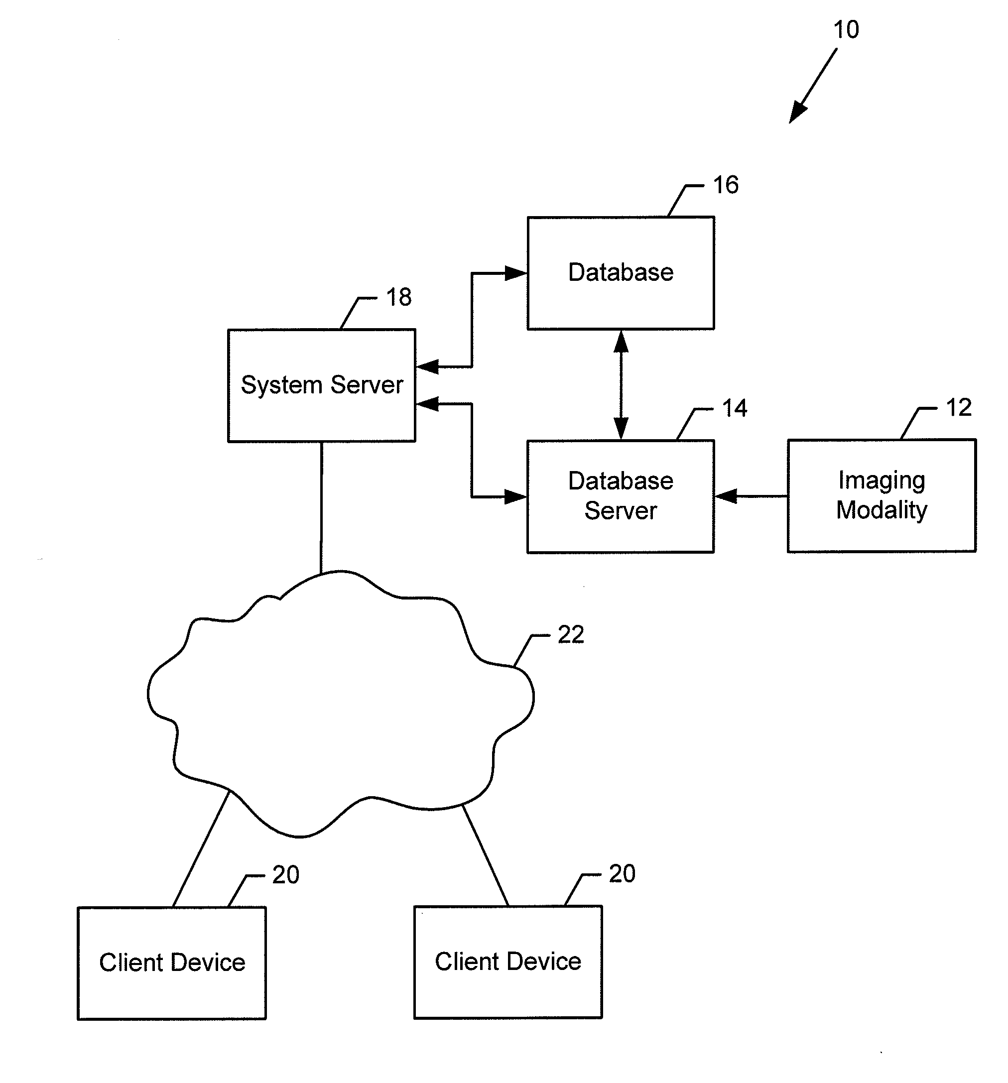
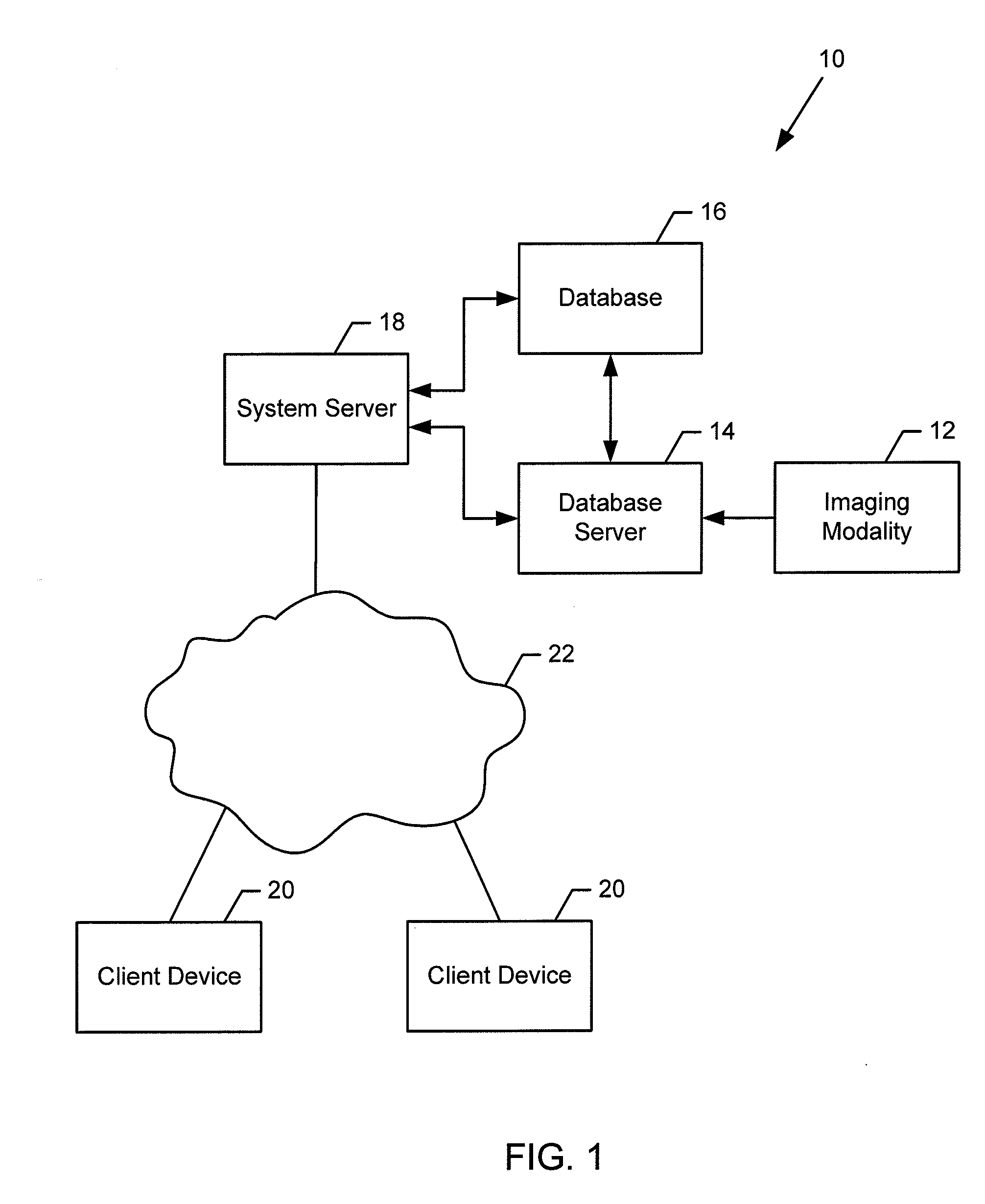
[0021]FIG. 1 illustrates a system 10 that may benefit from an example embodiment of the present invention....


PUM


Abstract

A method, apparatus and computer program product are provided to process a compressed image, such as a normalized DICOM image. A method may receive a compressed image having pixels that each have by a pixel value having a predefined number of bits in a grayscale format. The method may decompress the image by mapping the pixel value into two channels of a multi-channel output buffer, such as a red-green-blue (RGB) output buffer, such that each channel has fewer than the predefined number of bits. The method may render a scene of the image based upon camera coordinates and, for a rendered pixel, sample a plurality of RGB values, convert the plurality of RGB values to the grayscale format having the predefined number of bits, perform an interpolation of the RGB values following conversion to the grayscale format and perform a window level operation on the result of the interpolation.

Description

TECHNOLOGICAL FIELD[0001]Embodiments of the present invention relate generally to the display of medical images and, more particularly, to the display of normalized medical images to facilitate receipt and processing of the images by a client device.BACKGROUND[0002]Medical imaging often includes creating images of regions of the human body for clinical purposes, such as examination, diagnosis and / or treatment. These images may be acquired by a number of different imaging modalities including, for example, ultrasound (US), magnetic resonance (MR), positron emission tomography (PET), computed tomography (CT), mammograms (MG), digital radiology (DR), computed radiology (CR) or the like. In a number of example medical imaging workflows, such as in the case of a picture archiving and communications system (PACS), an image study for a patient may include one or more acquired images of the patient along with information that may reside with or otherwise accompany the images. This informati...

Claims


Application Information

Patent Timeline
IPC IPC(8): G06K9/36
CPCG06T3/4015
Inventor BOCIRNEA, RADU CATALIN
Owner CHANGE HEALTHCARE HLDG LLC
Learn More