Pelvic Implants having Perimeter Imaging Features

a technology of perimeter imaging and pelvis, which is applied in the field of surgically implantable mesh or sling device, can solve the problems of weakness or damage to the normal pelvic support system, dislocation of the vaginal apex outside of the vagina, and difficulty to determine the placement of the mesh within the body, so as to reduce the effectiveness of the support mesh, facilitate imaging, and eliminate the opportunity to damage tissue

Inactive Publication Date: 2012-11-08
BOSTON SCI SCIMED INC
View PDF18 Cites 9 Cited by
  • Summary
  • Abstract
  • Description
  • Claims
  • Application Information

AI Technical Summary

Benefits of technology

[0009]Disclosed is a feature and method to allow visual confirmation of how the mesh of an implant is lying in the body of the patient. A small platinum iridium wire, or a like structure or marker, is provided around the perimeter of the mesh. This creates a small visible line that can be seen under X-ray, fluoroscopy or like visual imaging means. As a result, the doctor can see if the mesh is flat, or if there are any bends, bunches, or general disruptions in the mesh. This will allow the physician to properly lay the mesh along its proper plane and orientation, thereby eliminating opportunities to damage tissue or reduce the effectiveness of the support mesh.
[0010]Various embodiments of the imaging feature can be woven along, molded to, or otherwise disposed along the perimeter of at least a portion of the implant to facilitate imaging. In those embodiments employing a wire or like imaging member, the wire can provide a level of increased rigidity at or near the edge of the implant to facilitate stability and to reduce bunching along the edge or perimeter of the implant.

Problems solved by technology

Pelvic disorders such as these can result from weakness or damage to normal pelvic support systems.
In its severest forms, vaginal vault prolapse can result in the distension of the vaginal apex outside of the vagina.
When any mesh is currently implanted (incontinence, prolapse or the like), it can be very difficult to impossible to determine how the mesh is laying within the body.

Method used


Image

  • Pelvic Implants having Perimeter Imaging Features
  • Pelvic Implants having Perimeter Imaging Features
  • Pelvic Implants having Perimeter Imaging Features

Examples


Embodiment Construction

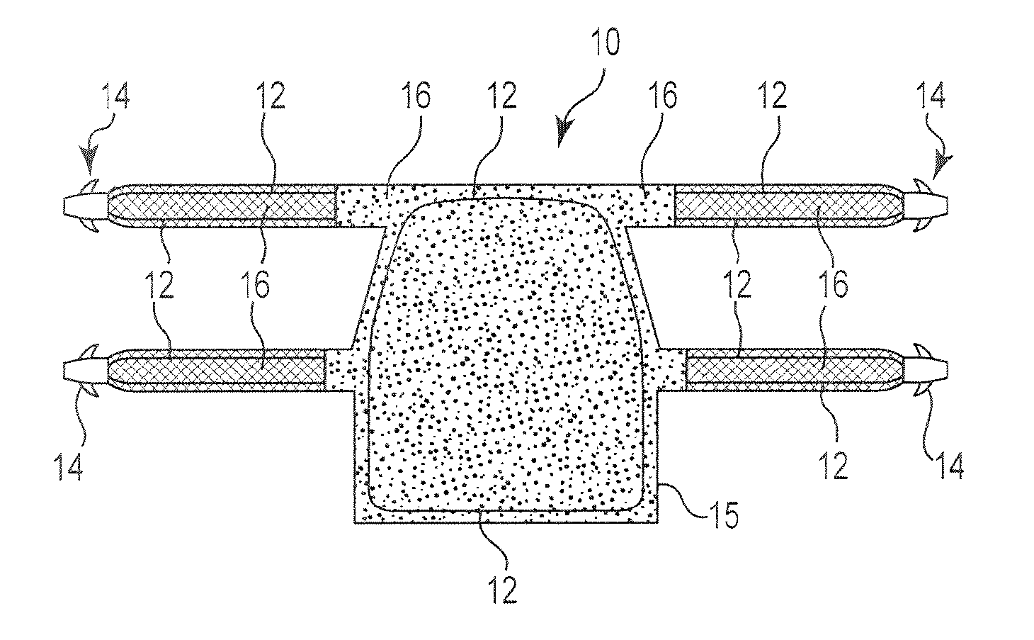
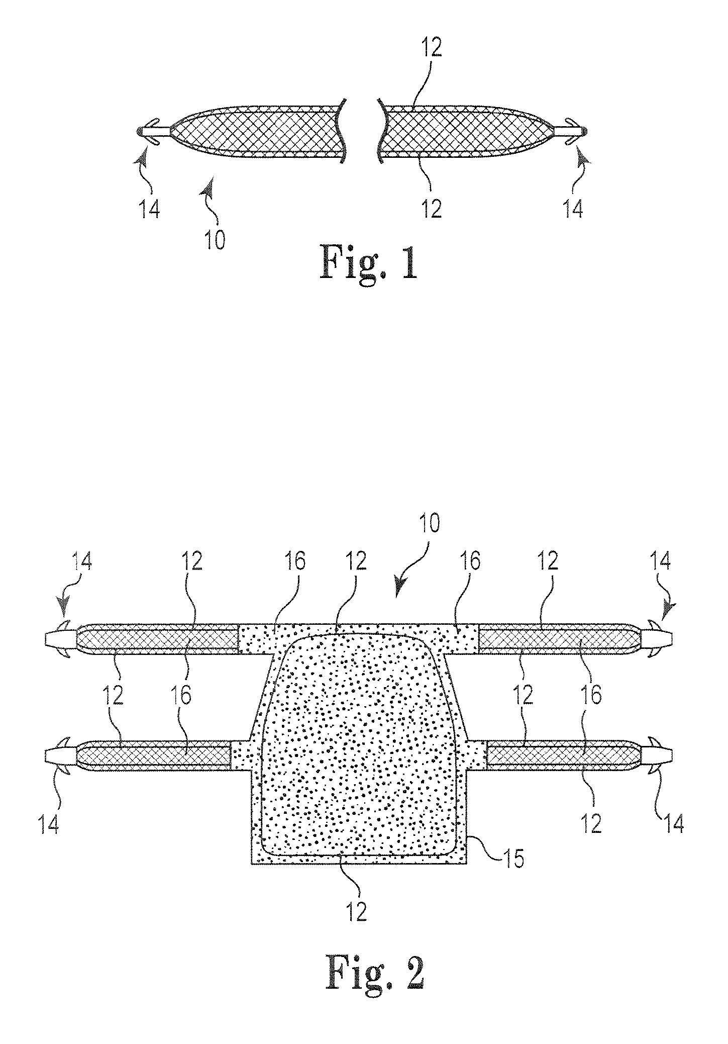
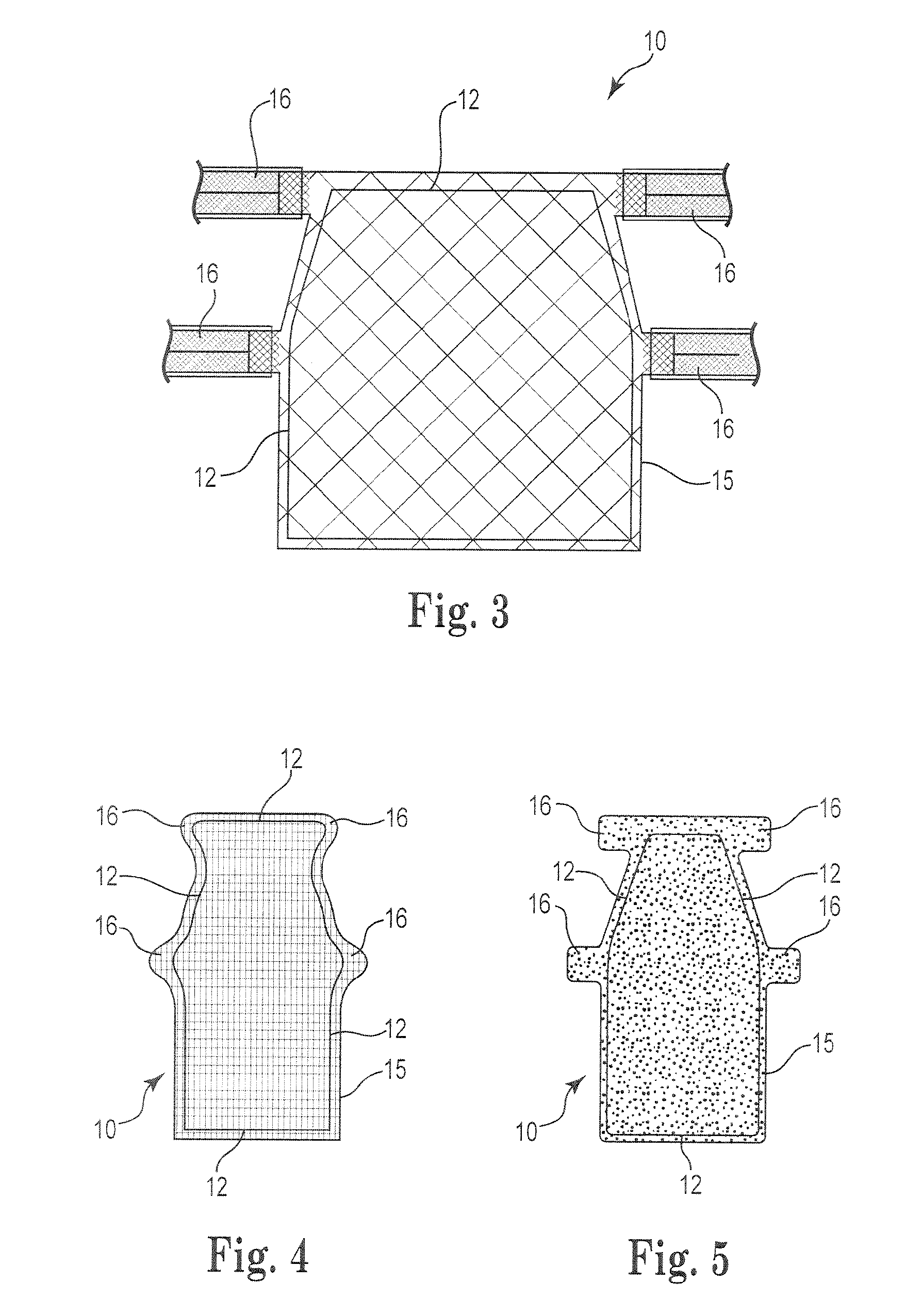
[0013]The present invention may be used in conjunction with any mesh or other implant or biologically-compatible graft 10 that is implanted and where the orientation, lay or plane of the implant is desired to be seen with imaging equipment. Examples of such implants 10 are found in implants used to treat pelvic conditions, including incontinence (fecal and urinary) and vaginal prolapse. Various exemplary implants, systems and methods are disclosed in U.S. Pat. Nos. 7,500,945, 7,407,480, 7,351,197, 7,347,812, 7,303,525, 7,025,063, 6,691,711, 6,648,921, and 6,612,977, International Patent Publication Nos. WO 2008 / 057261 and WO 2007 / 097994, and U.S. Patent Publication Nos. 2010 / 0261955, 2002 / 0151762 and 2002 / 0147382. Accordingly, the above-identified references are fully incorporated herein by reference in their entirety.

[0014]Referring generally to FIGS. 1-5, a distinguishable imaging feature 12 is placed or provided around the perimeter of the implant 10. The imaging feature can incl...


PUM


Abstract

An implant adapted to treat various pelvic disorders can include one or more imaging features constructed of a material adapted to provide visualization on an imaging machine (e.g., X-Ray, fluoroscopy, etc.). The imaging feature generally follows along the perimeter shape of at least a portion of the implant to visually confirm how the implant is lying in a patient's body during implantation. The imaging feature can be constructed of a wire (e.g., platinum-iridium), a radio-opaque material or substance, or like materials or constructs adapted to provide the desired visualization.

Description

PRIORITY[0001]This Application claims priority to and the benefit of U.S. Provisional Application No. 61 / 291,380, filed Dec. 31, 2009, which is incorporated herein by reference in its entirety.FIELD OF THE INVENTION[0002]The present invention relates generally to surgical methods and apparatus and, more specifically, to surgically implantable mesh or sling devices and methods for forming and using the same.BACKGROUND OF THE INVENTION[0003]Pelvic health for men and women is a medical area of increasing importance, at least in part due to an aging population. Examples of common pelvic ailments include incontinence (e.g., fecal and urinary), pelvic tissue prolapse (e.g., female vaginal prolapse), and conditions of the pelvic floor.[0004]Urinary incontinence can further be classified as including different types, such as stress urinary incontinence (SUI), urge urinary incontinence, mixed urinary incontinence, among others. Other pelvic floor disorders include cystocele, rectocele, enter...

Claims


Application Information

Patent Timeline
Patent Type & Authority Applications(United States)
IPC IPC(8): A61F2/08
CPCA61F2250/0098A61F2/0045
Inventor ALEXANDER, JAMES A.
Owner BOSTON SCI SCIMED INC
Why Eureka
Try Eureka