
Articular cartilage treatment method

a treatment method and cartilage technology, applied in the field of articular cartilage treatment methods, can solve the problems of cartilage degeneration, bone tissue deterioration, cartilage loss, etc., and achieve the effect of successfully treating articular cartilage degeneration

Inactive Publication Date: 2012-11-08
BRANNON JAMES K
View PDF2 Cites 4 Cited by
  • Summary
  • Abstract
  • Description
  • Claims
  • Application Information

AI Technical Summary

Benefits of technology

[0008]It has been found that in some patients articular cartilage degeneration is not a primary condition but a symptom of localized deterioration of the underlying bone. The present invention provides embodiments of a method to diagnose and treat an underlying articular bone condition to more successfully treat articular cartilage degeneration.
[0009]Bones are typically composed of a hard outer tissue referred to as cortical bone tissue, a spongy inner tissue referred to as cancellous bone tissue, and other types of tissues. At the various joints within the body, the ends of the bones are covered with cartilage which acts as connective tissue and also as padding. In healthy bone condition, the cortical and cancellous bone tissues cooperate to provide the necessary strength to support strenuous activities as well as resilience to absorb impacts. However, certain conditions can cause deterioration of bone tissue known as osteonecrosis. Osteonecrosis changes the character of the bone tissue resulting in a loss of resilience. Over time, the hardened bone tissue at areas of engagement with other bones increases compressive stress on the articular cartilage lining the bones of the joint, thereby causing the cartilage to deteriorate.
[0016]Many products exist to treat this surgical need. One example is autologous bone particles or segments recovered from the patient. When removed from the patient, it is wet and viscous from the associated blood. This works very well to heal the defect but requires significant secondary surgery resulting in lengthening the surgery, extending the time the patient is under anesthesia and increasing the cost. In addition, a significant increase in patient morbidity is attendant in this technique as the surgeon must take bone from a non-involved site in the patient to recover sufficient healthy bone, marrow and blood to perform the defect filling surgery. This leads to significant post-operative pain.

Problems solved by technology

However, certain conditions can cause deterioration of bone tissue known as osteonecrosis.
Osteonecrosis changes the character of the bone tissue resulting in a loss of resilience.
Over time, the hardened bone tissue at areas of engagement with other bones increases compressive stress on the articular cartilage lining the bones of the joint, thereby causing the cartilage to deteriorate.
There are around 6.5 million fractures per year in the United States, of which approximately 15% are difficult to heal.
For those fractures in which the healing is slow (delayed union) or does not occur (nonunion), there are few effective therapies at present.
Poor fracture healing is associated with chronic pain and prolonged ambulatory impairment and must often be treated by surgical intervention.
External fixation devices may stabilize fractures at risk from poor healing, although a lack of viable bone at the fracture site may result, at best, in the production of unstable bone that is prone to refracture.
Although bone grafting is generally successful, it suffers from the limited amount of donor tissue that may be available, and the patient may suffer side effects such as numbness or tingling at the donor site, infection, or prolonged pain.

Method used


Image

  • Articular cartilage treatment method
  • Articular cartilage treatment method
  • Articular cartilage treatment method

Examples


Embodiment Construction

[0027]As required, detailed embodiments of the present invention are disclosed herein; however, it is to be understood that the disclosed embodiments are merely exemplary of the invention, which may be embodied in various forms. Therefore, specific structural and functional details disclosed herein are not to be interpreted as limiting, but merely as a basis for the claims and as a representative basis for teaching one skilled in the art to variously employ the present invention in virtually any appropriately detailed structure.

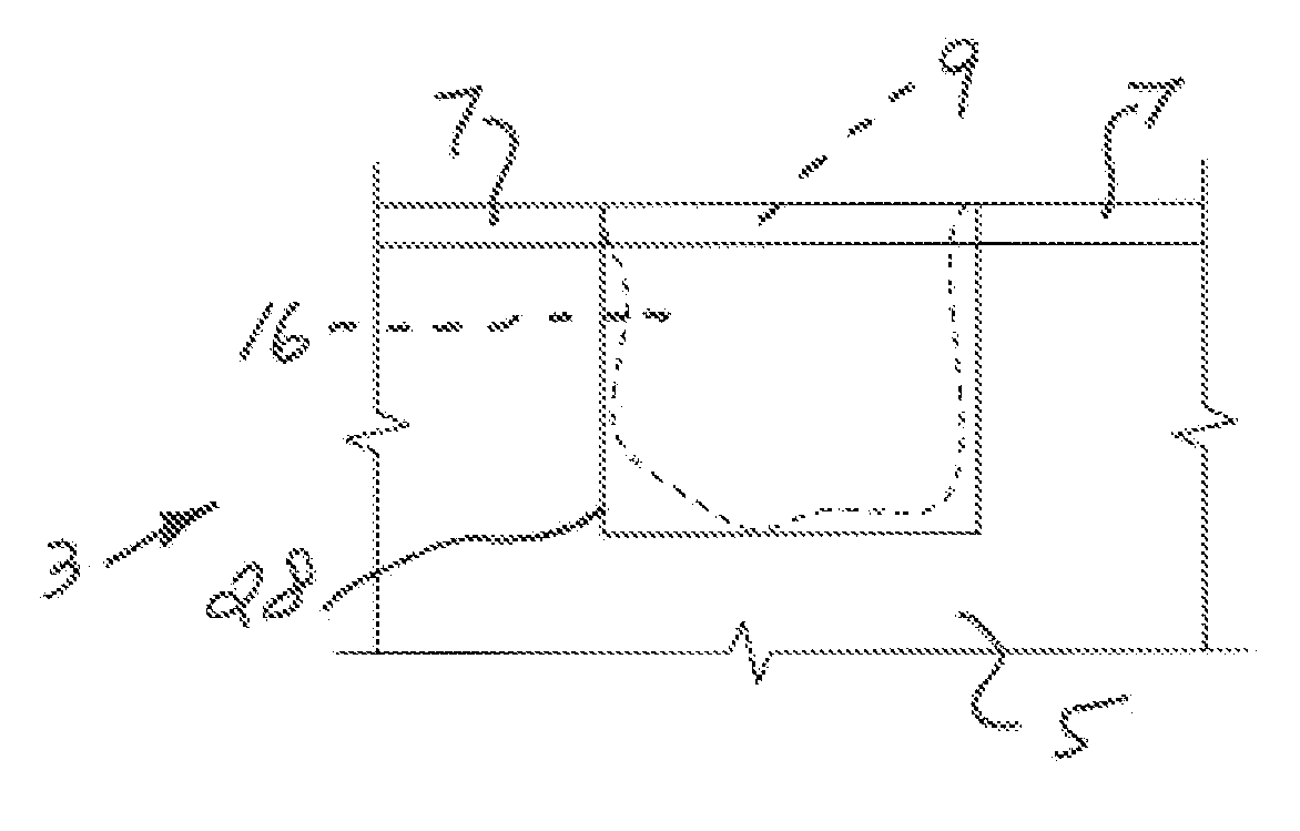
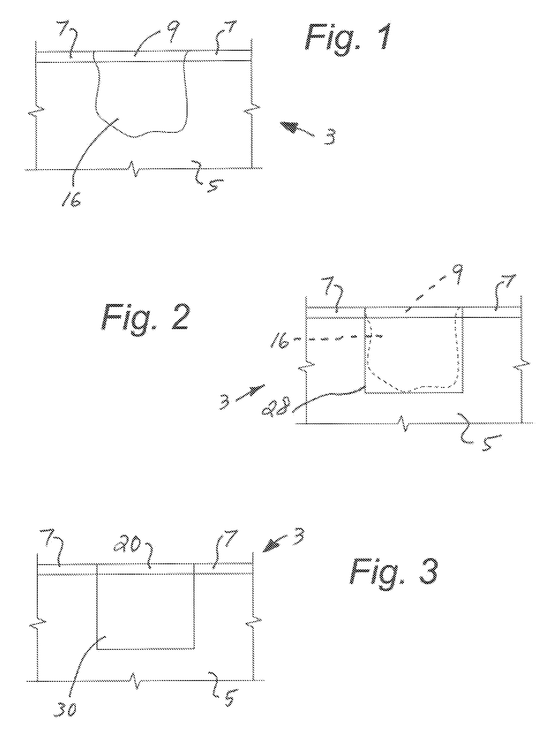
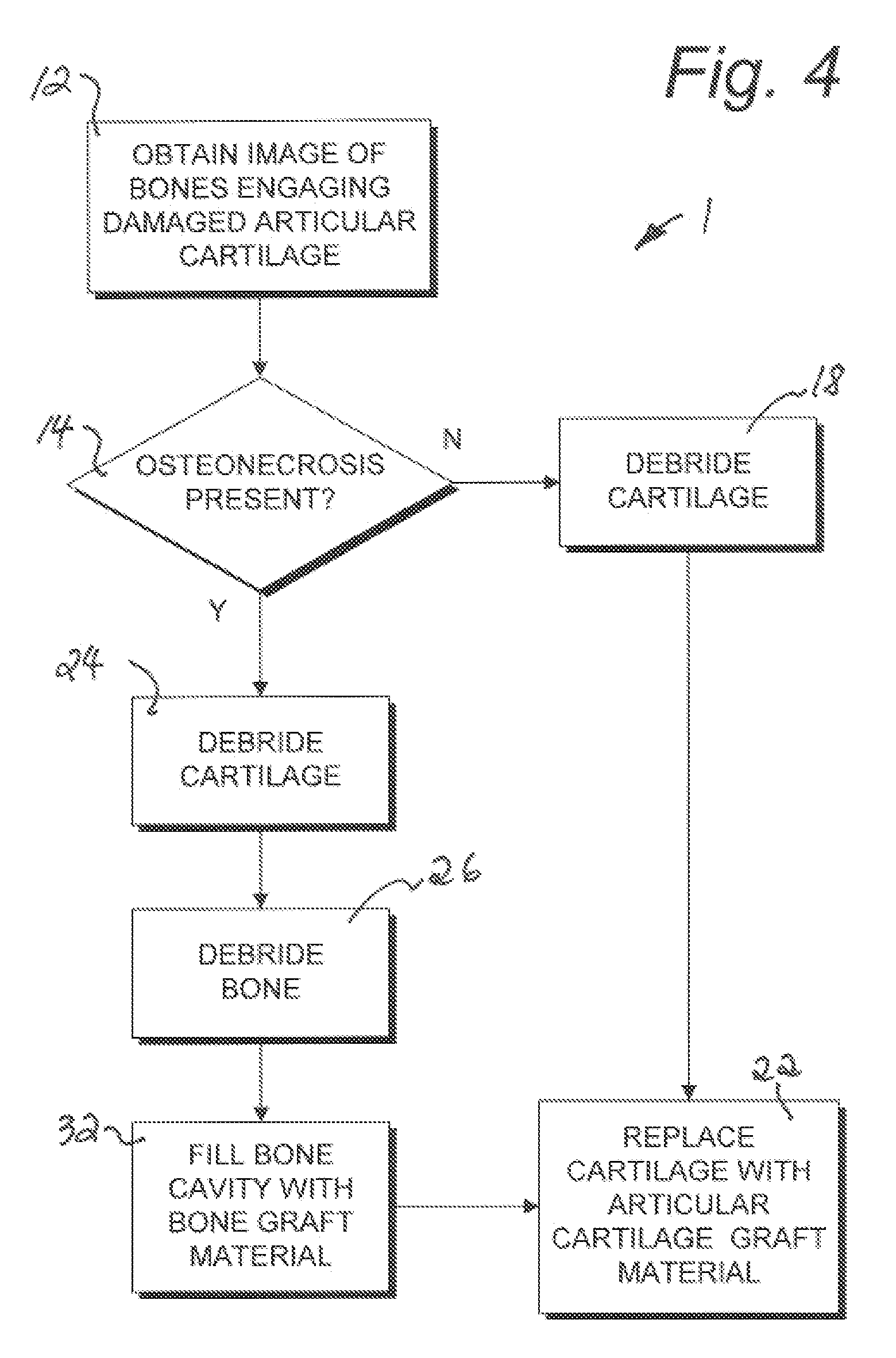
[0028]This application incorporates by reference applicant's prior pending U.S. patent application 61 / 218,757, attached hereto along with the referenced article by Kevin R. Stone. Referring to the drawings in more detail, the reference numeral 1 (FIG. 4) generally designates an embodiment of an articular cartilage treatment method according to the present invention. The method 1 is practiced to alleviate a deteriorated condition at an articular or bone joint ...


PUM


Abstract

The present invention provides an method to alleviate a deteriorated condition at an articular joint site and for repair of damaged cartilage the method including the steps of imaging for the presence of osteonecrosis in the underlying bone, forming a cylindrical plug comprising articular cartilage and osteonecrotic bone using an endoscopic trephine, inspecting the removed cylindrical plug and the joint site for additional articular cartilage and osteonecrotic bone, selectively debriding osteonecrotic bone material associated with the bone cavity to achieve desired vascular characteristics for receiving a prepared bone graft material within the bone cavity, and overlying the received bone graft material with cartilage tissue at the cartilage tissue receiving surface.

Description

CROSS-REFERENCE TO RELATED APPLICATION[0001]This application claims the benefit of the prior filed U.S. provisional application No. 61 / 266,900 filed Dec. 4, 2009 and prior filed U.S. nonprovisional application Ser. No. 12 / 820,133 filed Jun. 21, 2010 which are incorporated herein by reference.FIELD OF THE INVENTION[0002]The present invention is broadly concerned improvements in the treatment of articular cartilage damage, injury, or lesions and, more particularly, to a treatment method which includes treatment of bone conditions underlying the cartilage damage.BACKGROUND OF THE INVENTION[0003]Articular cartilage on the surface of bones in joints, most particularly the knee, ankle and hip joints, is susceptible to deterioration caused by injury, disease, and aging. Untreated articular cartilage lesions have a limited ability to heal and may promote degenerative changes in the joint. Damage to the structure and function of the articular cartilage leads to pain, loss of range of motion,...

Claims


Application Information

Patent Timeline
Patent Type & Authority Applications(United States)
IPC IPC(8): A61F2/28
CPCA61F2/28A61F2/30942A61F2/30756
Inventor BRANNON, JAMES K.
Owner BRANNON JAMES K
Learn More