
Method for removing motion from non-ct sequential x-ray images

a non-ct sequential, x-ray technology, applied in the field of x-ray machines, can solve the problems of motion blurring, limiting, and geometric blur of images, and achieve the effect of reducing x-ray exposur

Inactive Publication Date: 2012-11-22
GRADY JOHN K
View PDF3 Cites 8 Cited by
  • Summary
  • Abstract
  • Description
  • Claims
  • Application Information

AI Technical Summary

Benefits of technology

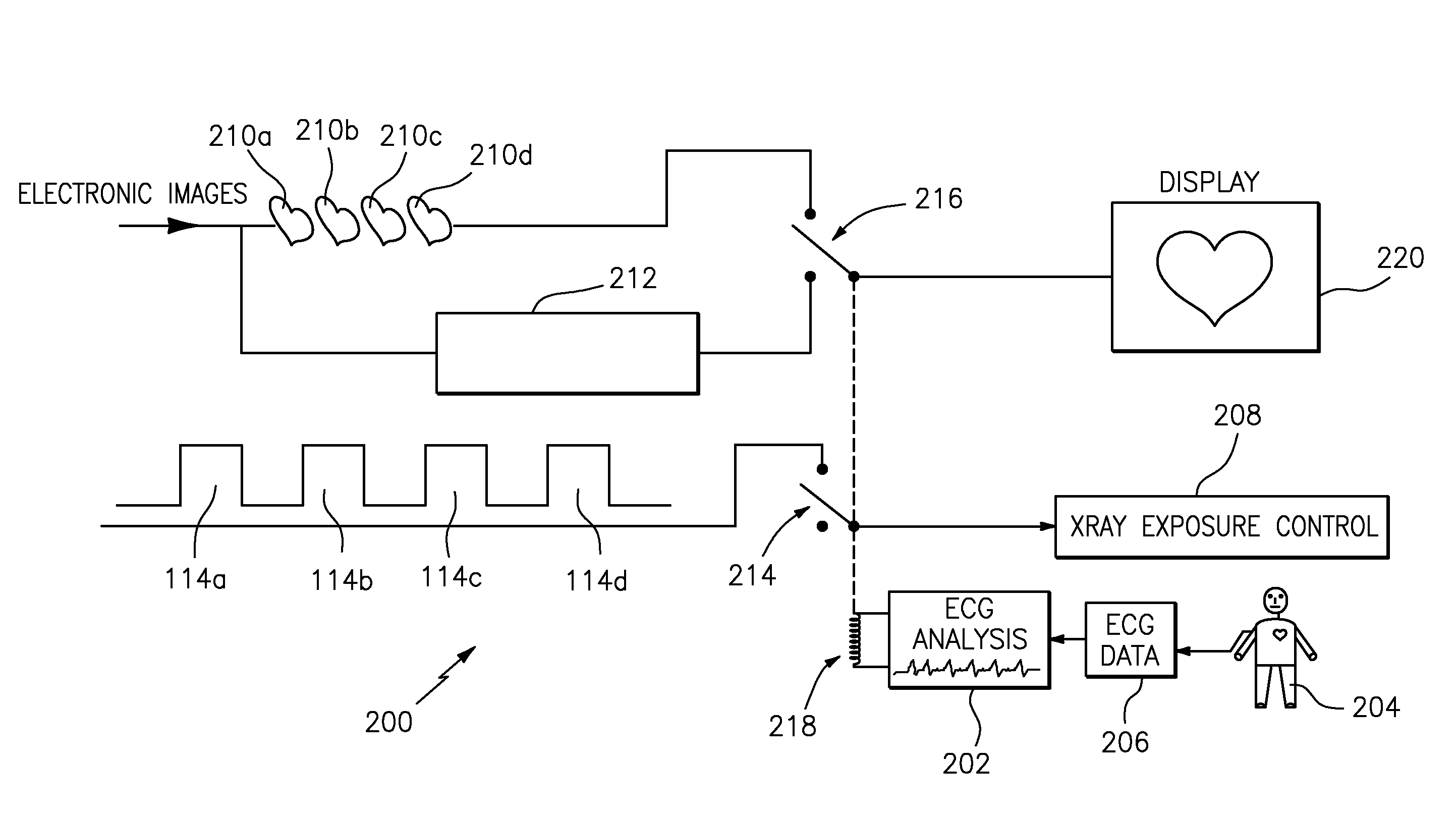
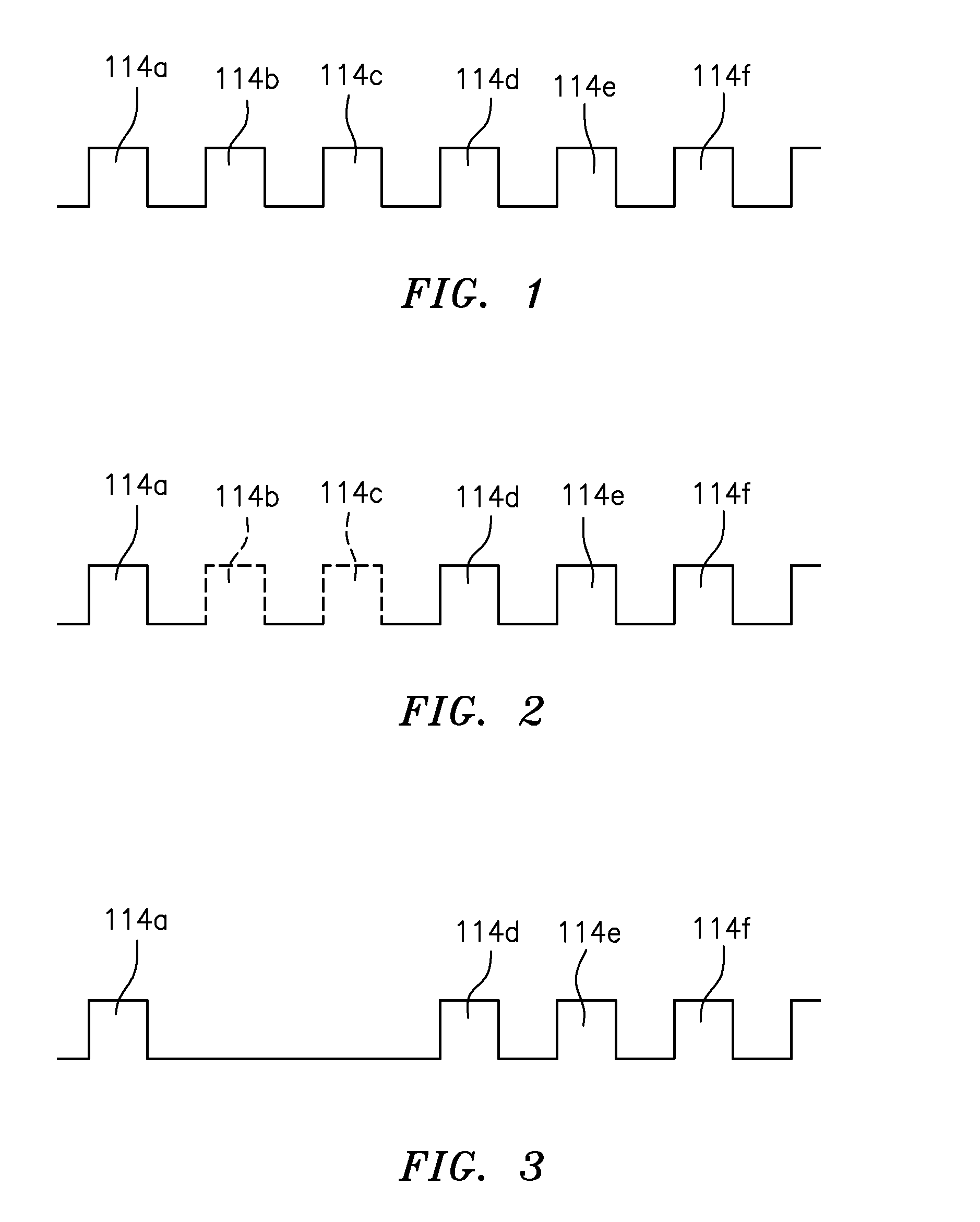
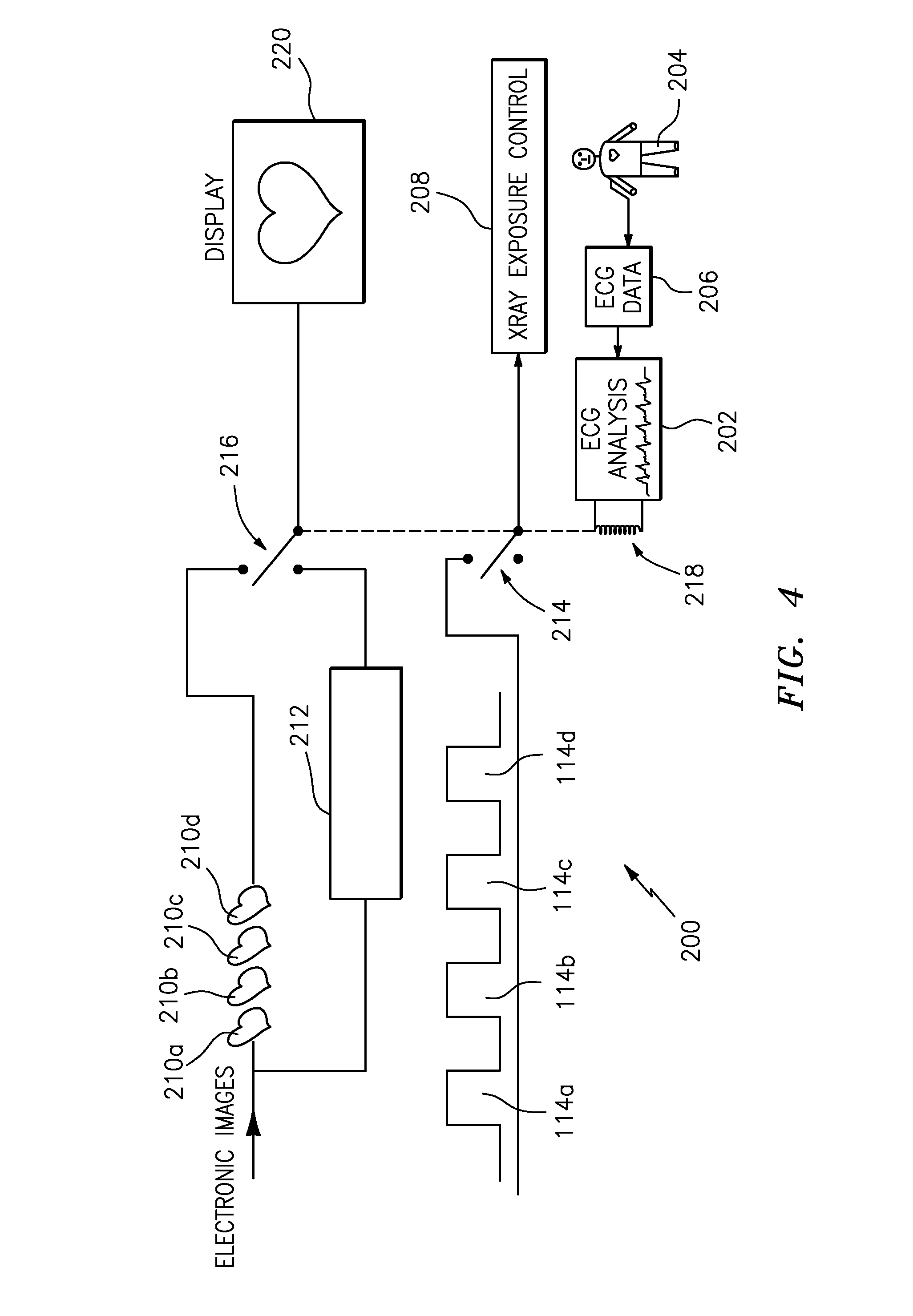
[0013]Applicant has disclosed a method for removing motion from non-CT cardiac angiographic or fluoroscopic x-ray 2-D sequential images, without necessarily using data prediction techniques as is done in sequential CT imagery. Applicant's results are achieved by actively deleting or skipping exposure of certain 2-D flash image acquisitions during rapid heart motion, the latter to reduce x-ray exposure.

Problems solved by technology

This rather large focus or x-ray source can cause geometric blurring of the images, even on a still heart, when used with improved higher resolution image receptors.
The tradeoff of a large x-ray focus was acceptable with rather low resolution (e.g., 2 lp / mm) detector systems, but as detectors have improved it has become more limiting, and will become more so in the future.
That however requires a long exposure (i.e., 10 or more msec) to get enough x-ray, causing motion blurring.
Another related problem involves an increasing use of rotational 2-D angiography and reconstruction of the acquired data sets into 3-D images; this process entails taking a series of 2-D images approximately every one degree on a rotating gantry and reconstructing the data as a volume.
CT devices present a totally different and inherent to CT problem, in that the acquisitions are long and will span significant gantry motion as well as heart motion.
Applicant's invention, described later in this application, is not related to CT devices at all, and prior art involving CT is not applicable.
This bad data propagates through the reconstruction; to correct this, elaborate correction algorithms are needed, e.g., to spatially shift the data.
Even more common, the bad 2-D data set from heart motion is presently deleted, wasting the radiation used on the patient.

Method used


Image

  • Method for removing motion from non-ct sequential x-ray images
  • Method for removing motion from non-ct sequential x-ray images
  • Method for removing motion from non-ct sequential x-ray images

Examples


Embodiment Construction

)

[0022]Applicant has disclosed a method for removing motion from non-CT cardiac angiographic or fluoroscopic x-ray 2-D sequential images by actively deleting or skipping exposure of certain 2-D flash image acquisitions during rapid heart motion, the latter to reduce x-ray exposure. Longer temporal exposures are possible, so that small focal spots can be used, resulting in geometrically sharper images. Such sharpness can be critical to correctly assessing stent deployment and cardiac vessel geometry.

[0023]As used herein, the term “non-CT” means “not computer tomography”.

[0024]Applicant's method is only applicable to flash 2-D acquisitions or conventional fluoroscopy. It is inapplicable to CT type x-ray devices. Such CT types already pause data acquisitions (i.e., sequential images) by synchronizing with the electrocardiograph (a.k.a. ECG or EKG), requiring data prediction by either predictive algorithms or estimated compensation of motion, as described in U.S. Pat. No. 7,672,490 to K...


PUM


Abstract

Applicant has disclosed a method for removing motion from non-CT cardiac angiographic or fluoroscopic x-ray 2-D sequential images, without using data prediction techniques in sequential CT imagery. Applicant's results are achieved by actively deleting or skipping exposure of certain 2-D flash image acquisitions during rapid heart motion (e.g., beating), the latter to reduce x-ray exposure. Applicant's preferred method comprises: positioning a person relative to a non-CT type x-ray machine, designed for fluoroscopy or angiography, with the person's heart between an x-ray source and a detector; monitoring rapid movement of the person's heart by electrocardiography; generating a series of x-ray pulses from the x-ray source; actively skipping any x-ray pulses by switching off the x-ray source during beating of the person's heart to prevent any images being generated from the skipped x-ray pulses; and generating sequential (i.e., either angiographic or fluoroscopic) 2-D cardiac images from the non-skipped x-ray pulses; wherein the motion is removed from the sequential images without using predictive algorithms and without using estimated compensation of motion.

Description

FIELD OF INVENTION[0001]The present invention relates generally to x-ray machines. More particularly, it relates to non-CT cardiac angiographic or fluoroscopic x-ray 2-D sequential images.BACKGROUND OF INVENTION[0002]The problem of acquiring sharp x-ray fluoroscopy images of the heart, to enable cardiac interventions via catheter (e.g., stents or diagnosis) has a long history of innovation geared toward sharper, non-blurred images.[0003]Because the heart can move up to 400 mm / second in its most rapid phase, normal continuous fluoroscopy in the early days of catheterization (approximately 1960-1970) resulted in blurred images during the rapid motion phase.[0004]This led to the development of the “grid control” pulse x-ray system, by Keleket X-Ray Corporation and General Electric Corporation, and the Philips®“cine pulse” system. Both systems allowed x-ray pulsed exposures as short as 1 millisecond. This “froze” the cardiac image similar to a fast shutter on a camera, by using a short ...

Claims


Application Information

Patent Timeline
IPC IPC(8): H05G1/60
CPCH01J35/045H05G1/60A61B6/481A61B6/487A61B6/542A61B6/504A61B6/5264A61B6/541A61B6/503
Inventor GRADY, JOHN K.
Owner GRADY JOHN K
Learn More