Methods for Microalification Detection of Breast Cancer on Digital Tomosynthesis Mammograms

a technology of digital tomosynthesis and breast cancer, applied in the field of breast cancer detection, can solve the problems of high recall rate of about 10%, limited sensitivity of dense breasts, and radiologists not detecting all carcinomas, so as to reduce the number of false positives, enhance the 3d calcification response function, and increase the visibility of microcalcification-like objects.

Active Publication Date: 2012-11-22
RGT UNIV OF MICHIGAN
View PDF11 Cites 30 Cited by
  • Summary
  • Abstract
  • Description
  • Claims
  • Application Information

AI Technical Summary

Benefits of technology

[0019]A CAD system may detect microcalcification clusters in reconstructed volumes of DBT 3D images. A method using the CAD system to detect microcalcification may generally include three stages: prescreening, clustering, and false-positive reduction. In the prescreening stage, the conspicuity of microcalcification-like objects may be increased by an enhancement-modulated 3D calcification response function. An iterative thresholding and 3D object growing method may then be used to detect seed objects. In the cluster detection stage, a second iterative thresholding procedure may be used to identify microcalcification candidates with a positive calcification response in the SNR-enhanced volume. Starting with each seed object as the initial cluster center, a dynamic clustering algorithm may form a cluster candidate by including microcalcification candidates within a 3D neighborhood of the seed object that satisfied the clustering criteria. The number, size, and SNR of microcalcifications in a cluster candidate and the cluster shape may be used to reduce the number of false-positives (FPs).
[0020]Using this system, the prescreening stage may detect a seed object in biopsied microcalcification clusters at a threshold of 100 seed objects per DBT volume. After clustering, the number of detected clusters may be reduced to 15 marks per DBT volume. The FP reduction stage may further reduce the number of FPs per DBT volume and the cluster detection sensitivity will depend on the decision threshold setting.

Problems solved by technology

Although mammography has a high sensitivity for detection of breast cancers when compared to other diagnostic modalities, studies indicate that radiologists do not detect all carcinomas that are visible upon retrospective analyses of the images.
One major problem in screening mammography is the limited sensitivity in dense breasts, due to the camouflaging effect of overlapping dense tissue.
Another problem in screening is the high recall rate of about 10%.
Finally, the specificity of screening mammography for differentiating lesions as malignant and benign is very low.
Recall and benign biopsies not only cause patient anxiety and morbidity, but also increase health care costs.
US scanning is time consuming and relatively operator-dependent and cannot detect microcalcifications (MCs).
Breast MR is expensive and has relatively low specificity, limiting its use to high-risk patients and diagnostic purposes.
Because of the lack of PVs at large angles, the spatial resolution in the direction perpendicular to the detector plane (the depth or Z-direction) is poor.
DBT is inferior to breast CT in terms of depth resolution and the need of compression.
They found that the recall rate could be reduced by 40% with the addition of DBT and the DBT image quality was equivalent or superior to the diagnostic mammogram in the vast majority of the cases but the conspicuity of MCs were inferior in many of the cases.
Second, the microcalcifications may appear blurred due to many factors that can cause blurring in DBT reconstruction, including inaccuracy in the system geometry and the projection model, a range of oblique incidence angles to the detector, focal spot motion and patient motion.
The fact that their computer-aided readings seem to track the performance of the CAD systems, especially the reduced cancer rate for invasive cancers, raised an alarm that the participating radiologists might have over-relied on the CAD system and did not maintain their vigilance in the first read.
The performance of these preliminary MC detection systems and methods are far from being practical for clinical use.
Although the conspicuity of masses may be improved in DBT, the chance of oversight may not be negligible because of the large number of images in a DBT volume.
In addition, the visibility of microcalcifications may be reduced, because of the separation of microcalcifications in a cluster into different slices, increased noise associated with multiple low-dose projection view acquisition, and potential blurring and artifacts arising from the tomosynthesis reconstruction, thereby making microcalcifications more subtle and difficult to visualize.

Method used


Image

  • Methods for Microalification Detection of Breast Cancer on Digital Tomosynthesis Mammograms
  • Methods for Microalification Detection of Breast Cancer on Digital Tomosynthesis Mammograms
  • Methods for Microalification Detection of Breast Cancer on Digital Tomosynthesis Mammograms

Examples


Embodiment Construction

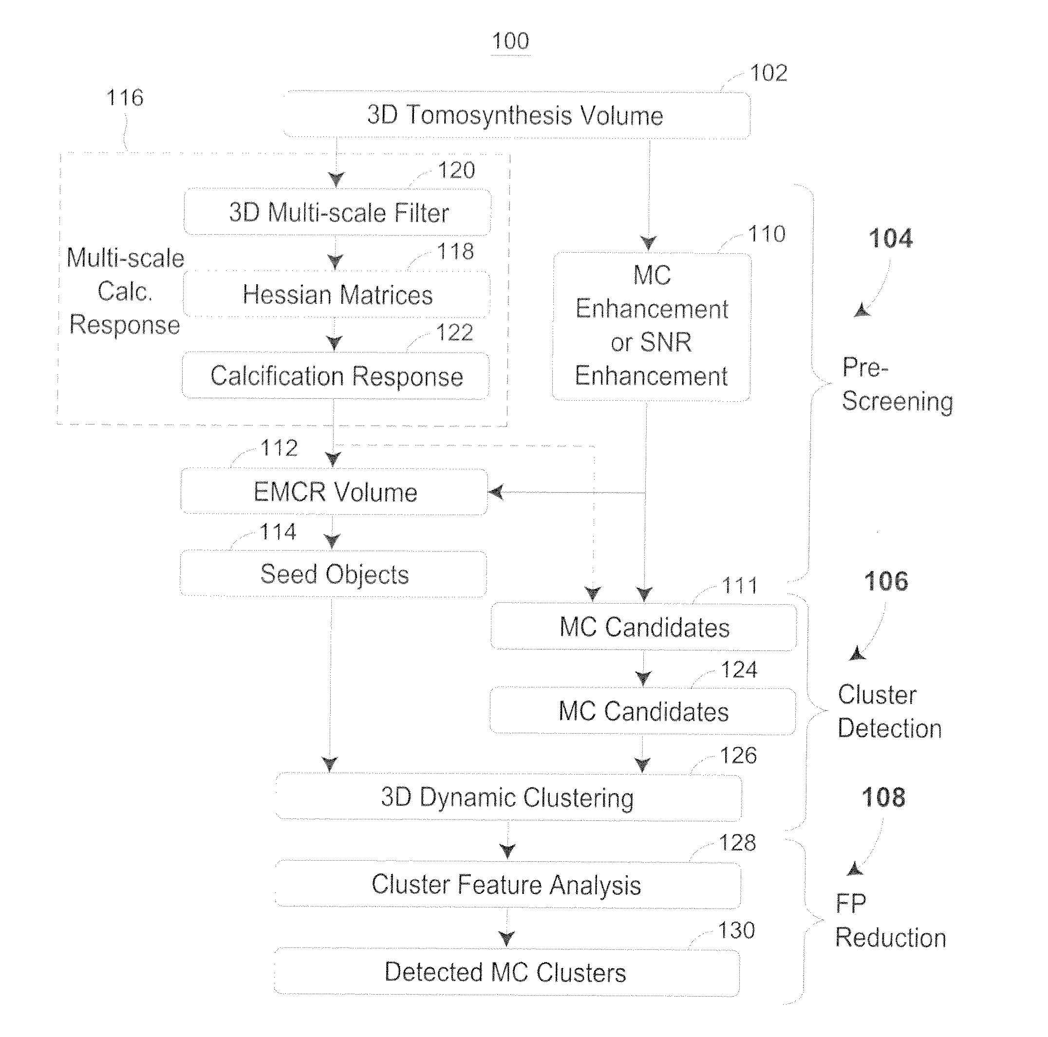
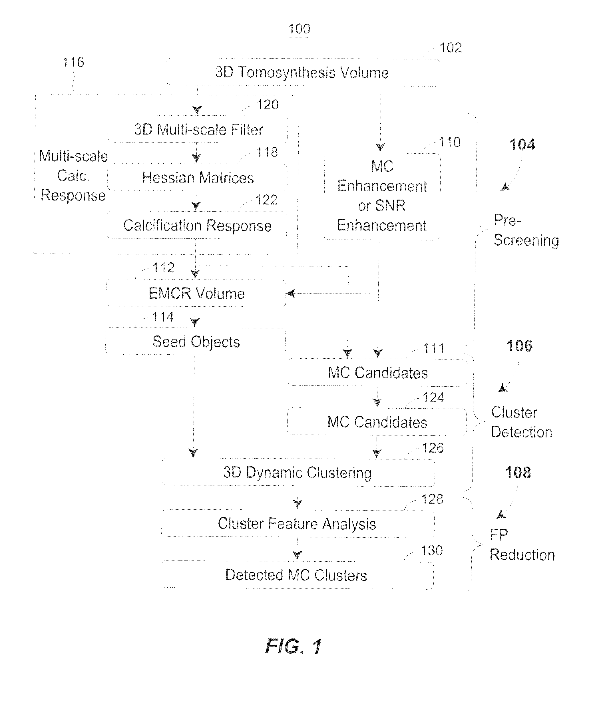
[0038]FIG. 1 discloses an exemplary flow chart of a computer-aided detection (CAD) method 100 for clustered microcalcifications in digital breast tomosynthesis (DBT). The method may begin with generation or collection of a plurality of DBT mammograms using a DBT mammography system. The DBT system may acquire, for example, 11 to 25 PVs over an arc of 15 to 60 degrees in various increments (e.g., 3 degree increments). The DBT system may use a full-field digital detector, for example, a CsI / a-Si flat-panel or an amorphous-Se flat panel detector. The digital detector may be stationary or moving during image acquisition. The breasts may be imaged in either the craniocaudal (CC), mediolateral oblique (MLO), or other views. The DBT volumes may be reconstructed at a 1-mm-thick or other slice interval using a simultaneous algebraic reconstruction technique (SART) or other tomosynthesis reconstruction technique.

[0039]With reference to FIG. 1, a method 100 uses the tomosynthesized DBT slices o...


PUM


Abstract

A computer-aided detection system to detect clustered microcalcifications in digital breast tomosynthesis (DBT) is disclosed. The system performs detection in 2D images and a reconstructed 3D volume. The system may include an initial prescreening of potential microcalcifications by using one or more 3D calcification response function (CRF) values modulated by an enhancement method to identify high response locations in the DBT volume as potential signals. Microcalcifications may be enhanced using a Multi-Channel Enhancement method. Locations detected using these methods can be identified and the potential microcalcifications may be extracted. The system may include object segmentation that uses region growing guided by the enhancement-modulated CRF values, gray level voxel values relative to a local background level, or the original DBT voxel values. False positives may be reduced by descriptors of characteristics of microcalcifications. Detected locations of clusters and a cluster significance rating of each cluster may be output and displayed.

Description

CROSS REFERENCE TO RELATED APPLICATION[0001]This is a national phase of International Patent Application No. PCT / US2011 / 024469 filed Feb. 11, 2011, which in turn claims the priority benefit of the benefit of U.S. Provisional Application No. 61 / 303,387 filed Feb. 11, 2010, and are entirely incorporated by reference herein.STATEMENT REGARDING FEDERALLY SPONSORED RESEARCH OR DEVELOPMENT[0002]This invention was made with government support under Grant number CA120234 awarded by the USPHS. The government has certain rights in the invention.FIELD OF TECHNOLOGY[0003]This relates generally to breast cancer detection and, more particularly, to a system and method for using computer-aided detection (CAD) for clustered microcalcifications in Digital Breast Tomosynthesis (DBT).DESCRIPTION OF THE RELATED ART[0004]Breast cancer is one of the leading causes of death among American women between 40 and 55 years of age. There is considerable evidence that early diagnosis and treatment significantly ...

Claims


Application Information

Patent Timeline
Patent Type & Authority Applications(United States)
IPC IPC(8): G06K9/62
CPCA61B6/025A61B6/502G06T2207/30068G06T7/0012G06T2207/10112A61B6/5211
Inventor CHAN, HEANG-PINGSAHINER, BERKMANHADJIISKI, LUBOMIR M.WEI, JUNHELVIE, MARK A.
Owner RGT UNIV OF MICHIGAN
Why Eureka
Try Eureka