Cancer detection markers

a detection marker and cancer technology, applied in the field of molecular classification of diseases, can solve the problems of poor prognosis, vexing aspects of cancer, and major health challenges, and achieve the effects of low cost, easy to use, and easy to us

Inactive Publication Date: 2012-12-06
MYRIAD GENETICS
View PDF3 Cites 21 Cited by
  • Summary
  • Abstract
  • Description
  • Claims
  • Application Information

AI Technical Summary

Benefits of technology

[0012]Another aspect of the invention provides exosome blocks comprising fixed (e.g., formalin-fixed) exosomes embedded in some medium (e.g., paraffin). Such exosome blocks allow for long-term storage of exosomes and also allow for analysis of the interior of exosomes.
[0013]These aspects of the invention provide a relatively quick and inexpensive screen for the presence of cancer. This can be used as a general population screen (e.g., yearly blood screen) whose ease, non-invasiveness, patient inclusivity, and sensitivity may provide vast improvements over existing general screens such as mammograms, colonoscopies, etc.

Problems solved by technology

Cancer is a major health challenge.
Despite recent advances in molecular and imaging diagnostics, one of the most vexing aspects of cancer remains early detection.
In fact, for certain types of cancer—e.g., pancreatic adenocarcinoma—detection often occurs so late as to practically preclude any good prognosis.

Method used


Image

  • Cancer detection markers
  • Cancer detection markers
  • Cancer detection markers

Examples


example 1

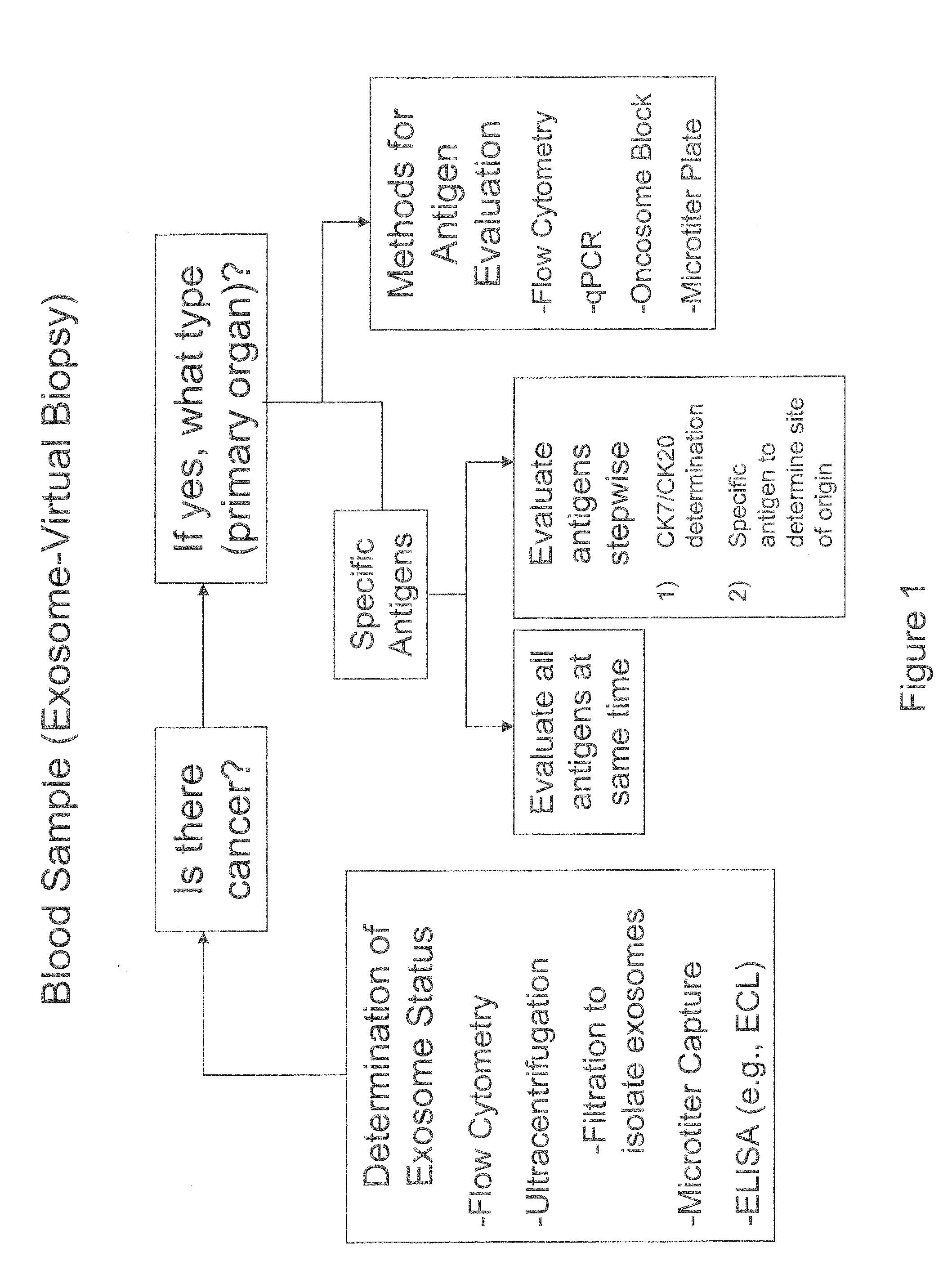
Detection / Quantitation / Characterization of Exosomes by Flow Cytometry

[0073]Exosomes may also be measured by flow cytometry. Flow cytometry is generally used to measure cell-surface antigen expression and thus may be readily adapted to measuring antigens on the surface of exosomes. In general, exosomes are isolated from a sample (e.g., serum) and then incubated with a labeled antibody (e.g., fluorescence-labeled) against some surface marker expected to be found on the exosomes (e.g., EpCAM). This solution is then passed through a cell sorter, which detects and “counts” the number of labeled exosomes as they flow through an optical / electronic detection apparatus. Multiple labeled antibodies may be used in the same sample as cell sorters are capable of detecting multiple wavelengths of light at once. Flow cytometry may also be used to detect and quantitate cytoplasmic antigens. For this it is generally necessary to fix and permeabilize cells to enable antibodies to gain access to them,...

example 2

Detection / Quantitation / Characterization of Exosomes by ELISA

[0094]ELISA (Enzyme-Linked immunosorbent assay) can be adapted to detect, quantify, and characterize exosomes according to the invention. The purpose of an ELISA is to determine if a particular protein is present in a sample and optionally, how much. EpCAM-bearing exosomes were detected and quantitated in the supernatant of epithelial cancer cell lines in the following experiment.

[0095]Reagents:[0096]1. Capture antibody is a mouse monoclonal (clone 158210, R&D systems catalog #MAB 960) raised against the extracellular domain of Human EpCAM.[0097]2. Detection antibody is a goat polyclonal (R&D systems catalog #BAF960) raised against recombinant extracellular domain of Human EpCAM.[0098]3. Quantification achieved by comparison with a standard curve generated using recombinant extracellular domain of Human EpCAM (recombinant EpCAM / Fc chimera, R&D catalog #960-EP-050).

[0099]Sample Preparation:[0100]1. Exosomes were isolated by ...

example 3

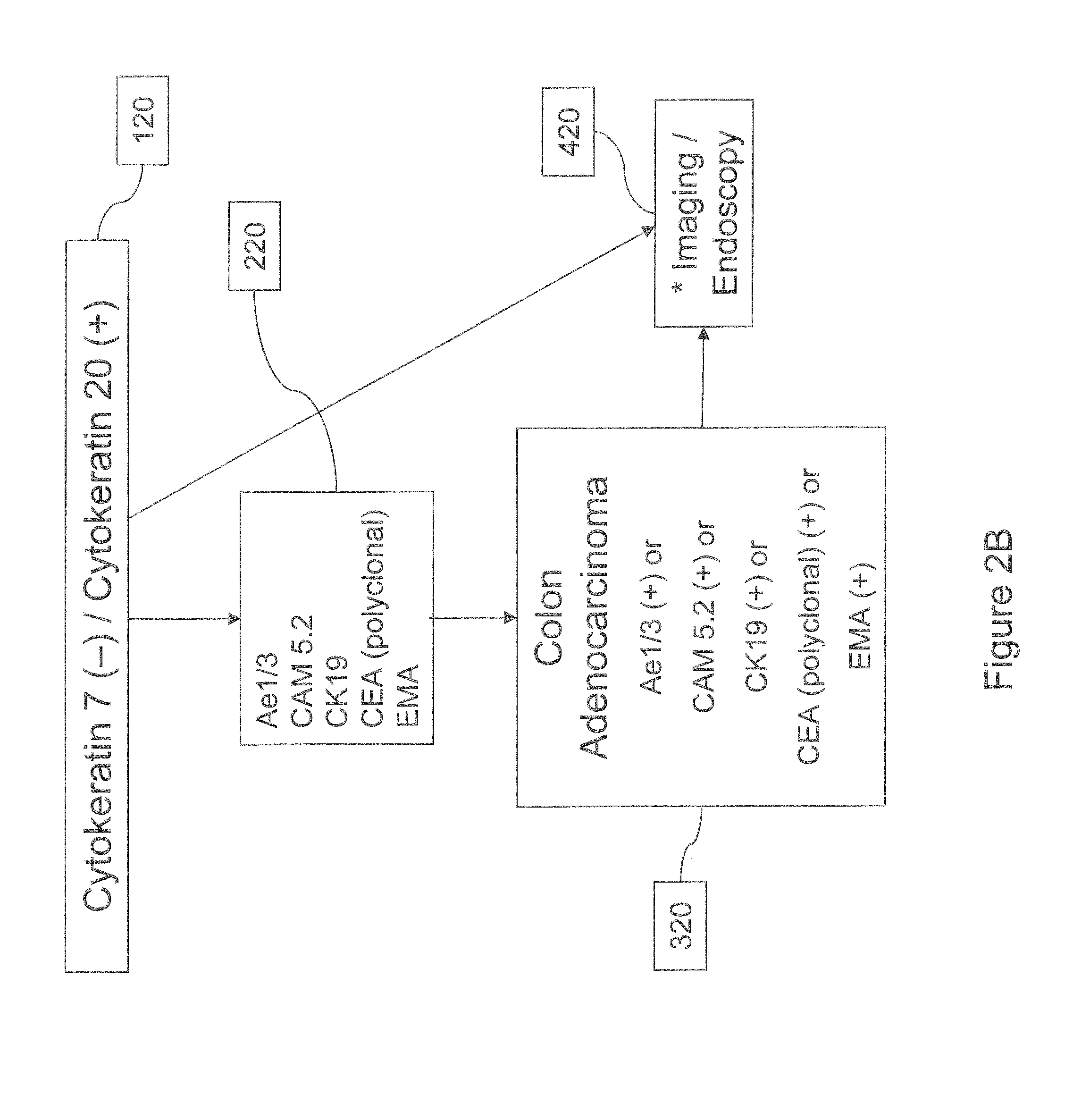
Detection / Quantitation / Characterization of Exosomes by IHC

[0110]Exosome IHC will generally be carried out on an exosome block. Exosome blocks are prepared by isolating exosomes from a patient sample (e.g., plasma sample), fixing the exosome pellet (e.g., with formalin), and then embedding the fixed pellet in some medium (e.g., paraffin) that allows for convenient long-term storage. Qualitative and quantitative IHC analysis may then be performed on thin slices of the exosome blocks using antibodies against proteins of interest on the exterior or interior of the exosomes.


PUM

PropertyMeasurementUnit
sizesaaaaaaaaaa
sizesaaaaaaaaaa
sizesaaaaaaaaaa

Abstract

Methods and compositions involving molecular markers for the detection and characterization of cancer in a patient are provided.

Description

CROSS-REFERENCE TO RELATED APPLICATIONS[0001]This application is the U.S. national phase of International Application No. PCT / US2009 / 067029 filed on Dec. 7, 2009, which designated the U.S. and claims priority to U.S. Provisional Patent Application Ser. No. 61 / 175,954 filed May 6, 2009, U.S. Provisional Patent Application Ser. No. 61 / 174,600 filed May 1, 2009, U.S. Provisional Patent Application Ser. No. 61 / 158,975 filed Mar. 10, 2009, U.S. Provisional Patent Application Ser. No. 61 / 120,259 filed Dec. 5, 2008, the entire contents of each of which are hereby incorporated by reference.FIELD OF THE INVENTION[0002]The invention generally relates to a molecular classification of disease and particularly to molecular markers for cancer and methods of use thereof.BACKGROUND OF THE INVENTION[0003]Cancer is a major health challenge. Nearly 560,000 people die from cancer annually in the United States alone, representing almost 23% of all deaths. See American Cancer Society, Cancer Facts &Figur...

Claims


Application Information

Patent Timeline
Patent Type & Authority Applications(United States)
IPC IPC(8): G01N33/574G01N33/577
CPCG01N33/57434
Inventor SKOLNICK, MARKLANCHBURY, JERRYTIMMS, KIRSTENNEFF, CHRISHOFF, RYANWANG, HUBERTWAGNER, SUSANNESANGALE, ZAINA
Owner MYRIAD GENETICS
Why Eureka
Try Eureka