Virtual microscopy

a microscopy and virtual technology, applied in the field of virtual microscopy, can solve the problems that the viewing software is not necessarily tailored to improve the efficiency with which a user can view slide images, and achieve the effect of reducing data retrieval time and reducing rendering tim

Active Publication Date: 2012-12-20
VENTANA MEDICAL SYST INC
View PDF13 Cites 26 Cited by
  • Summary
  • Abstract
  • Description
  • Claims
  • Application Information

AI Technical Summary

Benefits of technology

[0008]Hence, the first region provides an overview of all slides including material from the specimen currently being reviewed and the second region displays images at a higher level of magnification. The first region can therefore be used to more easily navigate amongst the higher magnification images being displayed in the second region.
[0019]Displaying an image of at least a part of at least one of the plurality of slides in a region of the display device can comprise reading only those portions, or tiles, of image data corresponding to parts of the slide which would be visible in the region. The region can be the first region and / or second region and / or third region of the display device. This reduces rendering time as only those parts of the data relating to the slide image which needs to be displayed are read for image rendering, rather than all of the image data for that slide.
[0020]The method can further comprise storing a pointer to a location in local memory at which the portion of image data is stored, for each of a plurality of portions of image data. This provides direct access to the area of memory in which the image data is stored thereby reducing data retrieval time.
[0025]By encoding information about the slides into the layout pattern, it is easier for a user to determine and select which slides to view, out of all of the slides available to view, more efficiently in order to carry out their assessment.

Problems solved by technology

While virtual microscopy is already established and virtual slide image viewing software is already available, such viewing software is not necessarily tailored to improve the efficiency with which a user may view slide images and carry out their work, for example, a pathologist making a diagnosis.
There can therefore be issues in handling such large data files in efficient ways both to improve efficiency of use of the image viewing software by a user and also how the image viewing software itself handles such large amounts of data.

Method used


Image

  • Virtual microscopy
  • Virtual microscopy
  • Virtual microscopy

Examples


Embodiment Construction

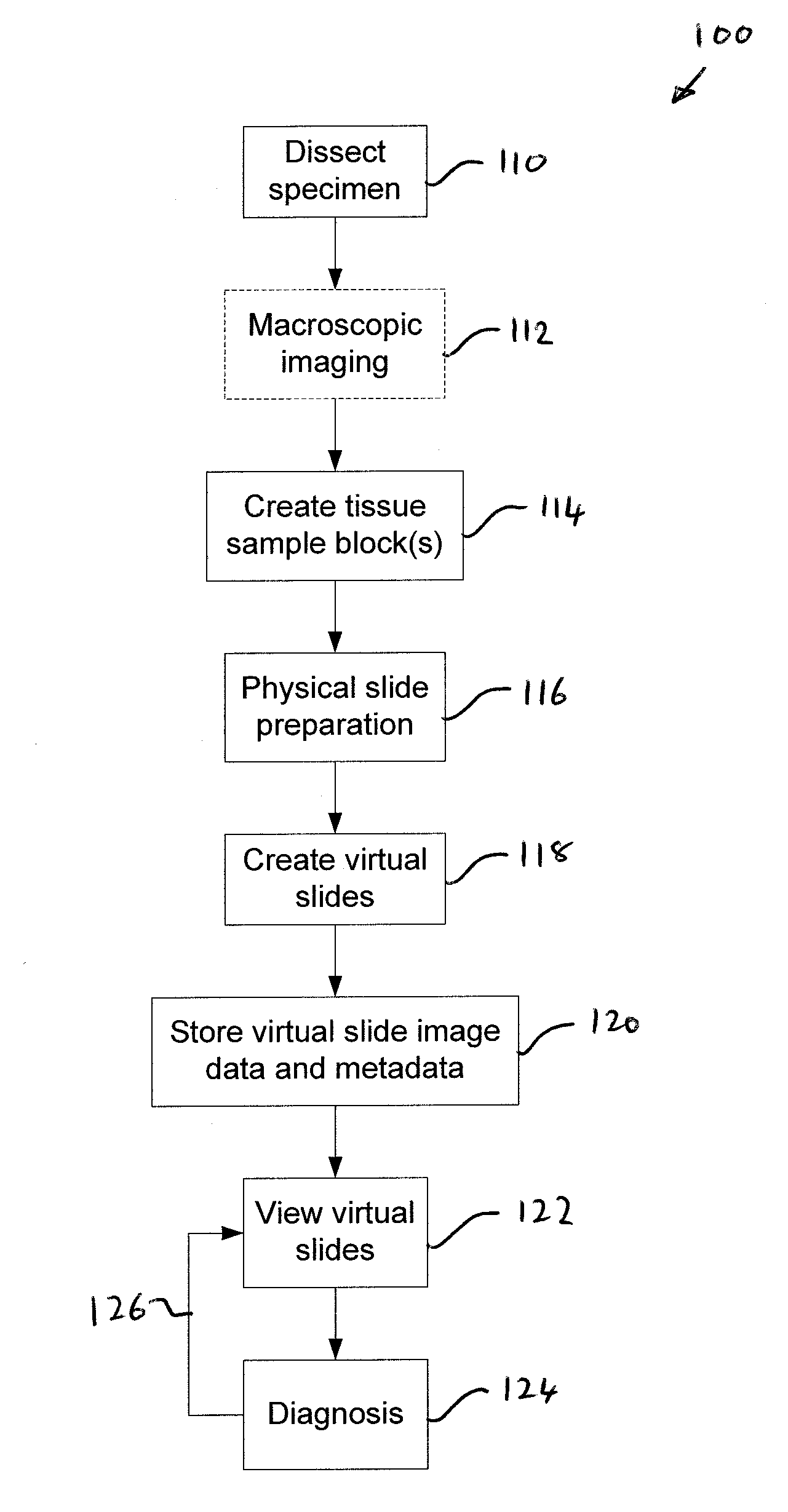
[0059]With reference to FIG. 1 there is shown a flow chart illustrating a pathology method 100, at a high level, in which the viewer of the invention may be used. It will be appreciated that the invention is not limited to use in such pathology applications. Rather, the viewer can be used in all virtual microscopy applications and indeed in all applications in which it is useful to be able to view a large number of related images in an efficient manner.

[0060]Although the invention will be described within the context of digital pathology, the invention is not limited to that field of application. Also, the embodiment will be described when applied to a single specimen. However, it will be appreciated that in some embodiments it may be useful to view slides having tissue from different specimens. Those different specimens may be from the same patient or from different patients. For example, a pathologist may want to look at different specimens from the same patient or look at the sam...


PUM


Abstract

Computer implemented methods and data processing apparatus are described for displaying virtual slide images. Images of a plurality of slides are automatically displayed in a first region of a display device at a first magnification. The slides comprise all the slides including material from a same specimen. An image of at least one of the slides is displayed in a second region at a second magnification greater than the first magnification. At least the image displayed in the second region is changed responsive to receiving user input. Methods for automatically determining a slide layout pattern and methods for virtually melding glass slide images into a single image are also described.

Description

CROSS REFERENCE TO RELATED APPLICATIONS[0001]This application claims benefit of U.S. Provisional Application No. 61 / 497,862, filed Jun. 16, 2011, which is hereby incorporated herein by reference.BACKGROUND OF THE INVENTION[0002]A number of virtual microscopy software packages and hardware systems, and parts thereof, are already known. For example Aperio Technologies, Inc. provide digital slide scanners, a viewing software application and information management software. Similarly digital pathology systems are provided by Omnyx LLC, Leica Microsystems and Koninklijke Philips Electronics N.V.[0003]While virtual microscopy is already established and virtual slide image viewing software is already available, such viewing software is not necessarily tailored to improve the efficiency with which a user may view slide images and carry out their work, for example, a pathologist making a diagnosis.[0004]Also, virtual slide images can be captured by scanners at very high resolution and so the...

Claims


Application Information

Patent Timeline
Patent Type & Authority Applications(United States)
IPC IPC(8): G09G5/373G16H30/20G16H30/40
CPCG02B21/367G06T2207/10056G06T2207/20092G06T2207/30024G06T7/30G16H30/40G16H30/20G06V20/695
Inventor RUDDLE, ROYTREANOR, DARREN
Owner VENTANA MEDICAL SYST INC
Why Eureka
Try Eureka