Use of Cytidine Deaminase-Related Agents to Promote Demethylation and Cell Reprogramming

Inactive Publication Date: 2013-01-10
THE BOARD OF TRUSTEES OF THE LELAND STANFORD JUNIOR UNIV
2 Cites 1 Cited by

AI-Extracted Technical Summary

Problems solved by technology

However, these processes are slow (2-3 weeks) and asynchronous, ...

Method used

[0057]Methods, compositions and kits for modulating the amount of methylation in a mammalian cell are provided. In one aspect of the invention, the amount of genomic DNA methylation in a mammalian cell is decreased by contacting a cell with one or more agents that promote cytidine deaminase activity. As discussed above, cytidine deaminase (CD) activity is an enzymatic activity in which amino groups are removed from cytosines or 5-methyl cytosines in DNA or RNA. Examples of agents that promote cytidine deaminase activity that find use in the present application are polypeptides and fragments of the AID/APOBEC class of cytidine deaminases and nucleic acids that encode these polypeptides and fragments.
[0064]To promote transport of CD activity polypeptides across the cell membrane, CD activity polypeptide sequences may be fused to a polypeptide permeant domain. A number of permeant domains are known in the art and may be used in the nuclear acting, non-integrating polypeptides of the present invention, including peptides, peptidomimetics, and non-peptide carriers. For example, a permeant peptide may be derived from the third alpha helix of Drosophila melanogaster transcription factor Antennapaedia, referred to as penetratin. As another example, the permeant peptide comprises the HIV-1 tat basic region amino acid sequence, which may include, for example, amino acids 49-57 of naturally-occurring tat protein. Other permeant domains include poly-arginine motifs, for example, the region of amino acids 34-56 of HIV-1 rev protein, nona-arginine, octa-arginine, and the like. (See, for example, Futaki et al. (2003) Curr Protein Pept Sci. 2003 April; 4(2): 87-96; and Wender et al. (2000) Proc. Natl. Acad. Sci. U.S.A 2000 Nov. 21; 97(24):13003-8; published U.S. Patent applications 20030220334; 20030083256; 20030032593; and 20030022831, herein specifically incorporated by reference for the teachings of translocation peptides and peptoids). The nona-arginine (R9) sequence is one of the more efficient PTDs that have been characterized (Wender et al. 2000; Uemura et al. 2002).
[0089]To promote reprogramming of demethylation-permissive cells into other types of cells, an additional step of contacting the demethylation-permissive cell with one or more agents that promote cell reprogramming may be performed. This step may be executed prior to contacting the demethylation-permissive cells with the agent that promotes CD activity, concurrently with contacting the demethylation-permissive cells with the agent that promotes CD activity, or subsequent to contacting the demethylation-permissive cells with the agent that promotes CD activity. The agents that promote cell reprogramming may be polypeptides, nucleic acid agents, or small molecule agents. Examples of agents that may be provided in this step include, but are not limited to, GSK-3 inhibitors, e.g. CHIR99021 and the like (Li, W. et al. (2009) Stem Cells, Epub Oct. 16 2009); HDAC inhibitors, e.g. Valproic Acid and the like (Huangfu, D. (2008) Nature Biotechnol 26(7):795-797; and as described in US20090191159, the disclosure of which is incorporated herein by reference); histone methyltransferase inhibitors, e.g. G9a histone methyltransferase inhibitors, e.g. BIX-01294, and the like (Shi, Y et al. (2008) Cell Stem Cel 3(5):568-574); agonists of the dihydropyridine receptor, e.g. BayK8644, and the like (Shi, Y et al. (2008) Cell Stem Cell 3(5):568-574); and inhibitors of TGFβ signaling, e.g. RepSox and the like (Ichida, J K. et al. (2009) Cell Stem Cell 5(5):491-503). Other examples of agents that may be provided in this step include reprogramming factors. As discussed above, reprogramming factors are biologically active factors that act on a cell to alter transcription, thereby reprogramming a cell to a new cell fate.
[0093]Neural stems derived from the pluripotent cells may be differentiated into neurons, oligodendrocytes, or astrocytes. Often, the conditions used to generate neural stem cells can also be used to generate neurons, oligodendrocytes, or astrocytes. For example, to promote differentiation into dopaminergic neurons, pluripotent cells or the neural stem cells derived therefrom may be co-cultured with a PA6 mouse stromal cell line under serum-free conditions, see, e.g., Kawasaki et al., (2000) Neuron, 28(1):3140. Other methods have also been described, see, e.g., Pomp et al., (2005), Stem Cells 23(7):923-30; U.S. Pat. No. 6,395,546, e.g., Lee et al., (2000), Nature Biotechnol., 18:675-679. Differentiation of the pluripotent cells or the neural stem cells derived therefrom into oligodendrocytes may be promoted by, e.g. co-culturing pluripotent cells or neural stem cells with stromal cells, see, e.g., Hermann et al. (2004), J Cell Sci. 117(Pt 19):4411-22, or by culturing the pluripotent cells or neural stem cells in the presence of a fusion protein, in which the Interleukin (IL)-6 receptor (GenBank Accession Nos. NM—000565.2 and NP—000556.1), or a derivative thereof, is linked to the IL-6 cytokine (GenBank Accession Nos. NM—000600.3 and NP—000591.1), or derivative thereof. Oligodendrocytes can also be generated from the pluripotent cells by other methods known in the art, see, e.g. Kang et al., (2007) Stem Cells 25, 419-424. Astrocytes may also be produced from the pluripotent cells or the neural stem cells derived therefrom by, e.g. culturing pluripotent cells or neural stem cells in the presence of neurogenic medium with bFGF and EGF (epidermal growth factor; GenBank Accession Nos. NM—001963.3 and NP—001954.2), see e.g., Brustle et al., (1999), Science, 285:754-756.
[0100]After contacting the demethylation-permissive cells with the agent(s) that promote CD activity, the contacted cells are cultured so as to promote the outgrowth of the desired cells. Methods for culturing cells to promote the growth of iPS cells or particular types of somatic cells as described above, for isolating iPS cell clones or clones of particular types of somatic cells as described above, and for culturing cells of those cell clones so as to promote the outgrowth of iPS cells or of particular types of somatic cells as described above are well known in the art, any of which may be used in the present invention to grow, isolate and reculture the desired cells from the reprogrammed demethylation-permissive cells.
[0101]Decreasing the amount of genomic DNA methylation in cells of a demethylation-permissive cell culture by contacting the cells with agent(s) that promote CD activity increases the efficiency of reprogramming those demethylation-permissive cells to the desired cell type relative to the efficiency observed in the absence of the agents that promote CD activity. In other words, somatic cells and cell cultures demonstrate an enhanced ability to give rise to the desired type of cell when contacted with one or more agents that promote CD activity in the presence of factors known in the art to promote reprogramming relative to cells that were not contacted with the one or more agents that promote CD activity. By enhanced, it is meant that the somatic cell cultures have the ability to give rise to the desired cell type that is at least about 50%, about 100%, about 200%, about 300%, about 400%, about 600%, about 1000%, at least about 2000% of the ability of the population of cells that were not contacted with the agent that promotes CD activity. In other words, the culture of demethylation-permissive cells produces about 1.5 fold, about 2-fold, about 3-fold, about 4-fold, about 6-fold, about 10-fold, about 20-fold, about 30-fold, about 50-fold, about 100-fold, about 200-fold the number of cells of the desired cell type that are produced by a population of demethylation-permissive cells that are not contacted with the one or more agents that promote CD activity. The efficiency of reprogramming may be determined by assaying the amount of methylation at promoters known in the art to become demethylated upon the acquisition of the desired cell type. In such cases, an enhanced efficiency of reprogramming due to the presence of an agent that promotes CD activity is observed when the amount of methylation at those promoters is about 1.5 fold, about 2-fold, about 3-fold, about 4-fold, about 6-fold, about 10-fold less than the amount of methylation observed in the absence of the agent that promotes CD activity. Alternatively or additionally, the efficiency of reprogramming may be determined by assaying the level of expression of gene known in the art to become more highly expressed upon the acquisition of the desired cell type. In such cases, an enhanced efficiency of reprogramming due to the presence of an agent that promotes CD activity is observed when the level of expression of these genes is about 1.5 fold, about 2-fold, about 3-fold, about 4-fold, about 6-fold, about 10-fold greater than the level of expression observed in the absence of the agent that promotes CD activity.
[0120]For some conditions, particularly central nervous system conditions, it may be necessary to formulate agents to cross the blood brain barrier (BBB). One strategy for drug delivery through the blood brain barrier (BBB) entails disruption of the BBB, either by osmotic means such as mannitol or leukotrienes, or biochemically by the use of vasoactive substances such as bradykinin. The potential for using BBB opening to target specific agents to brain tumors is also an option. A BBB disrupting agent can be co-administered with the therapeutic compositions of the invention when the compositions are administered by intravascular injection. Other strategies to go through the BBB may entail the use of endogenous transport systems, including caveoil-1 mediated transcytosis, carrier-mediated transporters such as glucose and amino acid carriers, receptor-mediated transcytosis for insulin or transferrin, and active efflux transporters such as p-glycoprotein. Active transport moieties may also be conjugated to the therapeutic compounds for use in the invention to facilitate transport across the endothelial wall of the blood vessel. Alternatively, d...

Benefits of technology

[0034]FIG. 17. Over-expression of human AID does not accelerate the onset of reprogramming in heterokaryons. Bisulfite sequencing analysis of methylation status of the human Oct4 and Nanog promoters in fibroblasts in heterokaryons on day 1 post fusion, with or without transient over-expression of human AID (hAID) (FIG. 5). a, hAID levels were assessed by real time PCR and found to be upregulated 2 and 4 fold respectively in two separate fusion experiments, in the day 1 heterokaryons. b,d, The extent of DNA demethylation of the human Oct4 and Nanog promoters does not increase upon hAID over-expression. Similar results were obtained for two independent fusion experiments. White circles indicate unmethylated and black circles indicate methylated CpG dinucleotides. At least 10 clones were analyzed in two independent fusion experiments; 10 representative clones are shown. c,e, Percent demethylation observed at the human Oct4 and Nanog promoters in heterokaryons on day 1 post fusion, with or without transient over-expression of hAID. DNA demethylation at the Oct4 and Nanog promoters does not increase when hAID is over-expressed.
[0035]FIG. 18. Over-expression of human AID rescues the initiation of reprogramming during transient knockdown of AID in heterokaryons. Rescue experiments were performed by over-expressing...

Abstract

Methods, compositions and kits for modulating demethylation in a mammalian cell are provided. Also provided are methods, compositions and kits for screening candidate agents for activity in modulating genomic DNA demethylation in mammalian cells. These methods, compositions and kits find use in producing induced pluripotent stem cells (iPS) and somatic cells in vitro and for treating human disorders including cancer and disorders arising from defects in genomic imprinting.

Application Domain

Peptide/protein ingredientsGenetic material ingredients +15

Technology Topic

Human Induced Pluripotent Stem Cellsgenomic DNA +14

Image

  • Use of Cytidine Deaminase-Related Agents to Promote Demethylation and Cell Reprogramming
  • Use of Cytidine Deaminase-Related Agents to Promote Demethylation and Cell Reprogramming
  • Use of Cytidine Deaminase-Related Agents to Promote Demethylation and Cell Reprogramming

Examples

  • Experimental program(2)

Example

Example 1
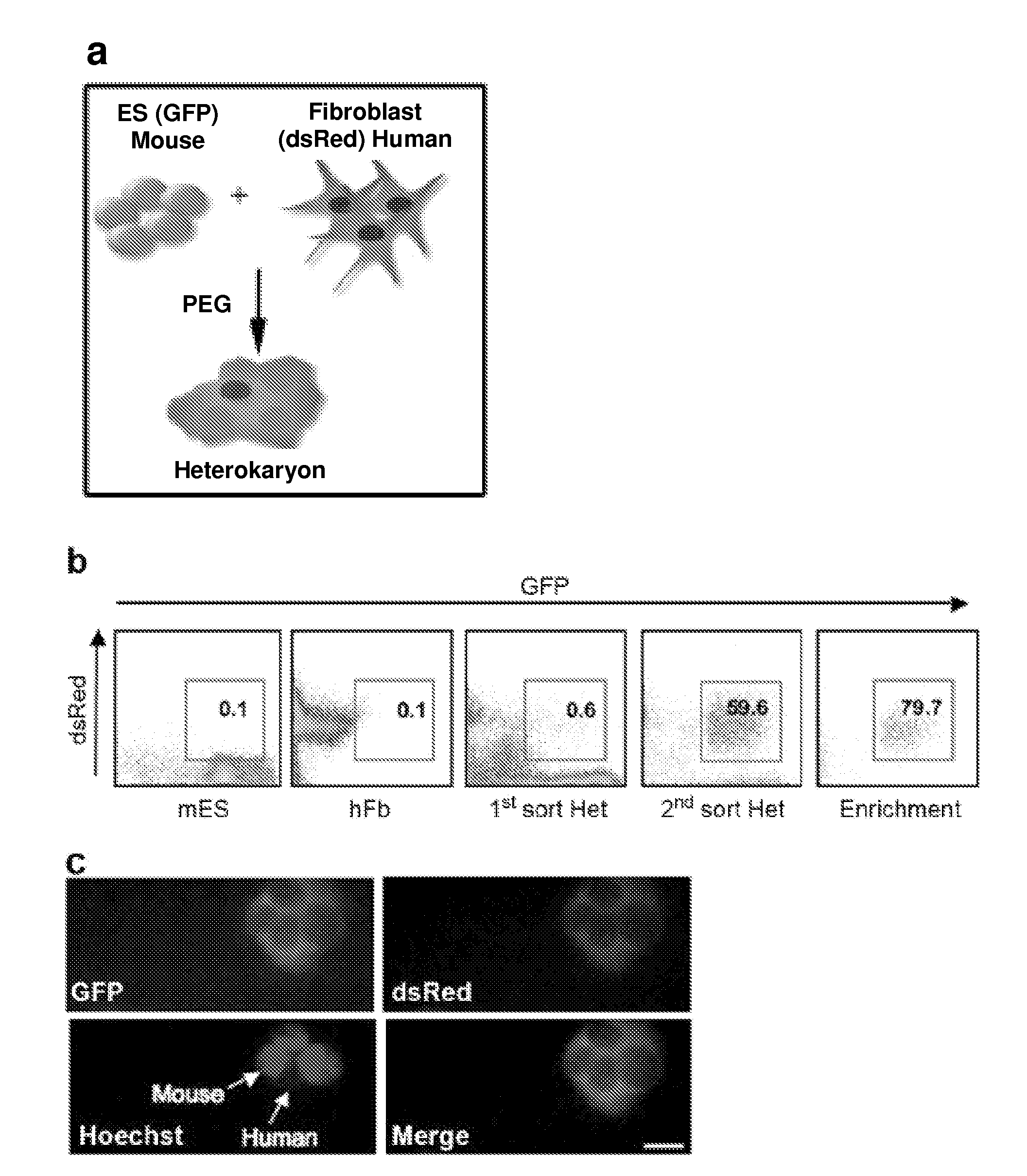
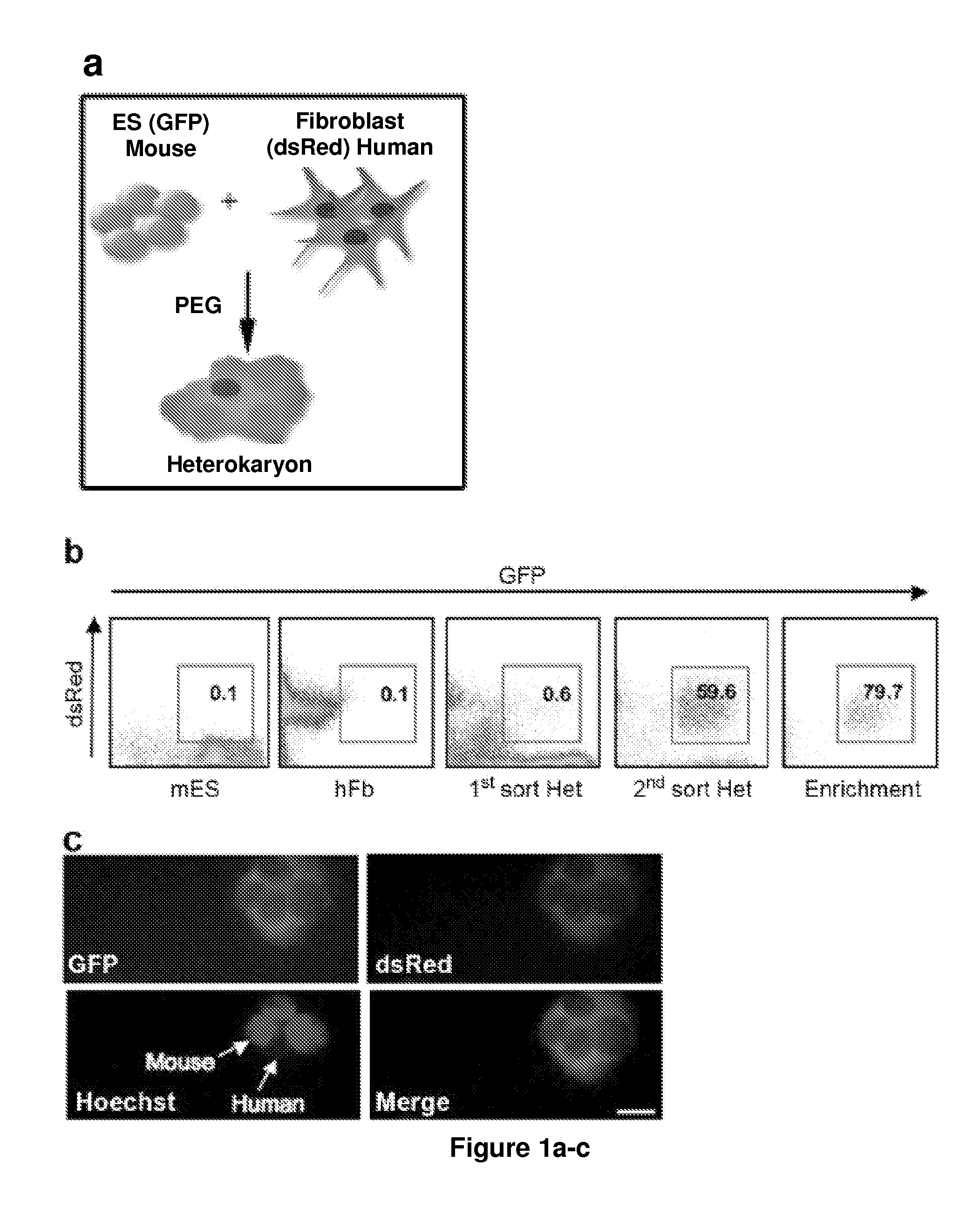
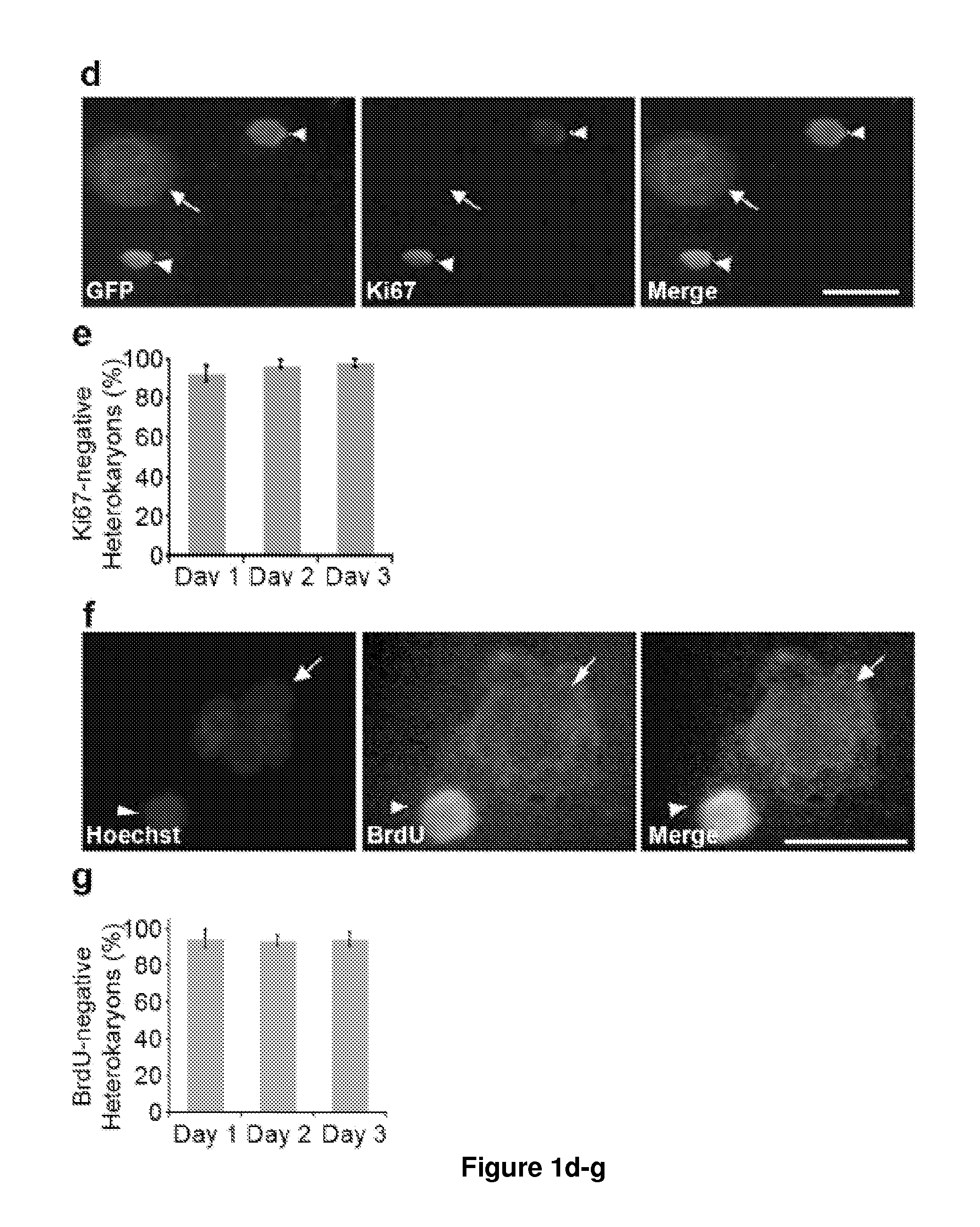
[0163]To identify novel early regulators essential to nuclear reprogramming towards pluripotency, we capitalized on our previous experience with heterokaryons that proved useful in elucidating the principles inherent to the maintenance of the differentiated state of somatic cells. Specifically, these earlier studies by us and others showed that the “terminally differentiated” state of human cells was not fixed, but could be altered and the expression of previously silent genes typical of other differentiated states induced (Blau, H. M., et al. (1983) Cell 32, 1171-801; Baron, M. H. & Maniatis, T. (1986) Cell 46, 591-602; Wright, W. E. (1984) Exp Cell Res 151, 55-69; Spear, B. T. & Tilghman, S. M. (1990) Mol Cell Biot 10, 5047-54; Chiu, C. P. & Blau, H. M. R (1984) Cell 37, 879-87). We reasoned that heterokaryons could be used to elucidate mechanisms and identify novel genes with a role at the onset of reprogramming towards pluripotency because: (1) reprogramming takes place in the presence of all ES cell factors, (2) the onset of reprogramming is synchronously initiated upon fusion, (3) reprogramming is assessed in fused, non-dividing cells, and (4) species differences distinguish the transcripts of the fused cell types.
Materials and Methods
[0164]Heterokaryon Generation and Isolation by Flow Cytometry.
[0165]GFP+ murine ES cells and DsRed+ human fetal lung primary fibroblasts were generated by transduction with retroviral constructs as previously described (Palermo, A. et al. (2009) Faseb J), and fused to form non-dividing, multinucleated heterokaryons. Cells were first co-cultured for 12 h in ES media and then treated with PEG 1500 (Roche) for 2 min at 37° C., followed by four successive washes with DMEM. ES media was replaced after washing and every 12 h thereafter. GFP+/DsRed+ heterokaryons were sorted twice by flow-cytometry (FACSVantage SE, BD) and analyzed for gene expression and methylation.
[0166]Immunofluorescence.
[0167]Heterokaryons were sorted twice in PBS with 2.5% v/v goat serum and 1 mM EDTA, and cytospun at 900 rpm for 5 min. The cytospun GFP+/DsRed+ heterokaryons were stained with Hoechst 33342, and imaged. For antibody staining, cytospun cells were fixed, permeabilized and blocked using 20% FBS in PBS. Cells were incubated with the primary antibody mouse anti-Ki-67 (Dako Denmark A/S) at 1:100 dilution in blocking buffer for 1 h, rinsed 3 times in PBS, and then incubated with a goat anti-mouse Cascade blue secondary antibody (Millipore) at 1:500 dilution for 30 min, rinsed 3 times and mounted with Fluoromount-G and imaged. Images were acquired using an epifluorescent microscope (Axioplan2; Carl Zeiss Microlmaging, Inc.), Fluar 20×/0.75 or 40×/0.90 objective lens, and a digital camera (ORCA-ER C4742-95; Hamamatsu Photonics). The software used for acquisition was OpenLab 4.0.2 (Improvision).
[0168]BrdU was added to mES and hFb co-cultures 3 hours after PEG-induced fusion. Labeling and antibody staining was performed using the BrdU Labeling and Detection Kit I (Roche).
[0169]Analysis of Gene Expression.
[0170]RNA was prepared from ES cells, fibroblasts and twice-sorted heterokaryons at different times post fusion or after siRNA treatment using the RNeasy micro kit (Qiagen). Total RNA for each sample was reverse transcribed using the Superscript First-Strand Synthesis System for RT-PCR (Invitrogen). The reverse transcribed material was subjected to PCR using Go GreenTaq DNA polymerase (Promega). Human specific primers were designed for analyzing the expression of Oct4, Nanog and GAPDH. Primers used for AID and GAPDH in the siRNA treatment experiments amplify both human and mouse transcripts to assess the total levels of AID and GAPDH in heterokaryons. Human-specific primers used for RT-PCR and quantitative PCR are: hOct4 F 5′-TCGAGAACCGAGTGAGAGGC-3′ (SEQ ID NO:45), R-5′-CACACTCGGACCACATCCTTC-3′ (SEQ ID NO:46); hNanog F 5′-CCAACATCCTGAACCTCAGCTAC-3′ (SEQ ID NO:47), R 5′-GCCTTCTGCGTCACACCATT-3′ (SEQ ID NO:48); hGAPDH F 5′-TGTCCCCACTGCCAACGTGTCA-3′ (SEQ ID NO:49), R 5′-AGCGTCAAAGGTGGAGGAGTGGGT-3′ (SEQ ID NO:50). Non-species specific primer sequences for assessing knockdown after siRNA treatment are as follows: GAPDH F 5′-ACCACAGTCCATGCCATCAC-3′ (SEQ ID NO:51), R 5′-TCCACCACCCTGTTGCTGTA-3′ (SEQ ID NO:52); AID F 5′-AAAATGTCCGCTGGGCTAAG-3′ (SEQ ID NO:53), R 5′-AGGTCCCAGTCCGAGATGTAG-3′ (SEQ ID NO:54).
[0171]Real Time PCR.
[0172]Real time PCR was performed using an ABI 7900HT Real time PCR system using the Sybr Green PCR mix (Applied Biosystems). Samples were cycled at 94° C. for 2 min, 40× (94° C. for 20 s, 58° C. for 45 s).
TABLE 1 Human-specific primers used for real time PCR Gene primer SEQ ID NO: Essrb 5′ GCCAGCGCCATGAGGAGC 55 Essrb 3′ GTATCCAGCCTGAGCAGTGC 56 TDGF1 5′ ATTGCCATTTTCGCTTTAGG 57 TDGF1 3′ ACACGCTGGGAAGACCGAGGC 58 Sox2 5′ CGACACCCCCGCCCGCCT 59 Sox2 3′ ACACCATGAAGGCATTCATGGGCC 60 Klf4 5′ ACCCCGACCCTGGGTCTT 61 Klf4 3′ GCCACTGACTCCGGAGGA 62 c-myc 5′ AAGGGAGATCCGGAGCGAATA 63 c-myc 3′ GGAGGCTGCTGGTTTTCCACT 64
[0173]Single cell RT-PCR.
[0174]Single heterokaryons were directly sorted by FACS (FACSVantage SE, BD) into PCR tubes containing 9-μl aliquots of RT-PCR lysis buffer. The buffer components included commercial RT-PCR buffer (SuperScript One-Step RT-PCR Kit Reaction Buffer, Invitrogen), RNase inhibitor (Protector RNase Inhibitor, Roche) and 0.15% IGEPAL detergent (Sigma). After a short pulse-spin, the PCR-tubes were immediately shock-frozen and stored at −80° C. for subsequent analysis.
[0175]For two-step multiplex nested single cell RT-PCR, cell lysates were first reverse-transcribed using the human and gene-specific primer pairs for Oct4, Nanog and GAPDH (Table 2, External primers; FIG. 5b) using SuperScript One-Step RT-PCR Kit (Invitrogen). Briefly, the RT-PCR was performed in the same PCR cell-lysis tubes by addition of an RT-PCR-reaction mix containing the genespecific primer pairs and RNase inhibitor. Genomic products were excluded by designing and using intron-spanning primer sets for the first and second round PCR and nested RT-PCR ensured greater specificity. In the first step, the reverse transcription reactions were carried out at 55° C. for 30 min, and followed by a 2-min step at 94° C. Subsequently, 30 cycles of PCR amplification were performed as follows: 94° C. for 30 s; 58° C. for 30 s; 68° C. for 30 s. In the final PCR step, the reactions were incubated for 3 min at 68° C. The completed reactions were stored at 4° C.
[0176]In the second step of the PCR protocol, the completed RT-PCR reaction from the first step was diluted 1:1 with water. One percent of these reactions were replica transferred into new reaction tubes for the second round of PCR, which was performed for each of the genes separately using nested gene-specific internal-primers, for greater specificity, in a total reaction volume of 20 μl (Platinum Taq Super-Mix HF, Invitrogen). Thirty cycles of PCR amplification were performed as follows: 94° C. for 30 s; 58° C. for 30 s; 68° C. for 30 s. In the final PCR step, the reactions were incubated for 3 min at 68° C. The completed reactions were stored at 4° C. The second-round PCR products were then subjected to gel electrophoresis using one fifth of the reaction volumes and 1.4% agarose gels.
TABLE 2 Primer sequences utilized for single cell nested PCR in heterokaryons Nested primer set SEQ ID SEQ ID External Primer [5′-3′] NO: Internal Primer [5′-3′] NO: Oct4 5′ GAAGGAGAAGCTGGAGCAAAAC 65 GAGAGGCAACCTGGAGAATT 66 Oct4 3′ CAAAAACCCTGGCACAAACT 67 CCAGAGGAAAGGACACTGGT 68 Nanog 5′ TGATTTGTGGGCCTGAAG 69 GATGCCTGGTGAACCCGA 70 Nanog 3′ AACCAGAACACGTGGTTTCC 71 TGCACCAGGTCTGAGTGTTC 72 GAPDH 5′ GCTCAGACACCATGGGGAAG 73 CCATGAGAAGTATGACAACAGC 74 GAPDH 3′ CCATGAGAAGTATGACAACAGC 75 TTCTAGACGGCAGGTCAGG 76
[0177]DNA Methylation Analyses.
[0178]FACS-sorted heterokaryons (2,000-10,000 cells) were collected in 20 uL PBS. DNA was extracted using the DNeasy Tissue Kit (Qiagen). Bisulfite treatment was performed using the Epitect Bisulfite Kit (Qiagen). Nested PCR for regions of the human Oct4 and Nanog promoters was performed using human and bisulfite specific primers (Table 3). Samples were cycled for the first and nested PCR at 94° C. for 2 min, 30× (94° C. for 20 s, 68° C. for 30 s, 68° C. for 30 s). PCR products from second-round bisulfite-specific PCR amplification were cloned and sequenced as described before (Zhang, F., et al. (2007) Proc Natl Acad Sci USA 104, 4395-400).
TABLE 3 Human and bisulfite specific primers for DNA methylation analyses SEQ ID SEQ ID External Primer [5′-3′] NO: Internal Primer [5′-3′] NO: Oct4 5′ GAGGAGTTGAGAGGGTGATTGG 77 GGAGAGGGGGTTAAGTATTTGG 78 TTTT GTTTT Oct4 3′ CGAAAAAACTACTCAACCCCT 79 TCCACTTTATTACCCAAACTAA 80 Nanog 5′ GGAAAATGGAGTTAGTTGAAATT 81 GGAATTTAAGGTGTATGTATTTT 82 TTTGTTT Nanog 3′ CCACCCCTATAATCCCAATAAAT 83 AACCAACCTAACCAACATAA 84 TAAAA B globin 5′ TGATTAAATAAGTTTTAGTTTTTT 85 CCATGAGAAGTATGACAACAGC 86 TTTAGTTTT B globin 3′ TAAGTATGAGTAGTTTTGGTTAG 87 TTCCATATCCTTATTTCATATTA 88 GTTT ATACATA
[0179]siRNA Transfection.
[0180]For siRNA transfection, ES cells and primary fibroblasts were plated at 50-60% confluence the day before transfection. siRNAs (Dharmacon) were transfected using silmporter (Millipore).
[0181]Chromatin Immunoprecipitation.
[0182]Chromatin immunoprecipitation was performed as previously described by Dahl and Collas ((2008) Nat Protoc 3, 1032-45) using primers provided in Table 4. ChIP data was presented as normalized to input DNA and the error bars represent standard error mean (sem).
TABLE 4 Primers used for ChIP experiments primer SEQ ID NO: Human primers [5′-3′] Thy1.1 5′ TCCCACAGACTCCTGAAGAATA 89 Thy1.1 3′ TTGTTCCCCTTTTAAGGCTTT 90 Nanog 5′ GAGTACAGTGGCGCGATATCG 91 Nanog 3′ CGGGAGAATCCCTTGAACCT 92 Oct4 5′ GTGGCTCACGCCTTTAATCA 93 Oct4 3′ CCAGGCTGGTCTTGAATTCC 94 Cμ 5′ ACCCCAATGCCACTTTCA 95 Cμ 3′ AGTCATCCTCGCAGATGCT 96 Mouse primers [5′-3′] Cdx2 5′ AGGTTAAAGTGCACCCAGGTT 97 Cdx2 3′ CAGGCCCTTCTTGCTAGCT 98 Nanog 5′ AACGCTGAGTGCTGAAAGGA 99 Nanog 3′ GTCAGACCTTGCTGCCAAAG 100 Oct4 5′ GGGTGGGTAAGCAAGAACT 101 Oct4 3′ AATGTTCGTGTGCCAATTA 102 p53 5′ ACGGCAGCTTGCACCTCTA 103 p53 3′ CTTTCTAGCAACCCGTTTGC 104
[0183]Statistical Analysis.
[0184]Data are presented as the mean±s.e.m. Comparisons between groups used the Student's t test assuming two-tailed distributions.
[0185]Thy1.1 (CD90) Enrichment of Heterokaryons.
[0186]GFP− (non-GFP) mES and DsRed+ hFb co-cultures treated with PEG were trypsinized and resuspended in 3 mL FACS buffer. Cells were incubated for 30 min at room temperature with biotin mouse anti-human CD90 (BD Pharmingen) at a dilution of 1:5000. The cells were washed once, resuspended in 3 mL FACS buffer incubated for 30 min at room temperature with 10 uL of Dynabeads Biotin Binder (Invitrogen). Beads were removed by magnetic isolation, washed twice and the enriched heterokaryons were cytospun.
[0187]Immunoprecipitation and Western Blots.
[0188]Mouse ES cells were lysed in IP buffer (20 mM Tris pH 7.5, 1 mM DTT, 0.5 mM EDTA, 350 mM NaCl, 10% (vol/vol) glycerol, 10 uM ZnCl. Whole cell lysates were pre-cleared for 30 min at room temperature followed by AID pull down using. Briefly, cell lysates and then AID was pulled down using Protein A Plus Agarose beads (Pierce) cross-linked to a rabbit polyclonal AID antibody. Immunoprecipitation was performed from 2 mg of cell lysates.
[0189]To visualize AID protein knockdown in mES, cell lysates were harvested 3 days posttransfection with siControl or si-1. Detection of AID in these samples was performed from 170 ug of whole cell lysate using anti mouse-AID (L7E7, Cell Signaling, dilution 1:500). The membrane was stripped and probed with ant-mouse α-tubulin (Sigma, dilution 1:20,000) for the loading control. Immunoprecipitation of AID was detected using the same L7E7 antibody.
Results
[0190]To produce interspecies heterokaryons, mouse embryonic stem cells (mES) transduced with a GFP reporter gene were co-cultured with primary human fibroblasts (hFb) transduced with a DsRed reporter gene, and fused using polyethylene glycol (PEG) (FIG. 1a; Scheme in FIG. 5). Fused GFP+DsRed+ heterokaryons, which were readily sorted by FACS (FIG. 1b) and identified using fluorescence microscopy, contained distinctly stained human and mouse nuclei when visualized with Hoechst 33342 or Hoechst 33258 (FIGS. 1c and 1f, respectively). Since the efficiency of PEG fusion is low (0.6 to 1.0%), GFP+DsRed+ heterokaryons were sorted twice and enriched to 80% purity (FIG. 1b). Using an antibody for Ki-67, a nuclear protein present only in proliferating cells, we determined that cell division did not occur in 98(±2) % of heterokaryons over the three day time period assayed post fusion (FIG. 1d,e). In addition, BrdU labeling was not detected in 94(±4) % of heterokaryons over the same time period, indicating that DNA replication did not occur (FIG. 1f,g; FIG. 6; FIG. 7). To favor reprogramming towards a pluripotent state, we skewed the ratio of the input cells so that ES cells outnumbered the fibroblasts (2:1), as gene dosage and the proportion of proteins contributed by each cell type determines the direction of nuclear reprogramming in somatic cells
[0191]To determine if ES cell-specific genes were induced in the human fibroblasts, the induction of human Oct4 and Nanog were assayed relative to ubiquitous GAPDH using species-specific primers (FIG. 8). mRNA isolated from sorted heterokaryons 1, 2 and 3 days post fusion was assessed by semi-quantitative RT-PCR and real time PCR (FIG. 2a,b). The day 0 controls used were either (a) human fibroblasts alone; (b) pre-PEG, unfused co-cultures of mES and hFb; or (c) human fibroblasts treated with PEG to control for the effects of PEG and fusion. All of the above day 0 controls gave similar results. Induction of both human Oct4 and Nanog transcripts was evident as early as day 1 post fusion in heterokaryons (FIG. 2a,b), but not in controls (FIG. 9), indicating that the onset of expression of two key human pluripotency genes is rapid in heterokaryons. By day 1, expression of human Oct4 and Nanog (normalized to GAPDH) in the same samples, had increased 5-fold relative to the unfused co-culture control (day 0) and persisted at 10-fold on days 2 and 3 (FIG. 2b). Human-specific primers were used to determine if other key pluripotency genes in addition to Oct4 and Nanog were induced using real time PCR. Essrb (Bhattacharya, B. et al. (2004) Blood 103, 2956-64) and TDGF1 (Bhattacharya, B. et al. (2004) Blood 103, 2956-64) (Cripto), which have been shown to be essential for maintaining ES cell self-renewal and are targets of Oct4 and Nanog were found to be upregulated 3-fold and 2.5-fold, respectively, in heterokaryons on day 2 post fusion (FIG. 10). Sox2 is already expressed in human fibroblasts and its promoter is extensively demethylated pre-fusion, in agreement with findings in mouse fibroblasts; its expression did not increase post fusion. Expression of Klf4 (Feng, B. et al. (2009) Nat Cell Biol 11, 197-203), which is functionally interchangeable with Essrb, did not change in heterokaryons at day 2 post fusion (FIG. 10).
[0192]To assess the efficiency of nuclear reprogramming in human fibroblasts following fusion, single FACS-sorted heterokaryons were analyzed by nested RT-PCR for the three human transcripts, Oct4, Nanog, and GAPDH (control), using two sets of human-specific primers in each case (FIG. 2c). No human gene products were detected in mouse ES cells (control) and only human GAPDH was detected in human fibroblasts (control) (FIG. 8). In contrast, 70% of single FACS-sorted heterokaryons from three independent fusion experiments on day 3 post fusion expressed both human Oct4 and Nanog (FIG. 2c,d; FIG. 11), showing that a high proportion of heterokaryons initiated reprogramming towards pluripotency. This is in marked contrast to the slow and inefficient induction of Oct4 and Nanog expression in iPS cells (<0.1%) of the total population in 2 to 3 weeks as observed in, for example, Takahashi, K. et al. (2007) Cell 131, 861-72; Takahashi, K. & Yamanaka, S. (2006) Cell 126, 663-76; Wernig, M. et al. (2007) Nature 448, 318-24; and Wernig, M. et al. (2008) Nat. Biotechnol.
[0193]Since DNA demethylation has been shown to be a major limiting step in reprogramming fibroblasts towards iPS cells, the time course and extent of demethylation of the human Oct4 and Nanog promoters in heterokaryons was analyzed relative to control. DNA was isolated from heterokaryons on days 1, 2 and 3 post-fusion and subjected to bisulfite conversion. Human Oct4 and Nanog promoters were amplified by PCR using human- and bisulfite-specific primers (Table 3, FIG. 8), and the products cloned and sequenced. DNA demethylation was evident at the human Oct4 and Nanog promoters and progressively increased through day 3 (FIG. 3a). By contrast, the β-globin HS2 locus remained methylated throughout, indicating that the DNA demethylation was specific. The time-course and progressive accumulation of demethylated CpG sites in the human Oct4 and Nanog promoters (FIG. 3b,c) parallels the progressive increase in transcript accumulation observed over the same three day time period using real time PCR (FIG. 2b). Notably, promoter demethylation and activation of pluripotency genes in human somatic cells takes place in the absence of Ki-67 or BrdU labeling (FIG. 1e,g); thus demethylation is active and independent of cell division and DNA replication.
[0194]Because is detected in mammalian pluripotent germ cells (Morgan, H. D., et al. (2004) Biol Chem 279, 52353-60) and implicated in active DNA demethylation in zebrafish post fertilization (Rai, K. et al. (2008) Cell 135, 1201-12), mouse ES cells and human fibroblasts were assayed for AID expression using real time PCR. Although AID expression in somatic cells is generally thought to be restricted to B lymphocytes, AID mRNA was detected in human fibroblasts as well as mouse ES cells, albeit at greatly reduced levels (5% and 15%, respectively) compared to Ramos, a B-lymphocyte cell line (FIG. 12). To investigate the role of AID in these cells, mouse and human AID mRNA levels were transiently knocked down by transfection of three distinct, non-overlapping siRNAs to different sequences within the AID coding region, and a fourth siRNA specific to the non-coding 3′UTR of AID, in order to rule out off-target effects and ensure that the results were specific to AID (FIG. 13). A fifth siRNA with 50% identity to the AID coding region was used as a control (siControl). The extent and timing of knockdown was first confirmed in control mouse ES cells in which siRNA-1, 2, 3 and 4 reduced AID transcripts by 81(±13) %, 79(±12) %, 70(±8) %, and 99(±0.1) %, respectively, at day 3 post-transfection as compared to the control siRNA (FIG. 14, top). AID protein was detected in mouse ES cells using immunoprecipitation followed by Western blot as well as in concentrated whole ES cell lysates (FIG. 15). AID knockdown by siRNA 1 was verified in ES cell lysates, and the reduction by 88% of the control protein levels correlated well with the mRNA reduction by 81% (FIG. 15). In human fibroblasts, AID transcripts were reduced by 46(±11)%, 72 (±23) %, 99(±0.1) % and 99(±0.1) % by siRNA 1, 2, 3 and 4, respectively (FIG. 14, bottom). These data show that AID is present and can be efficiently reduced by four distinct siRNAs in both ES cells and fibroblasts.
[0195]To assess the initiation of reprogramming in heterokaryons subjected to AID knockdown, expression of Oct4 and Nanog relative to GAPDH was assessed by real time PCR. For heterokaryon experiments, siRNAs were transfected into both the mouse ES cells and the human fibroblasts 24 hours prior to fusion (See FIG. 5 for scheme). A persistent knock-down of AID was detected by real time PCR in heterokaryons. Using siRNA 1 and 2, AID was reduced by 77(±6) % and 35(±2) % on day 3 post fusion relative to heterokaryons transfected with the control siRNA (FIG. 4a). The siRNAs 3 and 4 caused a stronger knockdown in heterokaryons with a reduction in AID by 96(±1) %, and 89(±3) % on day 2 post fusion relative to the control siRNA (FIG. 4a). Strikingly, Oct4 expression was reduced to 0.9(±0.6) % and 9(±2) % using siRNA 1 and 2 on day 3 post fusion as compared to the control siRNA (FIG. 4a). Similarly, using siRNA 1 and 2, Nanog expression was greatly reduced to 1.5(±0.4) % and 1.5(±0.1) % on day 3 post fusion relative to the control siRNA (FIG. 4a). In the presence of siRNA 3 and 4, Oct 4 expression was reduced to 8(±2) % and 4(±3) % relative to the control siRNA on day 2 post fusion while Nanog expression was reduced to 19(±12) % and 7(±4) %. All the 4 siRNAs used here had a similar effect in blocking the expression of Oct4 and Nanog by at least 80%. These observations indicate that the effect of AID is extremely dosage sensitive as 35% knockdown led to a comparable inhibition of pluripotency gene induction as a 96% knockdown. Together, these data show that all 4 siRNAs to AID used here had a similarly potent effect in blocking the Oct4 and Nanog activation by at least 80%.
[0196]To assess the effect of AID on promoter demethylation, we assayed the CpG methylation status of the human Oct4 and Nanog promoters in heterokaryons. In Day 3 heterokaryons subject to AID knockdown using siRNA 1 and siRNA 2, the extent of CpG demethylation in the human Oct4 promoter was reduced to 26% and 6%, respectively, as compared to the 82% in the control (FIG. 4b,c). For the Nanog promoter, CpG demethylation was reduced to 24% and 25%, respectively, as compared to 53% demethylation for the control (FIG. 4b,c). Using siRNA 3 and 4, the extent of CpG demethylation in the Oct4 promoter was reduced to 18% and 8%, respectively, as compared to 72% in the day 2 control sample, while for the Nanog promoter, the extent of CpG demethylation was reduced to 3% and <1%, respectively, compared to 48% in the control (FIG. 4b,c). A summary of the bisulfite sequencing data for all the siRNA knockdown experiments is shown in FIG. 16. In parallel with the reduction in demethylation of the Oct4 and Nanog promoters upon AID knockdown, the induction of Oct4 and Nanog transcripts was reduced by at least 80% on days 2 and day 3, relative to the control (FIG. 4a). These data show that promoter demethylation is critical to the expression of these two pluripotency genes and that AID is required for mammalian DNA demethylation in somatic cell reprogramming.
[0197]To further investigate the requirement of AID for initiating reprogramming, we tested its ability to rescue the DNA demethylation block caused by the siRNA knockdown in heterokaryons. hAID was transiently overexpressed in mouse ES cells prior to siRNA transfection in order to test whether the siRNA knockdown could be overcome by increasing AID levels (see scheme in FIG. 5). In two separate experiments, when hAID was over-expressed 2-fold or 4-fold relative to the control in heterokaryons in the absence of AID siRNA, there was no acceleration in promoter demethylation or reprogramming at day 1 post fusion (FIG. 17). This could possibly be due to the kinetics of human Oct4 promoter demethylation, which in heterokaryons may require at least 1 day to occur, or by the lack of additional factors that work in concert with hAID to accelerate reprogramming. However, upon overexpression of hAID in heterokaryons undergoing transient knockdown by siRNA-1, i.e., in the presence of siRNA, there was a complete rescue of Nanog promoter demethylation and gene expression and a partial rescue of Oct4 promoter demethylation and gene expression (FIG. 18). These data show that the added hAID is functional and rule out any non-specific effects of the siRNA, further confirming the specific and essential role of AID in DNA demethylation at the onset of reprogramming towards pluripotency.
[0198]To further validate the role of AID in DNA demethylation of human Oct4 and Nanog promoters, we tested whether AID specifically binds to their promoter regions by performing chromatin immunoprecipitation (ChIP) experiments using an anti-AID antibody. The promoter regions assessed in ChIP experiments were designed to be within the Oct4 and Nanog promoter regions that were analyzed for CpG demethylation by bisulfite sequencing (FIG. 4d; FIG. 19). In the human fibroblasts, the ChIP analyses showed significant binding of AID to both human Oct4 (6-fold) and human Nanog (8-fold) promoters (FIG. 4d). Thus, AID binds to the heavily methylated promoter regions of human Oct4 and Nanog in fibroblasts that undergo demethylation during reprogramming. As controls, AID binding to the promoter of the IgM constant region (Cμ) was significant, as expected (Okazaki, I. M., et al. (2002) Nature 416, 340-5), while no binding was observed for Thy1.1, which is expressed in fibroblasts.
[0199]In contrast to fibroblasts, no AID binding was observed at the promoter regions of mouse Oct4 and Nanog despite the higher levels of AID protein in ES cells, presumably because these promoters are expressed and demethylated (FIG. 4d). As controls, AID binding was detected at the promoter of Cdx2, a gene not expressed in undifferentiated ES cells, but was absent from the p53 promoter, as previously reported. Together, these findings provide strong support for a direct involvement of AID in DNA demethylation and the sustained expression of human Nanog and Oct4 leading to the onset of reprogramming towards pluripotency.
Discussion
[0200]DNA demethylation is essential to overcoming gene silencing and inducing temporally and spatially controlled expression of mammalian genes, yet no consensus mammalian DNA demethylase has been identified despite years of effort. Evidence of DNA demethylation via 5 methyl-cytosine DNA glycosylases has been shown in plants (Gong, Z. et al. (2002) Cell 111, 803-14; Choi, Y. et al. (2002) Cell 110, 33-42), but mammalian homologues such as Thymine DNA Glycosylase (TDG) or the Methyl-CpG-binding domain protein 4 (Mbd4) have not exhibited comparable functions (Cortazar, D., et al. (2007) DNA Repair (Amst) 6, 489-504; Millar, C. B. et al. (2002) Science 297, 403-5).
[0201]AID belongs to a family of cytosine deaminases (AID, Apobec 1, 2 and 3 subgroups) that have established roles in generating antibody diversity in B cells, RNA editing and antiviral response (Conticello, S. G., et al. (2007) Adv Immunol 94, 37-73). Both AID and Apobec1 are expressed in progenitor germ cells, oocytes and early embryos and have a robust 5-methyl cytosine deaminase activity in vitro (Morgan, H. D., et al. (2004) J Biol Chem 279, 52353-60), resulting in a T-G mismatch that is repaired through the Base Excision DNA Repair (BER) pathway, and could theoretically lead to DNA demethylation without replication. Recently in zebrafish embryos, AID was implicated as a member of a tri-partite protein complex along with Mbd4 and Gadd45a, effecting cytosine deamination and leading to base excision by Mbd4 (Rai, K. et al. (2008) Cell 135, 1201-12). The third component Gadd45a lacks enzymatic activity and its role in repair-mediated DNA demethylation and gene activation in Xenopus oocytes remains a matter of debate (Barreto, G. et al. (20070 Nature 445, 671-5; Jin, S. G., et al. (2008) PLoS Genet. 4, e1000013).
[0202]The data provide herein provides evidence implicating AID in active DNA demethylation in mammalian cells and demonstrating that AID-dependent DNA demethylation is an early epigenetic change necessary for the induction of pluripotency in human fibroblasts. Knockdown of AID in heterokaryons prevented DNA demethylation of the human Oct4 and Nanog promoters in fibroblast nuclei. Consistent with this, the expression of these pluripotency factors and the initiation of nuclear reprogramming towards pluripotency was inhibited in human somatic fibroblasts when AID-dependent DNA demethylation was reduced, providing strong evidence that AID is a regulator crucial to the onset of reprogramming. The inhibitory effects of AID reduction were rescued by hAID over-expression, with a complete rescue observed for Nanog and a partial rescue observed for Oct4. Moreover, AID binding was observed at silent methylated Oct4 and Nanog promoters in human fibroblasts but not in active unmethylated Oct4 and Nanog promoters in mouse ES cells, demonstrating its specific role in DNA demethylation.
[0203]The high efficiency of reprogramming in heterokaryons achieved here allowed the discovery of a regulator critical to the induction of five pluripotency genes including Oct4 and Nanog, the first known markers of stable reprogramming leading to the generation of iPS cells. The heterokaryon platform can now be exploited (a) to elucidate the other components of the mammalian DNA demethylation complex (glycosylase and other DNA repair enzymes) that are likely to work together with AID to mediate active DNA-demethylation (FIG. 4e) and (b) to perform an unbiased search for additional regulators of nuclear reprogramming by screening for human genes that are immediately expressed after cell fusion. Future studies will reveal whether expression of AID alone or in conjunction with these other molecules will accelerate the generation of iPS cells.

Example

Example 2
[0204]Mass spectrometry was used to identify the potential interactors of AID and understand the functional molecular players that orchestrate mammalian DNA demethylation. The following AID constructs were used: 1) human AID containing two tandem Flag tags at the N-terminus of the protein, cloned into the pHAGE-STEMCCA lentiviral vector, and 2) human AID containing two tandem Flag tags at the C-terminus of the protein, cloned into the pHAGE-STEMCCA lentiviral vector. Virus containing these constructs was subsequently used to infect mouse embryonic stem cells (CGR8), and stable cell lines overexpressing Flag-human AID were selected. As a control, the lentiviral vector containing only the 2× Flag tag was used.
[0205]The stable ES cell lines expressing AID and Control 2× Flag were fractionated into cytoplasmic and nuclear extracts for immunoprecipitating the AID protein using an antibody against the Flag tag. The resulting complex was subjected to mass spectrometric analyses. In the analyses, AID was found to be the most abundant protein, and a number of unique proteins associated with AID were identified (Table 5).
TABLE 5 After immunoprecipitation, AID and the interacting proteins were identified by mass spectrometry (MS). All proteins were subjected to trypsin digestion to break them down into smaller peptides, and run through a mass spectrometer. Column C indicates the number of unique peptides of a particular protein detected by MS analysis, to be associated with the AID-Flag protein. Column D represents the number of peptides associated with the Flag only protein. The higher the number of unique peptides (>3) that are associated with AID, but not with the Flag protein, the stronger the indication of the specificity of the interaction. Columns E and F represent the associated spectra i.e. the frequency of these peptides detected in association with the AID protein as a measure of the abundance of the associated protein. Nucleus peptide Nucleus spectra EXPERI- EXPERI- GENE NAME MENTAL CONTROL MENTAL CONTROL Tet1 14 1 41 Mdn1 54 5 119 5 Aicda 5 0 35 0 Aicda 8 1 44 2 Ncapd2 10 1 22 1 Dnaja2 12 1 43 2 Dnaja3 7 1 28 2 Nol9 6 1 14 1 Ranbp2 57 8 160 12 Hells 6 1 13 1 Bst2 4 1 12 1 Psmd2 5 1 12 1 Canx 7 2 23 2 Supt5h 6 1 11 1 Pfkl 6 1 10 1 Sall4 6 1 10 1 Emd 4 1 10 1 Dnaja1 13 3 49 5 Zfp281 14 3 49 5 Nasp 5 1 9 1 Dnajb6 5 1 17 2 Gm1040 5 1 8 1 Smarcad1 5 1 8 1 Zc3h18 2 1 8 1 Las1l 9 2 21 3 Psmc2 3 1 7 1 Ddx20 3 1 7 1 Mcm4 6 2 13 2 Rif1 37 8 109 17 Plekha7 4 1 6 1 Ywhae 3 1 6 1 Akr1b3 4 2 11 2 Mllt4 7 1 11 2 Lmnb2 10 3 27 5 Nars 7 3 16 3 Aars 10 3 21 4 Tubb6 3 1 5 1 Pum2 2 1 5 1 Col18a1 4 1 5 1 Hdlbp 3 1 5 1 Vdac1 4 1 5 1 Gcn1l1 14 3 24 5 Plec1 48 15 94 20 Smarca1 7 2 14 3 Ssrp1 9 2 18 4 Cttn 4 1 9 2 LOC100045999; 3 1 9 2 Ran Chd4 15 4 26 6 Pou5f1 4 2 13 3 Lmna 6 1 13 3 Cct2 8 2 17 4 Mcm2 9 4 21 5 Hspd1 16 5 44 11 Tex10 6 1 12 3 Cbx5 4 2 12 3 Smarca4 5 2 8 2 Sfrs17b 3 2 8 2 Tmem48 3 1 4 1 Ttf1 3 1 4 1 Abce1 7 3 11 3 Uba1 10 3 18 5 Smc4 5 1 7 2 Eno2 3 1 7 2 Ranbp1 2 1 7 2 Msh2 26 9 78 23 Zc3h18 5 3 20 6 Krt18 21 13 96 29 Kpnb1 15 5 43 13 Seh1l 5 2 13 4 Mcm3 9 5 22 7 Spna2 14 5 28 9 Ruvbl2 12 4 31 10 Cct8 12 6 31 10 Smarca5 16 8 45 15 Cbx1 2 1 12 4 Atxn2l 4 2 9 3 Dars 4 1 6 2 Prdx6 3 1 3 1 Pkm2 3 1 3 1 Ywhaz 6 1 17 6 Cnot1 6 3 11 4 Kif23 4 3 11 4 Eef2 13 6 24 9 Smc2 7 4 16 6 Utf1 7 4 16 6 Tmpo 4 2 16 6 Rcc1 7 3 16 6 Upf1 5 2 8 3 Nup214 13 7 29 11 Tip2 16 6 36 14 Alpl 12 5 25 10 Dnmt3l 4 2 10 4 Smarcc1 6 4 10 4 Prpf4 4 2 5 2 Peg10 3 2 5 2 Tcp1 3 1 5 2 Uba2 5 2 5 2 Nsd1 4 1 5 2 Mcm6 13 8 29 12 Spnb2 17 7 26 11 Pfn1 5 4 21 9 Rps6ka1 3 2 7 3 Racgap1 6 3 7 3 Gart 5 2 7 3 Eprs 5 3 7 3 Nup188 4 2 7 3 Hspa5 13 10 30 13 Atxn10 10 4 23 10 Tufm 9 6 25 11 Hmga1 5 1 18 8 Copa 5 1 9 4 Ctbp2 4 2 9 4 Cct6a 9 6 20 9 Nup160 12 6 24 11 Ruvbl1 13 9 37 17 Impdh2 12 8 37 17 Lars 9 4 13 6 Pcbp1 5 2 13 6 Rbm25 6 3 15 7 Smpd4 9 4 17 8 Wapal 18 0 41 0 Aicda 5 0 35 0 Lmo7 15 0 31 0 Rangap1 10 0 29 0 Aff4 13 0 25 0 Mtap1b 11 0 24 0 Smc3 12 0 23 0 Dync1h1 16 0 21 0 Kif23 7 0 18 0 Ahdc1 8 0 15 0 Dnmt3a 6 0 13 0 Etl4 7 0 13 0 Ogt 7 0 12 0 Akap8 4 0 12 0 Kars 6 0 9 0 Zfp655 4 0 9 0 Dnmt3b 4 0 9 0 Ssr1 3 0 9 0 Ncapg 4 0 9 0 Rfc2 3 0 8 0 Psmc5 5 0 8 0 Cpsf1 4 0 8 0 Cul4b 4 0 7 0 Ywhab 3 0 7 0 Prpf4b 3 0 7 0 Pcnt 5 0 6 0 Rad21 3 0 6 0 Aff1 3 0 6 0 Mdn1 3 0 6 0 Xpo1 4 0 6 0 Calu 3 0 6 0 Cct7 3 0 5 0 Rfc5 3 0 5 0 Pum1 2 0 5 0 Rpn1 3 0 5 0 Ints3 4 0 5 0 Cse1l 3 0 4 0 Kif11 3 0 4 0 Ddb1 3 0 3 0
[0206]The preceding merely illustrates the principles of the invention. It will be appreciated that those skilled in the art will be able to devise various arrangements which, although not explicitly described or shown herein, embody the principles of the invention and are included within its spirit and scope. Furthermore, all examples and conditional language recited herein are principally intended to aid the reader in understanding the principles of the invention and the concepts contributed by the inventors to furthering the art, and are to be construed as being without limitation to such specifically recited examples and conditions. Moreover, all statements herein reciting principles, aspects, and embodiments of the invention as well as specific examples thereof, are intended to encompass both structural and functional equivalents thereof. Additionally, it is intended that such equivalents include both currently known equivalents and equivalents developed in the future, i.e., any elements developed that perform the same function, regardless of structure. The scope of the present invention, therefore, is not intended to be limited to the exemplary embodiments shown and described herein. Rather, the scope and spirit of the present invention is embodied by the appended claims.

PUM

PropertyMeasurementUnit
Volume500.0L
Volume200.0L
Volume900.0L

Description & Claims & Application Information

We can also present the details of the Description, Claims and Application information to help users get a comprehensive understanding of the technical details of the patent, such as background art, summary of invention, brief description of drawings, description of embodiments, and other original content. On the other hand, users can also determine the specific scope of protection of the technology through the list of claims; as well as understand the changes in the life cycle of the technology with the presentation of the patent timeline. Login to view more.

Similar technology patents

LED flashlight with battery life indicator

InactiveUS8104915B1not be increase
Owner:OSTERHOUT DESIGN GROUP

Classification and recommendation of technical efficacy words

  • not be increase

LED flashlight with battery life indicator

InactiveUS8104915B1not be increase
Owner:OSTERHOUT DESIGN GROUP
Why Eureka
Try Eureka