Fluorescent probe for plasma cell identification and isolation, and plasma cell identification or isolation method using the probe

Active Publication Date: 2013-01-31
UNIVERSITY OF TOYAMA
View PDF0 Cites 11 Cited by
  • Summary
  • Abstract
  • Description
  • Claims
  • Application Information

AI Technical Summary

Benefits of technology

[0016]According to the present invention plasma cells and plasmablasts can be identified with paying attention to the staining selectivity of the fluorescent probe(s) for cell organelles and so on without using an antibody against a cell surface marker. Therefore, an antibody itself against a cell surface marker is unnecessary for identifying plasma cells and plasmablasts, and it is possible to identify and isolate plasma cells and plasmablasts without limiting to particular animal species. Therefore, accor

Problems solved by technology

However, because plasma cells and plasmablasts are heterogeneous cell population by consisting of several sub

Method used


Image

  • Fluorescent probe for plasma cell identification and isolation, and plasma cell identification or isolation method using the probe
  • Fluorescent probe for plasma cell identification and isolation, and plasma cell identification or isolation method using the probe
  • Fluorescent probe for plasma cell identification and isolation, and plasma cell identification or isolation method using the probe

Examples


example 1

Identification of Plasma Cells and Plasmablasts by Using Fluorescent Proves Staining Endoplasmic Reticulum

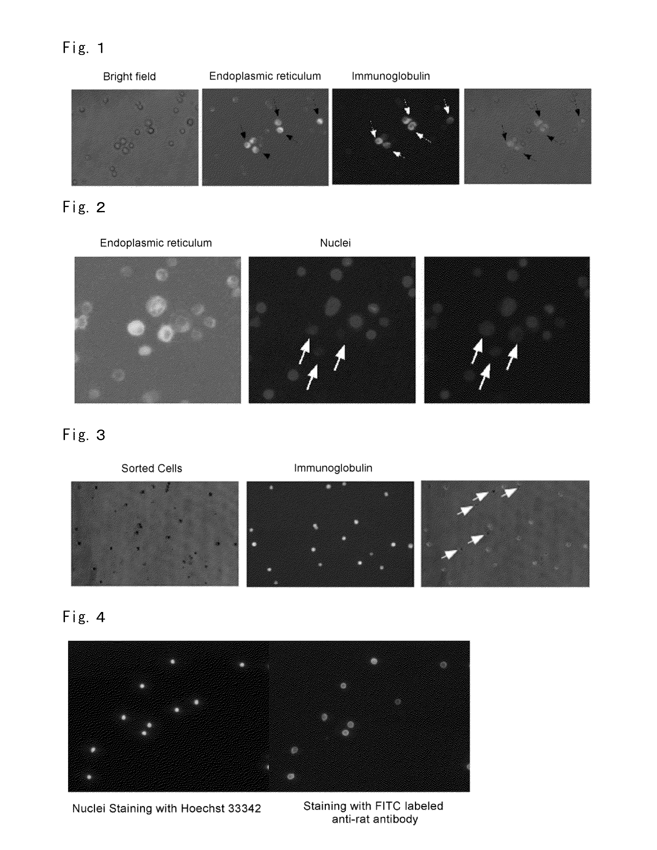
[0082]The cell group, which was CD49b negative, CD45R negative and CD138 positive, was obtained from the GFP immunized mouse lymph node according to the method described in the experimental method 1. These cells were stained with ER-Tracker (registered trademark) Blue-White DPX / lipid, and were subject to the fluorescence microscope observation. The result is shown in the FIG. 1. About 20% of cells emitted strong fluorescence with this probe, and these cells had the strongly stained area of the developed endoplasmic reticulum throughout cellular cytoplasm. Most of the remaining cells emitted weak fluorescence, and these cells had cell membrane area and small size of endoplasmic reticulum both of which were weakly stained. The fluorescent intensity ratio of cells emitting strong fluorescence to the remaining cells emitting weak fluorescence was about 5:1. In order to clarify wheth...

example 2

Identification of Plasma Cells and Plasmablasts Using Fluorescent Probe Having Affinity to Endoplasmic Reticulum and Fluorescent Probes Having Affinity to Euchromatin

[0083]The cell group, which was CD49b negative, CD45R negative and CD138 positive, obtained by the method described in the experimental method 1 was subject to the cell staining with SYTO (registered trademark) 59 and ER-Tracker (registered trademark) Blue-White DPX / lipid according to the method described in the experimental methods 2-2 and 2-3. The result is shown in the FIG. 2. Most of cells strongly stained with ER-Tracker (registered trademark) Blue-White DPX / lipid were weakly stained with SYTO (registered trademark) 59. The ratio of the fluorescent intensity of SYTO58 between cells having strongly stained cell nuclei and cells having weak stained cell nuclei was about 4:1.

[0084]The above results of cell staining were well corresponded to the specific morphology of plasma cells and plasmablasts (developed endoplasmi...

example 3

[0085]The ratio of plasma cells and plasmablasts among a lymph node cell group is equal to or less than about 0.1%. In order to obtain a high purity of plasma cells and plasmablasts from the cell group without cell surface antibodies, the inventors tried the separation and isolation of plasma cells and plasmablasts using the fluorescent probe described above and a cell sorter. Cell suspension prepared from the GFP immunized mouse lymph node was used and subject to the double staining of the endoplasmic reticulum and the nuclei according to the experimental method 2-3, and an experiment was conducted according to the preparation method of plasma cells and plasmablasts using a cell sorter described in the experimental method 3.

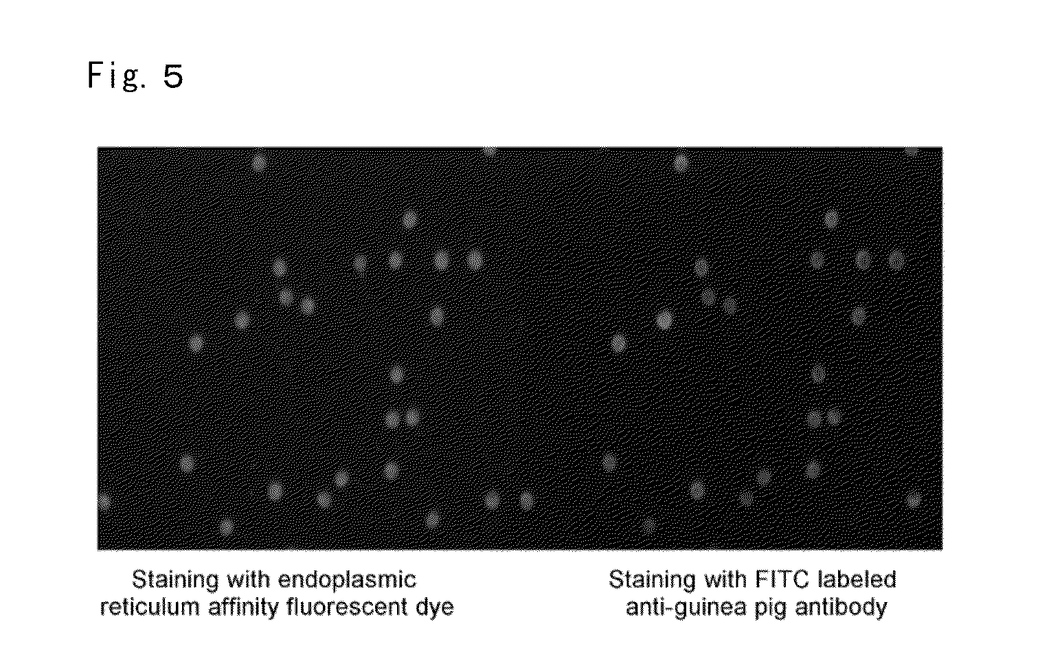
[0086]The cell group which was Blue-White DPX / lipid strong positive and SYTO (registered trademark) 59 weak negative were sorted by a cell sorter. For the sorted cells which were Blue-White DPX / lipid strong positive and SYTO (registered trademark) 59 weak negati...


PUM

PropertyMeasurementUnit
Molar densityaaaaaaaaaa
Amphiphilicityaaaaaaaaaa

Abstract

A method which can isolate plasma cells and plasmablasts efficiently and with high purity, from mammals and birds, without using a cell surface marker is provided.
Further disclosed is a fluorescent probe wherein the staining selectivity for the endoplasmic reticulum of cells is higher than the staining selectivity for cell organelles other than the endoplasmic reticulum. Also disclosed is a method for identifying plasma cells and plasmablasts which includes staining cells derived from lymph node tissue or similar by using this probe, and identifying plasma cells and plasmablasts on the basis of the fluorescence intensity from the stained cells. Also disclosed is a fluorescent probe wherein the staining selectivity for cell nuclei is higher than the staining selectivity for cell organelles other than the cell nuclei. Also disclosed is a method for identifying plasma cells and plasmablasts in lymph node tissue or similar which includes staining cells derived from lymph node tissue or similar by using this probe, and identifying plasma cells and plasmablasts on the basis of the fluorescence intensity from the stained cells.

Description

CROSS-REFERENCE TO RELATED APPLICATION(S)[0001]This application is based upon and claims the benefits of priority from JP Patent Application 2010-70730, filed on Mar. 25, 2010, the entire disclosure of which is incorporated herein by reference.TECHNICAL FIELD[0002]The present invention relates to the fluorescent probe for identifying and isolating plasma cells and plasmablasts and the method for identifying or isolating plasma cells and plasmablasts by using the probe.BACKGROUND ART[0003]An antibody plays important roles in the biophylaxis by specifically binding to a target antigen. Recently, antibody drugs which utilize its specific binding properties against its antigen have been developed as a therapeutic medication for autoimmune diseases or intractable diseases such as cancers, and its utility has high appraisal. In response to the development needs in effective manufacturing techniques for monoclonal antibodies having high specificity and high affinity which become antibody d...

Claims


Application Information

Patent Timeline
IPC IPC(8): G01N21/64C07D313/00C07C311/63C12N5/071C07K14/00
CPCG01N33/582G01N33/56966C12Q1/04G01N21/64G01N33/48G01N33/483
Inventor KUROSAWA, NOBUYUKIISOBE, MASAHARU
Owner UNIVERSITY OF TOYAMA
Why Eureka
Try Eureka