Tissue system with undifferentiated stem cells derived from corneal limbus

a stem cell and tissue technology, applied in the field of tissue systems comprising mammalian undifferentiated stem cells, can solve the problems of amniotic membrane transplantation, insufficient approaches to repair damage related to or resulting from the loss of limbal stem cells, and unclear vision or ocular discomfor

Inactive Publication Date: 2005-08-25
RELIANCE LIFE SCI PVT
View PDF14 Cites 32 Cited by
  • Summary
  • Abstract
  • Description
  • Claims
  • Application Information

AI Technical Summary

Benefits of technology

[0016] The present disclosure describes a tissue system, wherein the tissue system comprises limbal stem cells, wherein at least about 30-90% of the cells in the tissue system are undifferentiated stem cells (USCs). Preferably, the USCs comprise at least about 30%, 40%, 50%, 60%, 70%, 80%, or 90% of the limbal stem cells in the tissue system. In various embodiments, USCs express one or more stem cell marker genes selected from the group consisting of SSEA-4, SSEA-3, Oct-4, Nanog, Rex 1, Sox2, Tra-1-60, Tra-1-81, and Stem Cell Factor. In a preferred embodiment, USCs are positive for stage specific embryonic antigen marker 4 (SSEA-4). In preferred embodiments, at least about 50%, 60%, 70%, 80%, 90%, or 95% of the USCs in the tissue system are positive for SSEA-4. In other preferred embodiments, cells in the tissue system express one or more cell surface markers selected from the group consisting of K3 / K12, K19, and p63. In other preferred embodiments, the tissue system is derived from comeoscleral limbus tissue. While the corneoscleral limbus tissue may be obtained from any suitable mammal, human tissue is a particularly preferred source. Preferably the tissue system is a multi-layered tissue system.

Problems solved by technology

Without proper replenishment, an unhealthy or abnormal ocular surface can result in symptoms such as unclear vision or ocular discomfort.
Several approaches have been used to attempt to restore normal vision after corneal surface impairments, however these approaches are generally not sufficient to repair damage related to or resulting from the loss of limbal stem cells.
Amniotic membrane transplantations, however, have the disadvantage of not being uniformly successful, with the final outcome often not much different then the patient's starting point (Prabhasawat et al., (1997) Arch.
This technique also has had limited success in restoring a normal population of corneal limbal stem cells (Shimazaki et al., (1997) Ophthalmology 104:2068-2076; Tseng et al., (1997) Am. J. Ophthalmol. 124:765-774).
Methods for isolating human amniotic epithelial cells and differentiating them into corneal surface epithelium, such as disclosed by Hu et al (WO 00 / 73421), also have the disadvantage that they are very labor intensive and the yield of limbal stem cell is low.
Another approach to treating limbal stem cell deficiency involves corneal transplantation, which is also problematic for the treatment of chronic ocular surfaces because the ultimate success of the therapy is dependent on the gradual replacement of the donor's corneal epithelium with the recipient's corneal epithelium, which may have a poor prognosis due to a deficiency of limbal stem cells in the recipient's eye.
However, one major concern associated with this procedure is that it requires removal of large amounts of limbal stem cells from the patient's healthy eye, which may put the healthy eye at risk for a limbal stem cells deficiency, as well as other complications that may arise out of such a severe loss of limbal stem cells in the otherwise healthy eye.
Attempts have also been made to grow the limbal biopsies on amniotic membrane (Koizumi et al., (2000) Invest. Ophthalmol. Vis. Sci. 41:2506-2513; Koizumi et al., (2000) Cornea 19:65-71; Dua et al., (2000) Surv. Ophthalmol. 44:415-425), however, limbal cells isolated and transplanted from these cultures were unstable in long term follow up, and failed to show satisfactory ocular surface repair, particularly in patients with severe limbal stem cell deficiencies (Jun Shimazaki et al., (2002) Opthalmol.
The method for generating the surgical graft does not employ any kind of isolation step for the exclusive selection of limbal stem cells, and the distribution of the stem cell layer is non-uniform throughout the graft.
These characteristics of the grafts may lead to a lack of stability of the transplanted epithelial cells, particularly in patients with limbal stem cell deficiencies.
This method, however, may fail to correct damage in patients with severe limbal stem cell deficiencies because of limited supplies of limbal stem cells in the transplanted differentiated epithelial cells.
In addition, in patients with severe ocular or corneal surface damage, the insertion of a contact lens is not necessarily desirable because the presence of a contact lens on an already damaged surface may cause irritation and discomfort, and may further aggravate the condition of the eye, leading to other complications.
The methods has several drawbacks, including the uncertainty of whether limbal stem cells will be constantly released after being seeded on the lens to help heal the damaged eye, and the contact lens is seeded with a cell populations that may have as few as 10% limbal stem cells, which may not be adequate for successful corneal repair in patients with severe limbal stem cell deficiencies.
Again, these grafts of differentiated epithelial cells will have limited populations of undifferentiated limbal stem cells, which are necessary for successful ocular repair in patients severely deficient in limbal stem cells.
While these markers are known for indicating the corneal nature of cells, they are not particularly specific for determining whether the isolated cells are undifferentiated.
Therefore, these grafts do not appear to be well suited for repairing ocular damage in patients with severe limbal stem cell deficiencies.
Therefore, this method may preferentially select for blood cells that may contaminate the corneal tissue biopsy, and the undifferentiated status of cells isolated using this method was not evaluated.
The donor epithelium in such transplants or grafts will survive generally for only a short period of time due to the limited supply of limbal stem cells.
Alternatively, the transplants may yield a clear corneal epithelium, but the lack of sufficient limbal stem cells results in abnormal epithelial surfaces and poor healing, resulting in a failure to repair the ocular surface and improve vision.
Therefore, these approaches which are intended to supply limbal stem cells to an eye with a limbal stem cell deficiency have serious limitations, which may be due to the limited supply of undifferentiated limbal stem cells with self-regenerative capacity present in the transplants or grafts.

Method used


Image

  • Tissue system with undifferentiated stem cells derived from corneal limbus
  • Tissue system with undifferentiated stem cells derived from corneal limbus
  • Tissue system with undifferentiated stem cells derived from corneal limbus

Examples


example 1

[0068] 1) Collection of Limbal Tissue Biopsies

[0069] Prior to initiating the collection of limbal tissue biopsies from human patients, Institutional Review Board approval was obtained. Informed consent was obtained from each patient and donor, and all human subjects were treated according to the Helsinki Accord. A 0.8 mm2to 2 mm2limbal biopsy was surgically removed from the donor eye from superior or temporal quadrants of the corneal surface by lamellar keratectomy. These regions are particularly rich in limbal stem cells. After excision, the biopsy was immediately placed in a 2 ml transport vial filled with transport medium. The transport medium consisted of Dulbecco's Modified Eagles Medium (DMEM) and Ham's F-12 Medium (DMEM:F-12; 1:1) supplemented with 5% fetal bovine serum (FBS) or 5% human serum collected from cord blood, 0.5% dimethyl sulphoxide (DMSO), 2 ng / ml recombinant human epidermal growth factor (rhEGF), 5 μg / ml insulin, 5 μg / ml transferrin, 5 μg / ml sodium selenite, 0....

example 2

[0084] Analysis and Characterization of the Tissue System Containing Undifferentiated Stem Cells

[0085] As outlined in Example 1, a tissue system comprising USCs was derived from limbal tissue biopsies. To better understand the cell population of the tissue system derived from limbal tissue, cells in the tissue system were analyzed using flow cytometry, immunofluorescence and immunoperoxidase assays, and molecular analysis for the presence or absence of various cellular markers of undifferentiated cells.

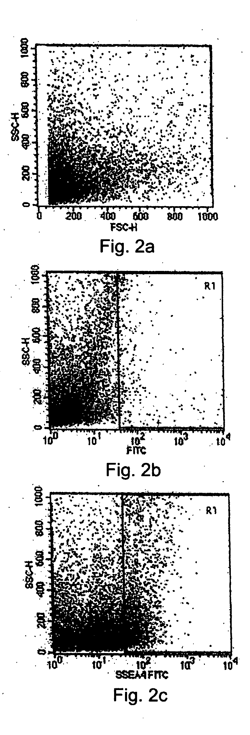
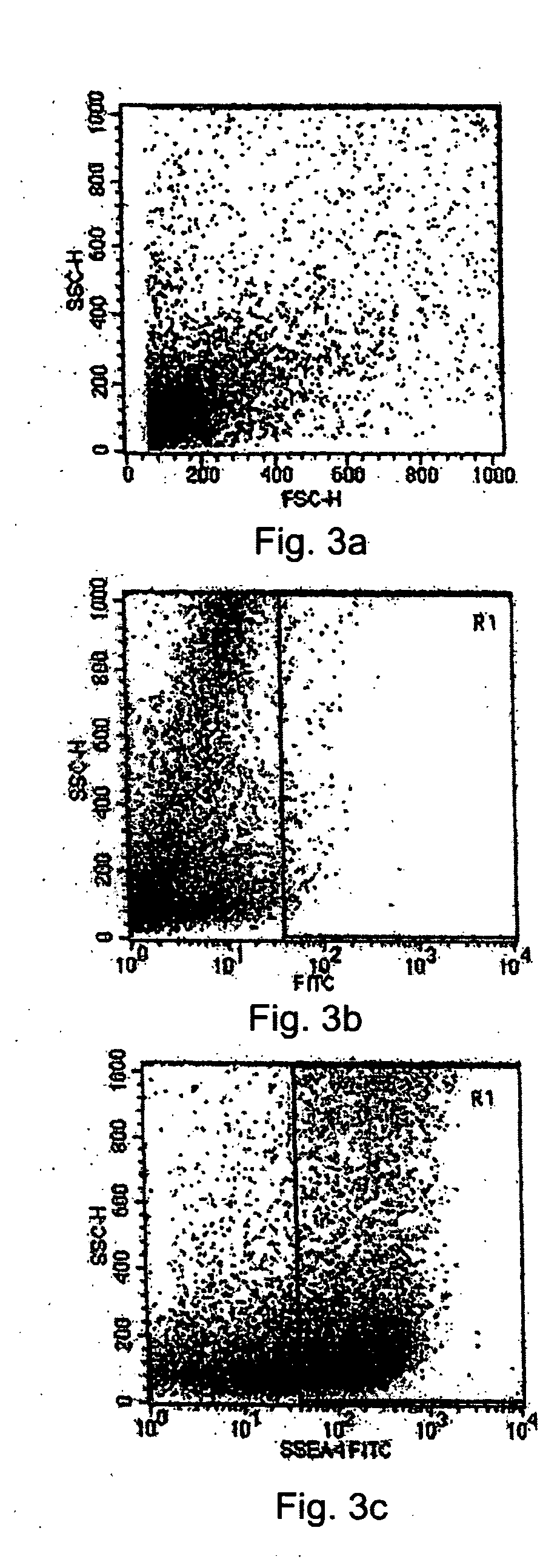
[0086] 1) Flow Cytometry Analysis

[0087] The cell population of the limbal biopsy cultures was compared to the cell population in the tissue system using flow cytometry analysis to detect the presence of the SSEA-4 marker. First, cells were collected from the limbal biopsy cultures at a semi-confluent stage and from the tissue system grown on an amniotic membrane tissue base after sorting and selecting cells by MACS as set forth above. The two populations of cells were analyzed sepa...

example 3

[0096] Viability Studies of the Tissue System Comprising Undifferentiated Stem Cells:

[0097] Limbal tissue biopsies were evaluated to determine how long the biopsies could remain in transport prior to in vitro culturing of a viable tissue system with USCs. The limbal tissue biopsies were transported or stored in the following culture media for 12, 24, 48, and 72 hours after surgery at 4° C.: DMEM and Ham's F-12 (ratio 1:1), supplemented with human cord blood serum (3-5%), DMSO (0.5%), rhEGF (2 ng / ml), insulin (5 μg / ml), transferrin (5 μg / m), sodium selenite (5 μg / ml), hydrocortisone (0.5 μg / ml), cholera toxin A (0.1 nmol / l ), gentamycin (50 μg / ml), and amphotericin B (1.25 μg / ml). After this period of time, the biopsies were cultured to generated tissue systems with USCs as described in Example 1. FIG. 13 shows the percent success in developing a tissue system from biopsies cultured approximately 12, 24, 48 and 72 hours after surgical collection from a subject. As shown in FIG. 13, ...


PUM

PropertyMeasurementUnit
limbal arc lengthaaaaaaaaaa
temperatureaaaaaaaaaa
cell surfaceaaaaaaaaaa

Abstract

The present disclosure describes a tissue system with self-regenerating limbal stem cells, wherein the limbal stem cells are primarily undifferentiated stem cells (USCs). The tissue system is derived from isolated corneal limbal tissue, and is suitable for restoring ocular surface impairments, particularly those that result from limbal stem cell deficiencies. The tissue system is generated by selectively augmenting the tissue system for USCs, for example by selecting and sorting cells that express stem cell-specific surface markers, such as stage specific embryonic antigen marker 4 (SSEA-4). After isolation, the USCs are cultured on a tissue base in the presence of enriched medium to generate the tissue system, which is suitable for transplantation, implantation, or grafting.

Description

STATEMENT REGARDING FEDERALLY SPONSORED RESEARCH OR DEVELOPMENT [0001] Not applicable. BACKGROUND OF THE INVENTION [0002] 1. Field of the Invention [0003] The present disclosure relates to tissue systems comprising mammalian undifferentiated stem cells, preferably human undifferentiated stem cells, which are derived from corneal limbus tissue and suitable for restoring damaged or diseased ocular surfaces. [0004] 2. Description of Related Art [0005] Stem cells are responsible for cellular replacement and tissue regeneration throughout the life of an organism. Stem cells are cells that have extensive proliferation potential, and may, depending on the stem cell, differentiate into several cell lineages, and / or repopulate tissue upon transplantation. Embryonic stem (ES) cells are quintessential stem cells with unlimited self-renewal and pluripotent potential. ES cells are derived from the inner cell mass of the blastocyst stage embryo. Adult stem cells are also specialized undifferentia...

Claims


Application Information

Patent Timeline
Patent Type & Authority Applications(United States)
IPC IPC(8): C12N5/071C12N5/0797
CPCC12N5/0621C12N5/0623C12N2500/25C12N2502/02C12N2501/115C12N2501/235C12N2501/11A61P27/02A61K35/44C12N5/0607
Inventor MAHADEORAO, TOTEYDEVI, KASHYAP
Owner RELIANCE LIFE SCI PVT
Why Eureka
Try Eureka