
Apparatus and method for detecting lesion and lesion diagnosis apparatus

Inactive Publication Date: 2013-01-31
SAMSUNG ELECTRONICS CO LTD
View PDF8 Cites 22 Cited by
  • Summary
  • Abstract
  • Description
  • Claims
  • Application Information

AI Technical Summary

Benefits of technology

The patent is about a device that can remove noise from an image of a specific tissue region. This improves the quality of the image and allows for better analysis of the tissue.

Problems solved by technology

However, it may be difficult to precisely extract a lesion since the images produced by theses medical imaging equipment are typically of poor quality.

Method used


Image

  • Apparatus and method for detecting lesion and lesion diagnosis apparatus
  • Apparatus and method for detecting lesion and lesion diagnosis apparatus
  • Apparatus and method for detecting lesion and lesion diagnosis apparatus

Examples


Embodiment Construction

[0039]The following detailed description is provided to assist the reader in gaining a comprehensive understanding of the methods, apparatuses, and / or systems described herein. Accordingly, various changes, modifications, and equivalents of the systems, apparatuses and / or methods described herein will be suggested to those of ordinary skill in the art. Also, descriptions of well-known functions and constructions may be omitted for increased clarity and conciseness.

[0040]Hereafter, examples will be described with reference to accompanying drawings.

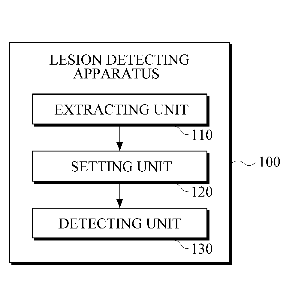
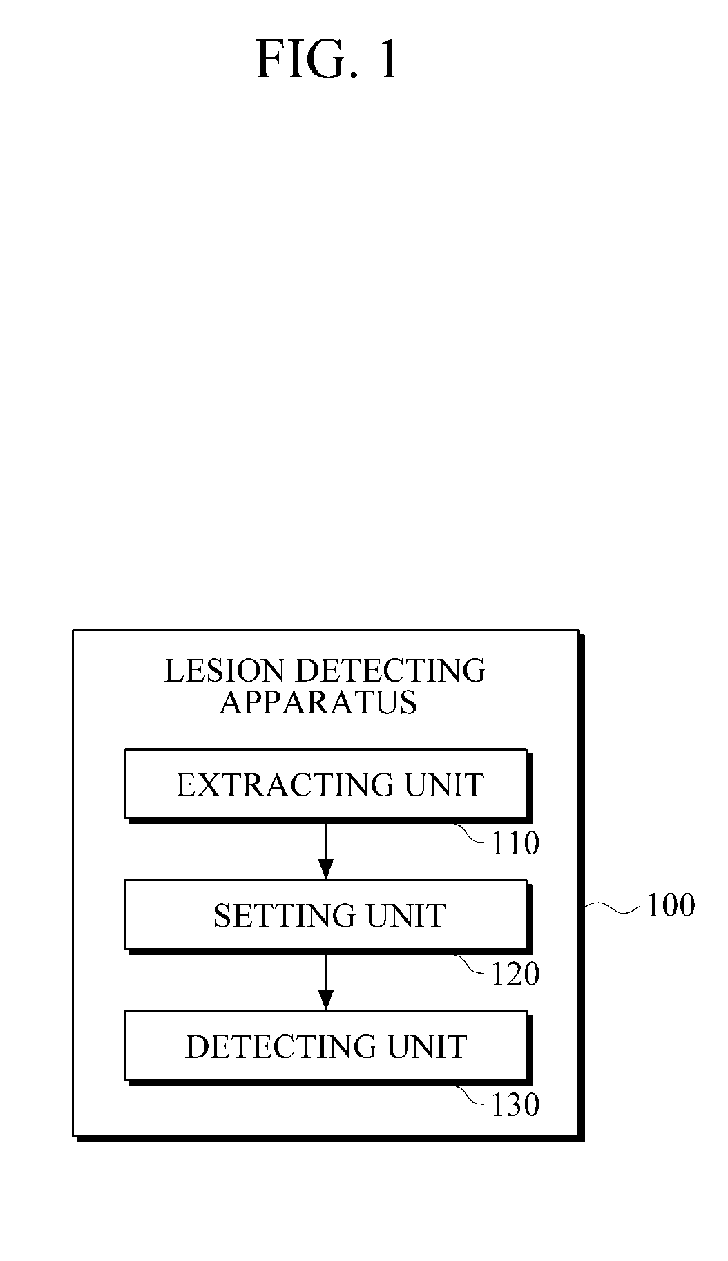
[0041]FIG. 1 illustrates an example of a lesion detecting apparatus.

[0042]Referring to FIG. 1, a lesion detecting apparatus 100 includes an extracting unit 110, a setting unit 120 and a detecting unit 130.

[0043]The extracting unit 110 may extract at least one tissue region from an image. The image may capture the inside of an organism and the image may include a plurality of tissue regions.

[0044]For example, the extracting unit 110 may use ...


PUM


Abstract

An apparatus for detecting a lesion is provided. The apparatus includes an extracting unit configured to extract at least one tissue region from an image of tissue regions, a setting unit configured to set at least one of the at least one extracted tissue region as a lesion detection candidate region, and a detecting unit configured to detect a lesion from the lesion detection candidate region.

Description

CROSS-REFERENCE TO RELATED APPLICATION[0001]This application claims the benefit under 35 U.S.C. §119(a) of Korean Patent Application No. 10-2011-0073398, filed on Jul. 25, 2011, the entire disclosure of which is incorporated by reference for all purposes.BACKGROUND[0002]1. Field[0003]The following description relates to an apparatus and a method for detecting lesions and a lesion diagnosis apparatus.[0004]2. Description of the Related Art[0005]With the advancement of surgery techniques, different kinds of minimum invasive surgeries have been developed. A minimum invasive surgery process represents a surgery method in which a medical operation may be performed by approaching a lesion using surgical instruments without incising skin and muscle tissues. The surgical instruments may include a syringe or a catheter. The medical operation may include a medicine injection, removal of lesions, appliance insertion etc. In order to perform the minimum invasive surgery process, doctors needs t...

Claims


Application Information

Patent Timeline
IPC IPC(8): A61B5/05
CPCA61B5/0091G06T7/0012G06T2207/30096G06T2207/10132G06T2207/30068G06T2207/10072A61B6/03G06T5/00
Inventor SEONG, YEONG-KYEONGPARK, MOON-HO
Owner SAMSUNG ELECTRONICS CO LTD
Learn More