
Method and system for determining a stage of fibrosis in a liver

a fibrosis and liver technology, applied in the field of methods and systems for determining the stage of fibrosis in the liver, can solve the problems of difficult to obtain high reproducibility,/or the failure of an organ with the disease, and the pathological features used in these systems are usually not clearly defined and ambiguous,

Active Publication Date: 2013-01-31
AGENCY FOR SCI TECH & RES
View PDF1 Cites 20 Cited by
  • Summary
  • Abstract
  • Description
  • Claims
  • Application Information

AI Technical Summary

Benefits of technology

This patent describes a new and useful method for determining the stage of fibrosis in a liver using multiple measurements based on morphological features. This method involves using a second harmonic generation based imaging system to obtain input data relating to the liver, identifying a plurality of morphological features from the input data, and generating a plurality of measurements based on the identified morphological features. The stage of fibrosis in the liver can then be determined based on the generated measurements. This invention can be used as a computer system integrated with a device for capturing images of a patient's liver, or as a computer program product.

Problems solved by technology

The stage of a disease is a measure of how far the disease has progressed in its natural history, with the end stage usually resulting in the death of a patient with the disease and / or the failure of an organ with the disease.
However, pathological features used in these systems are usually not clearly defined and are somewhat ambiguous.
Therefore, using these systems, inter- and intra-observer variations can be as high as 35%, making it difficult to obtain highly reproducible results [7-9].
However, all of the systems in the studies shown in Table 1 require stained biopsy samples and thus, are faced with the problem of staining variations.
Furthermore, although some correlations between the fibrosis area and the semi-quantitative scores were reported in the studies, the amount of fibrosis is not specifically addressed in any of the scoring systems.

Method used


Image

  • Method and system for determining a stage of fibrosis in a liver
  • Method and system for determining a stage of fibrosis in a liver
  • Method and system for determining a stage of fibrosis in a liver

Examples


Embodiment Construction

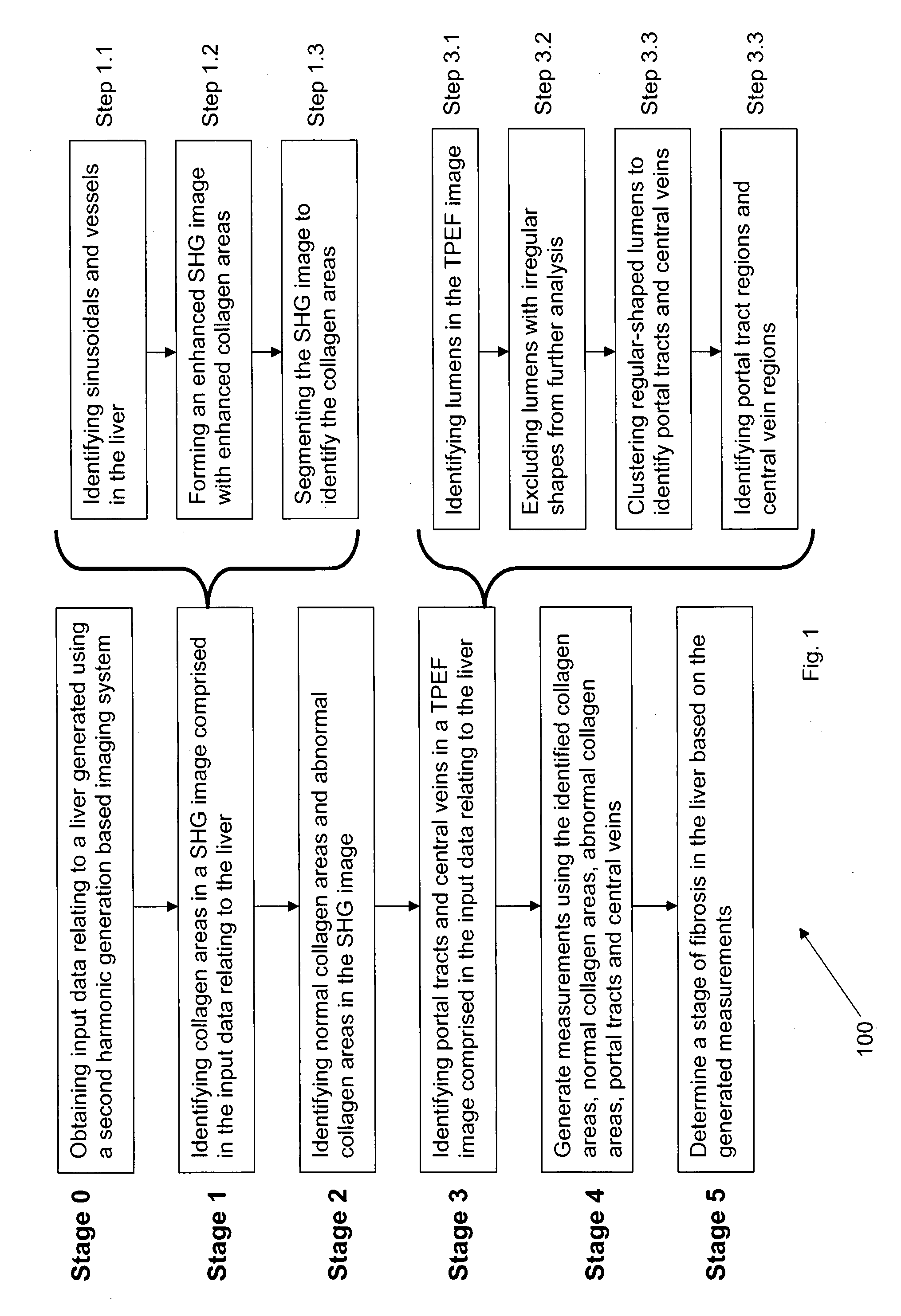
[0020]Referring to FIG. 1, the steps are illustrated of a method 100 which is an embodiment of the present invention, and which determines a stage of fibrosis in the liver.

[0021]Method 100 comprises 6 stages. In Stage 0, input data relating to a liver (comprising liver tissue) is obtained. The input data is generated using a second harmonic generation based imaging system. For example, the input data may be acquired using an endoscope (in other words, using reflective mode imaging). The input data may also be acquired without staining the liver tissue. Furthermore, the input data comprises two images: a first image in the Two-Photon Excitation Fluorescence (TPEF) channel (i.e. “TPEF image”) and a second image in the Second Harmonic Generation (SHG) channel (i.e. “SHG image”). The TPEF and SHG images respectively comprise information relating to the tissue structure in the liver and information relating to the collagen content in the liver. Furthermore, each of these images comprises...


PUM


Abstract

A method for determining a stage of fibrosis in a liver is disclosed. The method comprises the steps of: (1a) obtaining input data relating to the liver, the input data being generated using a second harmonic generation based imaging system; (1b) identifying a plurality of morphological features of the liver from the input data relating to the liver; (1c) generating a plurality of measurements based on the identified plurality of morphological features; and (1d) determining the stage of fibrosis in the liver based, on the generated plurality of measurements.

Description

FIELD OF THE INVENTION[0001]The present invention relates to a method and system for determining a stage of fibrosis in a liver. The present invention utilizes a method for generating a plurality of measurements relating to a plurality of morphological features in the liver.BACKGROUND OF THE INVENTION[0002]The stage of a disease is a measure of how far the disease has progressed in its natural history, with the end stage usually resulting in the death of a patient with the disease and / or the failure of an organ with the disease. In other words, the grade (or stage) of a disease reflects how quickly the disease is progressing to the end stage [1].[0003]In most forms of chronic liver diseases, the end stage involves a large amount of fibrosis or cirrhosis, whereas lower amounts of fibrosis or cirrhosis are present in the liver during the earlier stages. Descriptive or semi-quantitative scoring systems may be used to grade and stage liver biopsy samples. For example, a traditional meth...

Claims


Application Information

Patent Timeline
IPC IPC(8): A61B6/00
CPCA61B5/0084A61B5/4244G06T7/0012A61B5/7267G06T2207/10068G06T2207/20036G06T2207/30056G06T2207/10064
Inventor YU, HANRYTAI, DEANHE, YUTINGXU, SHUOYU
Owner AGENCY FOR SCI TECH & RES
Learn More