
Method and System for Segmentation and Removal of Pulmonary Arteries, Veins, Left Atrial Appendage

a technology of pulmonary arteries and segments, applied in the field of cardiac imaging, can solve the problems of reducing blood flow to the heart muscle, damage to the myocardium,

Inactive Publication Date: 2013-04-04
SIEMENS HEATHCARE GMBH
View PDF5 Cites 22 Cited by
  • Summary
  • Abstract
  • Description
  • Claims
  • Application Information

AI Technical Summary

Benefits of technology

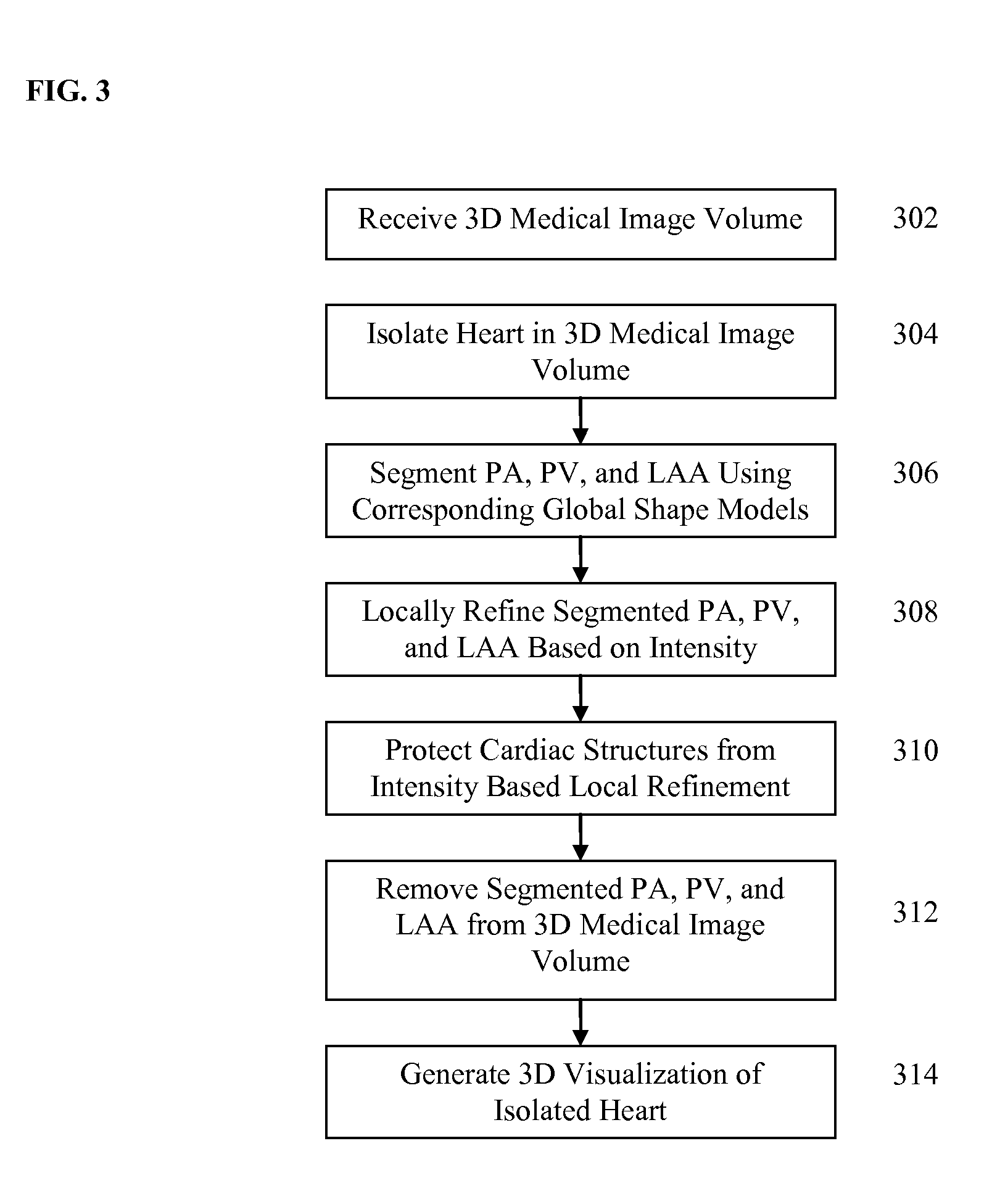
The present invention provides a method and system for segmenting the pulmonary arteries, pulmonary veins, and left atrial appendage in a 3D heart isolation image. This is useful for better visualizing coronary arteries and bypass arteries. The method uses global shape models and local region growing to accurately segment the pulmonary arteries, pulmonary veins, and left atrial appendage. The invention also generates masks for these segments based on local voxel intensities. This technology improves the accuracy and efficiency of segmenting and masking the pulmonary arteries, veins, and left atrial appendage in 3D heart isolation images.

Problems solved by technology

Eventually such plaque blocks or reduces blood flow to heart muscles.
Deprived of oxygen, the myocardium becomes damaged and other heart diseases may develop, as well.
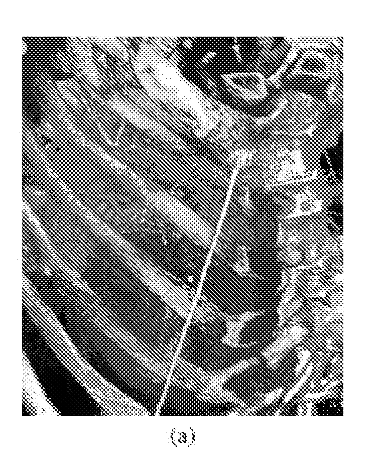
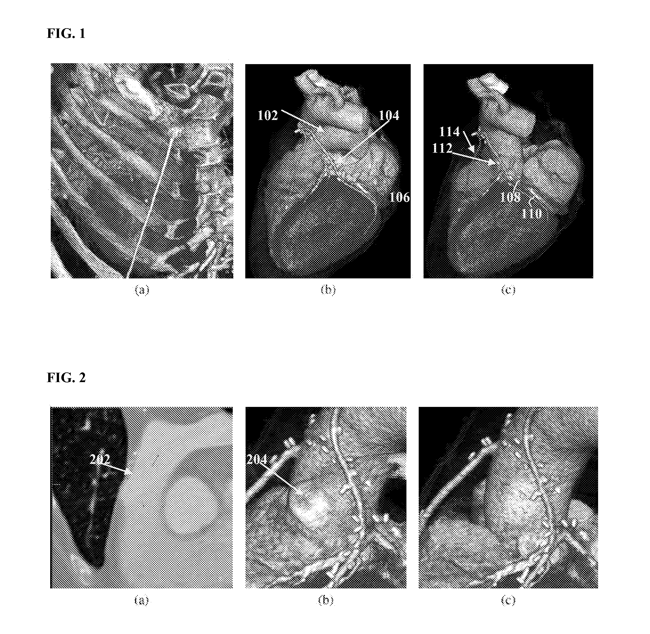
However, heart structures like the pulmonary arteries, pulmonary veins, and left atrial appendage may still partially occlude the coronary arteries in the isolated heart visualization.

Method used


Image

  • Method and System for Segmentation and Removal of Pulmonary Arteries, Veins, Left Atrial Appendage
  • Method and System for Segmentation and Removal of Pulmonary Arteries, Veins, Left Atrial Appendage
  • Method and System for Segmentation and Removal of Pulmonary Arteries, Veins, Left Atrial Appendage

Examples


Embodiment Construction

[0017]The present invention is directed to a method and system for segmentation and removal of pulmonary arteries (PA), pulmonary veins (PV), and the left atrial appendage (LAA) from 3D medical images, such as 3D computed tomography (CT) volumes, in order to visualize coronary arteries and bypass arteries. Embodiments of the present invention are described herein to give a visual understanding of the segmentation methods. A digital image is often composed of digital representations of one or more objects (or shapes). The digital representation of an object is often described herein in terms of identifying and manipulating the objects. Such manipulations are virtual manipulations accomplished in the memory or other circuitry / hardware of a computer system. Accordingly, it is to be understood that embodiments of the present invention may be performed within a computer system using data stored within the computer system.

[0018]Various algorithms have been developed to isolate the heart f...


PUM


Abstract

A method and system for segmentation and removal of pulmonary arteries, pulmonary veins, and a left atrial appendage from 3D medical image data, such as 3D computed tomography (CT) volumes, is disclosed. A global shape model is segmented for each of pulmonary arteries, pulmonary veins, and a left atrial appendage in a 3D volume. The segmented global shape model for each of the pulmonary arteries, pulmonary veins, and left atrial appendage is locally refined based in local voxel intensities in the 3D volume, resulting in a respective mask for each structure. The mask is used to remove voxels belonging to the pulmonary arteries, pulmonary veins, and left atrial appendage from the 3D volume in order to better visualize coronary arteries and bypass arteries.

Description

[0001]This application claims the benefit of U.S. Provisional Application No. 61 / 542,913, filed Oct. 4, 2011, the disclosure of which is herein incorporated by reference.BACKGROUND OF THE INVENTION[0002]The present invention relates to cardiac imaging, and more particularly, to segmentation of pulmonary arteries, veins, and the left atrial appendage for improved visualization of coronary arteries and bypass arteries.[0003]Coronary Artery Disease (CAD) or Coronary Heart Disease (CHD) is the leading cause of death in the world. CAD / CHD is caused by accumulation of plaque in coronary arteries. Eventually such plaque blocks or reduces blood flow to heart muscles. Deprived of oxygen, the myocardium becomes damaged and other heart diseases may develop, as well. A typical early symptom for CAD / CHD is chest pain, which can easily be mistaken for other less serious diseases until a patient experiences a heart attack.[0004]Cardiac computed tomography (CT) is often used for diagnosis and treat...

Claims


Application Information

Patent Timeline
IPC IPC(8): G06K9/00
CPCG06K2009/366G06T7/0081G06T7/0087G06T2200/04G06T2207/30101G06T2207/20076G06T2207/20081G06T2207/30048G06T2207/10081G06T7/11G06T7/143G06V10/248
Inventor ZHONG, HUAZHENG, YEFENGFUNKA-LEA, GARETHVEGA-HIGUERA, FERNANDOCOMANICIU, DORIN
Owner SIEMENS HEATHCARE GMBH
Learn More