Oral scope system with image sensor and method for visual examination of oral cavity and upper airway

a technology of image sensor and scope system, which is applied in the field of oral cavity and upper airway visual examination with image sensor, can solve the problems of hampered and restricted vision of mouth and throat, difficult inspection and examination of oral cavity and upper airway, and inability to see the mouth and throa

Inactive Publication Date: 2013-04-18
QSCOPE
View PDF12 Cites 24 Cited by
  • Summary
  • Abstract
  • Description
  • Claims
  • Application Information

AI Technical Summary

Benefits of technology

[0011]Another embodiment of the oral scope instrument contemplates that the image sensor may be selectively pivoted at a selected angle relative to the long axis of the flexible arm of the scope. To accomplish this functionality, an actuator is mounted on the handle operatively connected to the image sensor to selectively pivot and position the image sensor at a selected angle. This can be done by the person operating the oral scope when the instrument is in the mouth. This feature affords special benefit when the flexible arm is fully inserted; here the image sensor, if tilted upwardly can zoom in on the uvula and upper airway, if tilted downwardly can focus on the epiglottis, vestibular fold and adjacent anatomies.

Problems solved by technology

However, thorough inspection and examination of the oral cavity and upper airway are difficult even in the hand of experienced professionals, where a wood or metal tongue depressor is often used.
This is because clearly being able to see the mouth and throat is hampered and restricted by insufficient lighting, angles of the tongue depressor and / or deeper location of infection.
Most importantly, noxious stimulation (gag reflex) by a tongue depressor may make the visual examination impossible, especially in the situation where mouth and throat pain from infection and inflammation severely limit adequate opening of the mouth.
This is especially true in pediatrics, where an upper airway infection such as streptococcus throat is common and children are hardly cooperative when they are sick, making it very difficult for a physician to visualize the throat and tonsil.
Furthermore, the direct view through the naked eye simply cannot overcome the curvature of a normal airway to see the deeper tissue and the traditional examination with a tongue blade, even if done correctly, relies on visualization, memory and recollection of the person doing the examination, and that can vary significantly from person to person, and from time to time.
Furthermore, self-examination and inspection of one's own mouth and throat is even more difficult or if not impossible in some situations.
Misdiagnosis often occurs by using a traditional tongue blade, because of the visual difficulty described above.
Consequently, correct treatments cannot be established, which result in continued problems, leading to serious complication such as rheumatic heart diseases caused by streptococcal throat.
On other hand, the widespread misuse / overuse of antibiotic drugs worldwide is due to incorrect diagnoses, and has contributed to the rise of multiple antibiotic resistance bacteria that endanger populations as a whole.
In some cases, a sore throat is often a sign of upper airway irritation from air pollution, smoking, drinking and talking, where the correct treatment is simply rest, symptomatic relief and to refrain from the hazardous environment.
When other conditions exist, the symptom may be due to viral infection and insensitive bacterial infection in which case antibiotics are ineffective.

Method used


Image

  • Oral scope system with image sensor and method for visual examination of oral cavity and upper airway
  • Oral scope system with image sensor and method for visual examination of oral cavity and upper airway
  • Oral scope system with image sensor and method for visual examination of oral cavity and upper airway

Examples


Embodiment Construction

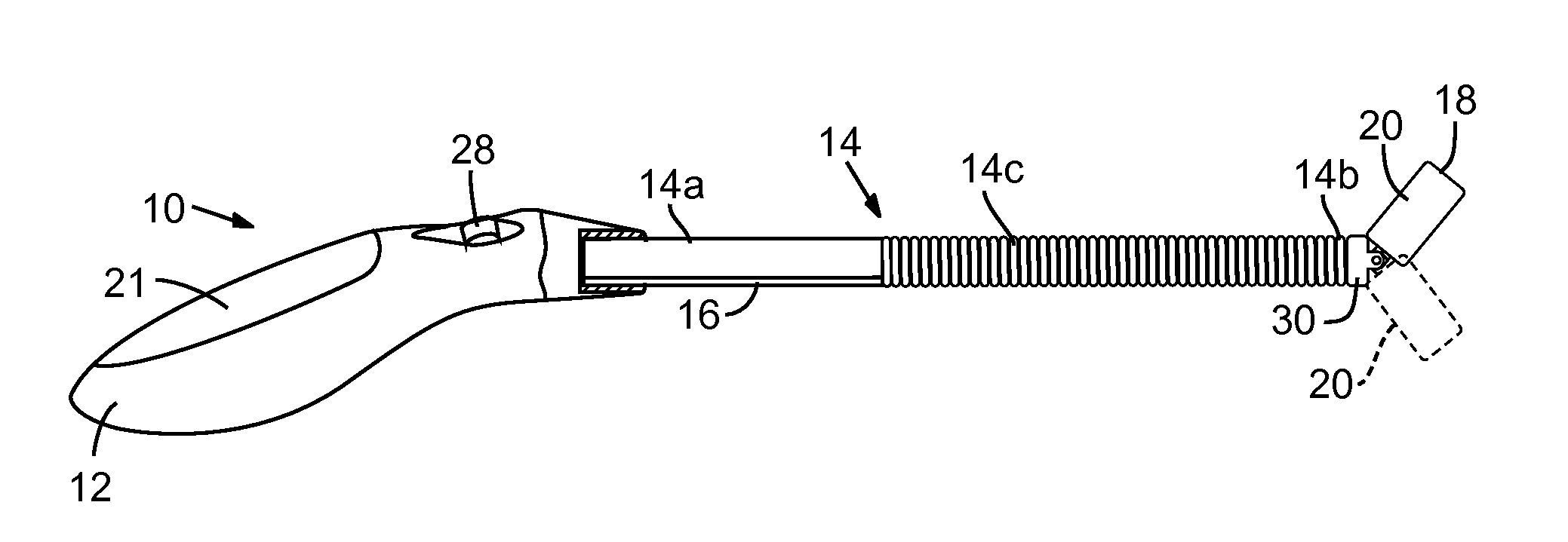
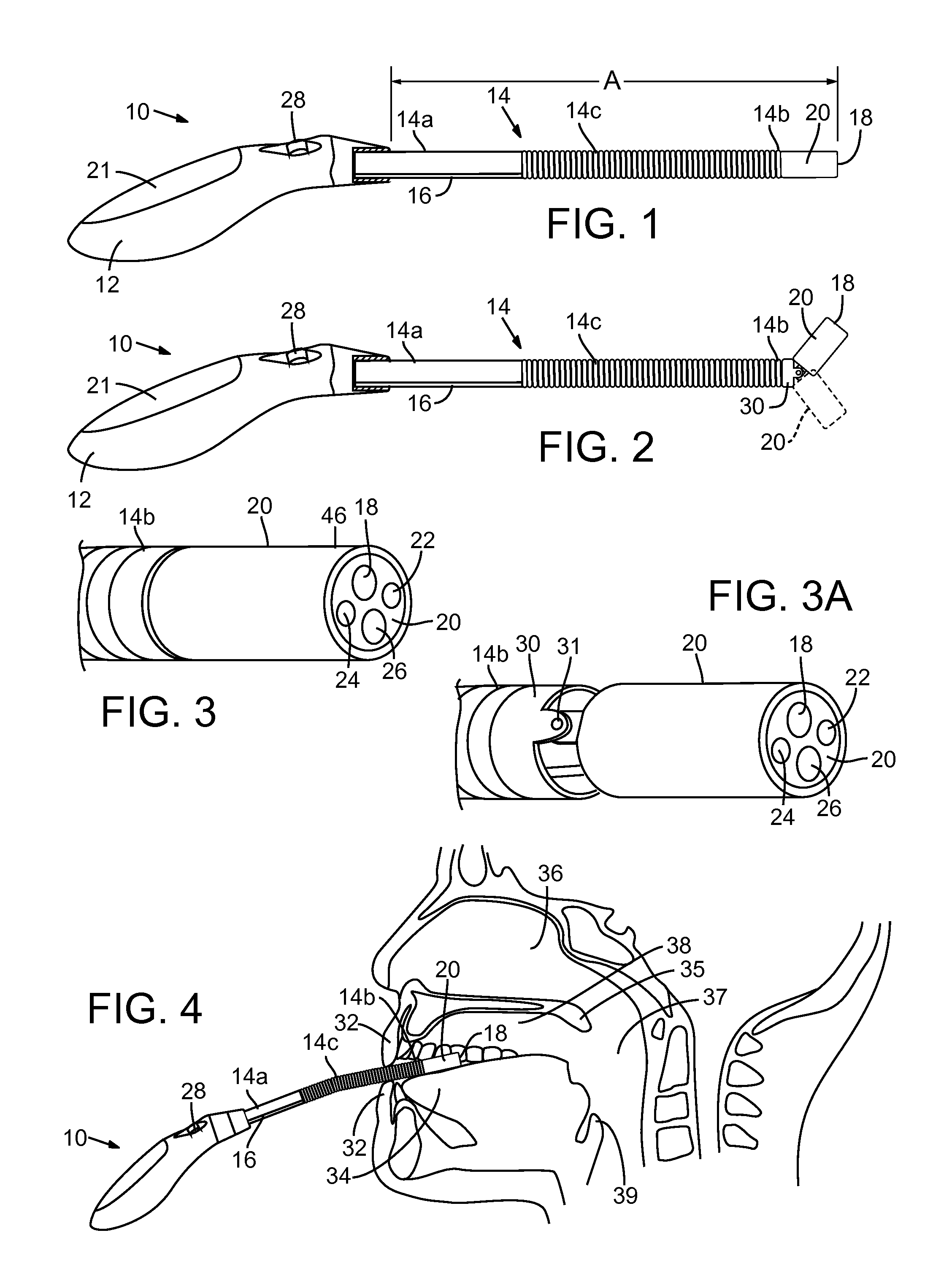
[0028]As described in more detail below, the present disclosure includes several embodiments of an oral scope used in a system for facilitating visual examination and inspection of the oral cavity and upper airway of a person's mouth. The oral scope can be used by a person for self-examination, to determine if a symptom in their oral cavity or upper airway is abnormal or pathological. The oral scope can also be used by an adult, such as a parent to examine the condition of children, to determine visually the location and condition of an area of the mouth or throat that a child appears to have symptoms of some abnormality. The oral scope, in its various embodiments, may utilize an image sensor, such as a still image camera, a video camera or a thermal image camera to capture or record an image, which may then be compared to normal anatomy, thereby to provide an initial diagnosis, and provide guidance for further treatment, if deemed necessary.

[0029]A first embodiment of an oral scope...


PUM


Abstract

An oral scope instrument for facilitating visual examination and inspection of a person's oral cavity and upper airway includes an elongate, flexible arm having first and second ends, a temperature sensor connected to the flexible arm adjacent the first end, an image sensor mounted adjacent the second end, a processor in electrical communication with the image sensor and an image display device. An illumination unit located adjacent the second end provides illumination for the image sensor. A handle connected to the first end enables manual insertion of the flexible arm into the mouth along a portion of the top of the tongue to engage the temperature sensor therewith and position the image sensor for viewing and recording anatomical features of the oral cavity or the upper airway. The processor stores normal anatomical images and may transmit images of recorded anatomical features to the image display device.

Description

TECHNICAL FIELD AND BACKGROUND[0001]Humans have the innate ability to know their abnormal feelings (i.e. symptoms) and to locate their abnormal findings (i.e signs). Because of this unique survival skill, a person is often the first to know if he or she needs to seek medical attention, and a doctor is often the first to use the symptoms and signs to make correct diagnosis. Needless to say, self-recognition of body abnormalities plays an essential role for maintaining one's health as well as battling against diseases. It is indeed the first line of human defense against wide range of diseases.[0002]Human eyes are capable of recognizing gross abnormalities, such as lumps and bumps, skin discoloration and swelling. Thus, diagnosis based on visual examination becomes a routine procedure for some health problems. For example, in traditional Chinese medicine and modern western medicine, the examination of a patient's tongue, gums, throat and upper airways is a routine medical procedure. T...

Claims


Application Information

Patent Timeline
Patent Type & Authority Applications(United States)
IPC IPC(8): A61B5/01A61B1/012A61B1/24A61B1/04A61B1/005
CPCA61B1/267A61B5/01A61B1/00108A61B5/0088A61B1/0055A61B1/247A61B1/00183
Inventor QIU, CHUNYUANWANG, ZAIQI
Owner QSCOPE
Why Eureka
Try Eureka