Radiological image radiographing display method and system thereof

Active Publication Date: 2013-05-02
FUJIFILM CORP
View PDF7 Cites 14 Cited by
  • Summary
  • Abstract
  • Description
  • Claims
  • Application Information

AI Technical Summary

Benefits of technology

The patent is about a method and system for taking and displaying radiological images. The system can accurately aim at a specific lesion using a stereoscopic biopsy device. The method involves receiving a position in a stereoscopic image and displaying the relevant image as a two-dimensional image. A mark is displayed on the two-dimensional image based on the position information in the stereoscopic image. The observer can then easily target the lesion in the two-dimensional image with higher accuracy. Overall, the method and system improve the accuracy and ease of targeting abnormalities in the body.

Problems solved by technology

However, by merely individually targeting positions in two two-dimensional images as described above, it is difficult to check whether the same lesion is targeted in the two two-dimensional images.
Particularly, when plural calcified portions exist in a radiological image of a breast, it is difficult to accurately select a corresponding calcified portion using two radiological images.
In addition, when calcified portions different in the lateral direction are selected, a biopsy needle may pierce an improper position.
For these reasons, the stereoscopic biopsy device of the related art takes a time of about 30 minutes to 1 hour, thereby causing a decrease in diagnostic efficiency.
For example, JP2000-500031T suggests that a position is targeted in a stereoscopic image while observing the stereoscopic image displayed on a display, but it is difficult to perform high-accuracy targeting in a stereoscopic image.

Method used


Image

  • Radiological image radiographing display method and system thereof
  • Radiological image radiographing display method and system thereof
  • Radiological image radiographing display method and system thereof

Examples


Embodiment Construction

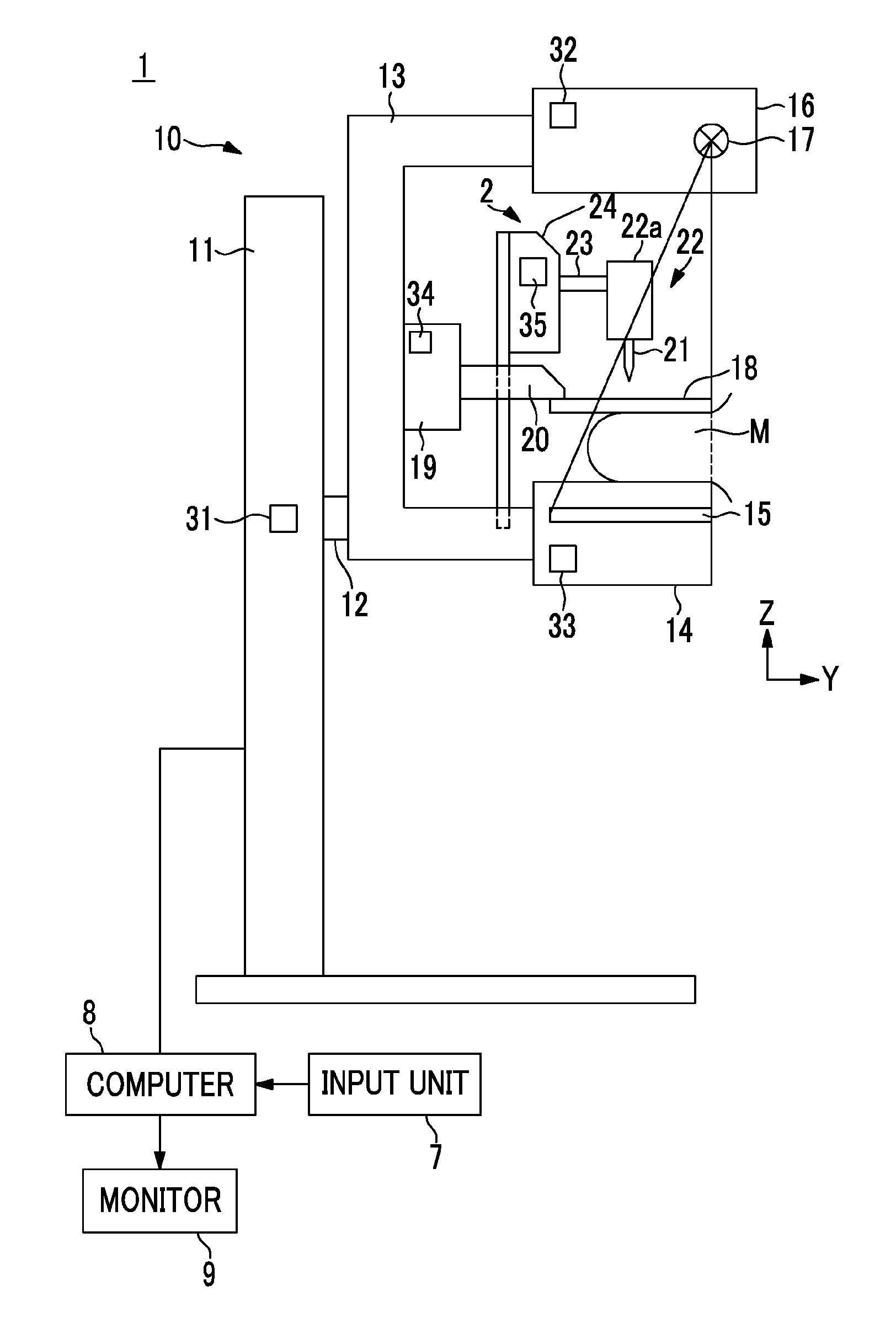
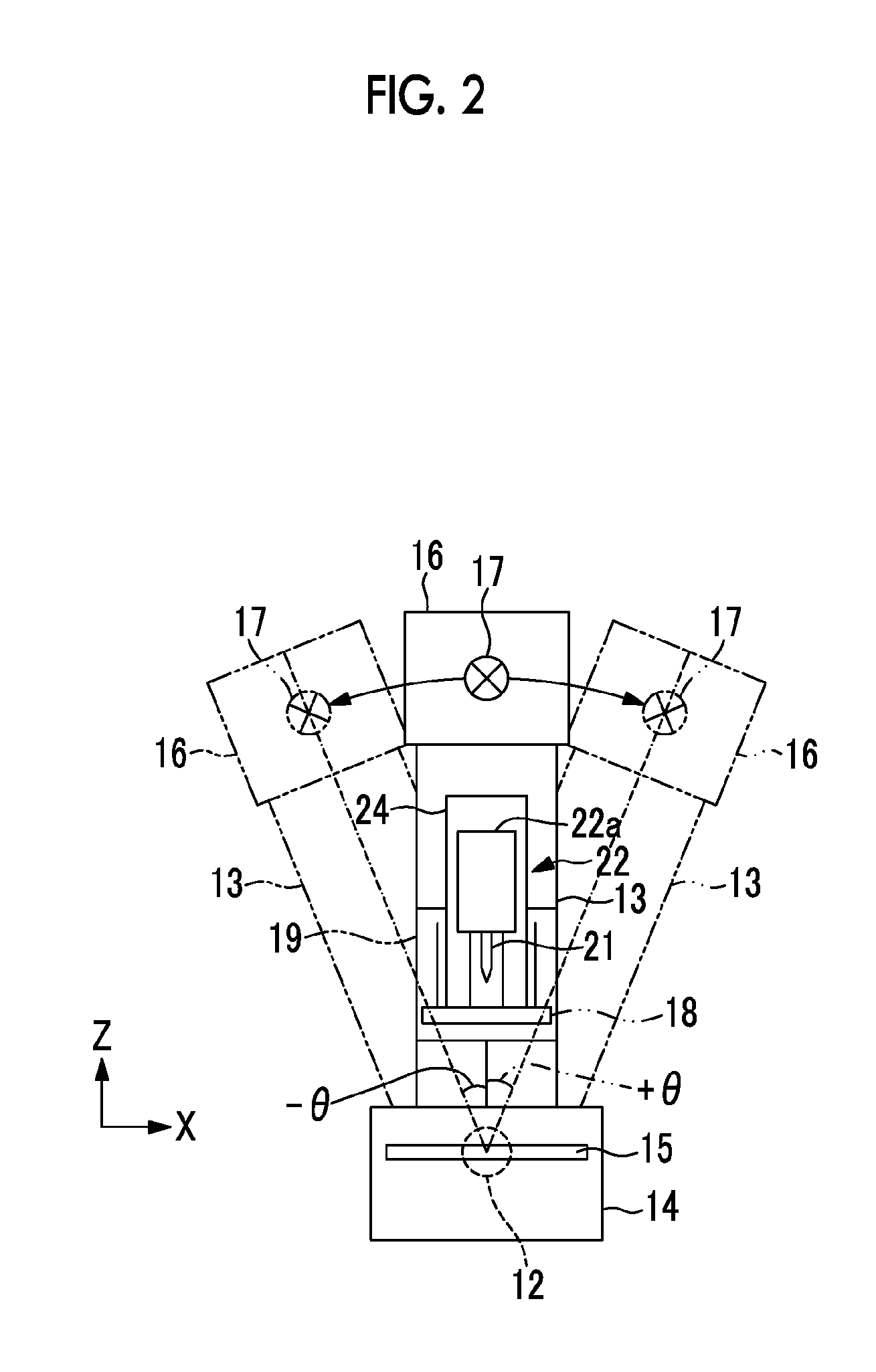
[0032]Hereinafter, a stereoscopic breast image radiographing and displaying system employing an embodiment of a radiological image radiographing and displaying system according to the present invention will be described with reference to the accompanying drawings. The breast image radiographing and displaying system according to this embodiment is a system also serving as a breast stereoscopic biopsy device by having a detachable biopsy unit attached thereto. First, the schematic configuration of the entire breast image radiographing and displaying system according to this embodiment will be described. FIG. 1 is a diagram schematically illustrating the configuration of the breast image radiographing and displaying system having a biopsy unit attached thereto.

[0033]As shown in FIG. 1, the breast image radiographing and displaying system 1 according to this embodiment includes a breast image radiographing unit 10, a computer 8 connected to the breast image radiographing unit 10, and a...


PUM


Abstract

Targeting of a lesion which is performed by a stereoscopic biopsy device or the like is performed simply and highly accurately. Designation of a predetermined position in the stereoscopic image is received to acquire position information when a stereoscopic image is displayed, radiological images of radiographing directions are displayed as two-dimensional images, a mark based on the position information, which is designated in the stereoscopic image, is displayed in the two-dimensional images, designation of a predetermined position in the two-dimensional images is further received to acquire the position information after the mark is displayed.

Description

BACKGROUND OF THE INVENTION[0001]1. Field of the Invention[0002]The present invention relates to radiological image radiographing and displaying method and system that displays a stereoscopic image using radiological images detected by a radiological image detector through irradiation of a subject with radiation from different directions, and more particularly, to use in a stereoscopic biopsy device that specifies a position of a lesion or the like in a stereoscopic image and collects a tissue fragment at the position.[0003]2. Description of the Related Art[0004]Tissue fragments around a lesion are often collected in hospital examinations. Recently, biopsy of piercing a patient with a tissue-collecting needle having a cavity (hereinafter, referred to as biopsy needle) and collecting a tissue filled in the cavity of the needle has attracted attention as a method of collecting a tissue fragment without giving a large burden to the patient. A stereoscopic biopsy device has been suggest...

Claims


Application Information

Patent Timeline
IPC IPC(8): G06T7/00
CPCA61B6/022G06T7/0014A61B10/0233A61B6/502A61B6/0414A61B6/466
Inventor NAKAYAMA, HIROKIHASEGAWA, AKIRAROMSDAHL, HARLAN
Owner FUJIFILM CORP
Why Eureka
Try Eureka