Radiation tomography apparatus

a tomography apparatus and radiation tomography technology, applied in the field of radiation tomography apparatus, can solve the problems of difficult to adjust the various conditions of radiography, the conventional radiation tomography apparatus b>51/b> has difficulty in obtaining tomographic images immediately after, and the tomographic image cannot be obtained immediately after, etc., to achieve high visibility, take time and effort for image processing, and high visibility

Inactive Publication Date: 2013-06-13
SHIMADZU CORP
View PDF6 Cites 20 Cited by
  • Summary
  • Abstract
  • Description
  • Claims
  • Application Information

AI Technical Summary

Benefits of technology

[0024]As mentioned above, the display device displays the preview image having the components on the high-frequency side extracted therefrom, thereby displaying the preview image with high visibility. In the method of generating the preview image by superimposing the fluoroscopic images having the components on the high-frequency side extracted therefrom, image processing has to be performed to every fluoroscopic image. Accordingly, it takes time and effort for image processing. On the other hand, according to the configuration mentioned above, it is only needed to perform image processing to the preview image. Consequently, the preview image with high visibility can be provided through simpler calculations.
[0025]The examples of this invention generate a tomographic image for diagnosis having a sectional image of a subject in any cut surface appearing therein by reconstructing the fluoroscopic images that are taken while a radiation source and a radiation detecting device are move in a direction of movement along a top board synchronously in opposite directions to each other. It takes some time to generate the tomographic image for diagnosis. Consequently, although blurred sectional image of the subject appears in the tomographic image for diagnosis, radiography cannot be conducted again for the fluoroscopic images until the tomographic image for diagnosis is generated. Then, according to the examples of this invention, the preview image used for previewing the sectional image of the subject is generated besides the tomographic images for diagnosis. The preview image is a sectional image for the subject same as that for the tomographic image for diagnosis, but has a limited cut surface in a particular position. As a result, the preview image can be obtained by simpler computing process. Consequently, the operator sees once the preview image displayed on the display device prior to the tomographic image for diagnosis, thereby determining immediately blurred conditions of the subject image appearing in the tomographic image generated from a series of fluoroscopic images. As a result, reconducting of radiography can be determined quickly.

Problems solved by technology

Moreover, it is supposed that low doses of radiation upon taking the fluoroscopic images cause a dark tomographic image.
The conventional configuration, however, has the following problem.
That is, there arises a problem that the conventional radiation tomography apparatus 51 has difficulty in adjusting various conditions of radiography.
Thus, the tomographic image cannot be obtained immediately after the fluoroscopic images are taken.
In the case of taking the tomographic image, however, it takes longer time from the first radiography until conducting the radiography again.
On the other hand, although the fluoroscopic images as an intermediate image in tomographic-image generation are visible, it is difficult to expect blur conditions of the subject in a tomographic image to be generated.
This increases a burden on the subject.
In addition, the doctor cannot determine not only blur conditions of the tomographic image but also underexposure / overexposure of the tomographic image, i.e., an extent of exposure before generating the tomographic image.
Also in this case, radiography cannot be conducted again rapidly.
Consequently, although blurred sectional image of the subject appears in the tomographic image for diagnosis, radiography cannot be conducted again for the fluoroscopic images until the tomographic image for diagnosis is generated.
Accordingly, it takes time and effort for image processing.
Consequently, although blurred sectional image of the subject appears in the tomographic image for diagnosis, radiography cannot be conducted again for the fluoroscopic images until the tomographic image for diagnosis is generated.

Method used


Image

  • Radiation tomography apparatus
  • Radiation tomography apparatus
  • Radiation tomography apparatus

Examples


example 2

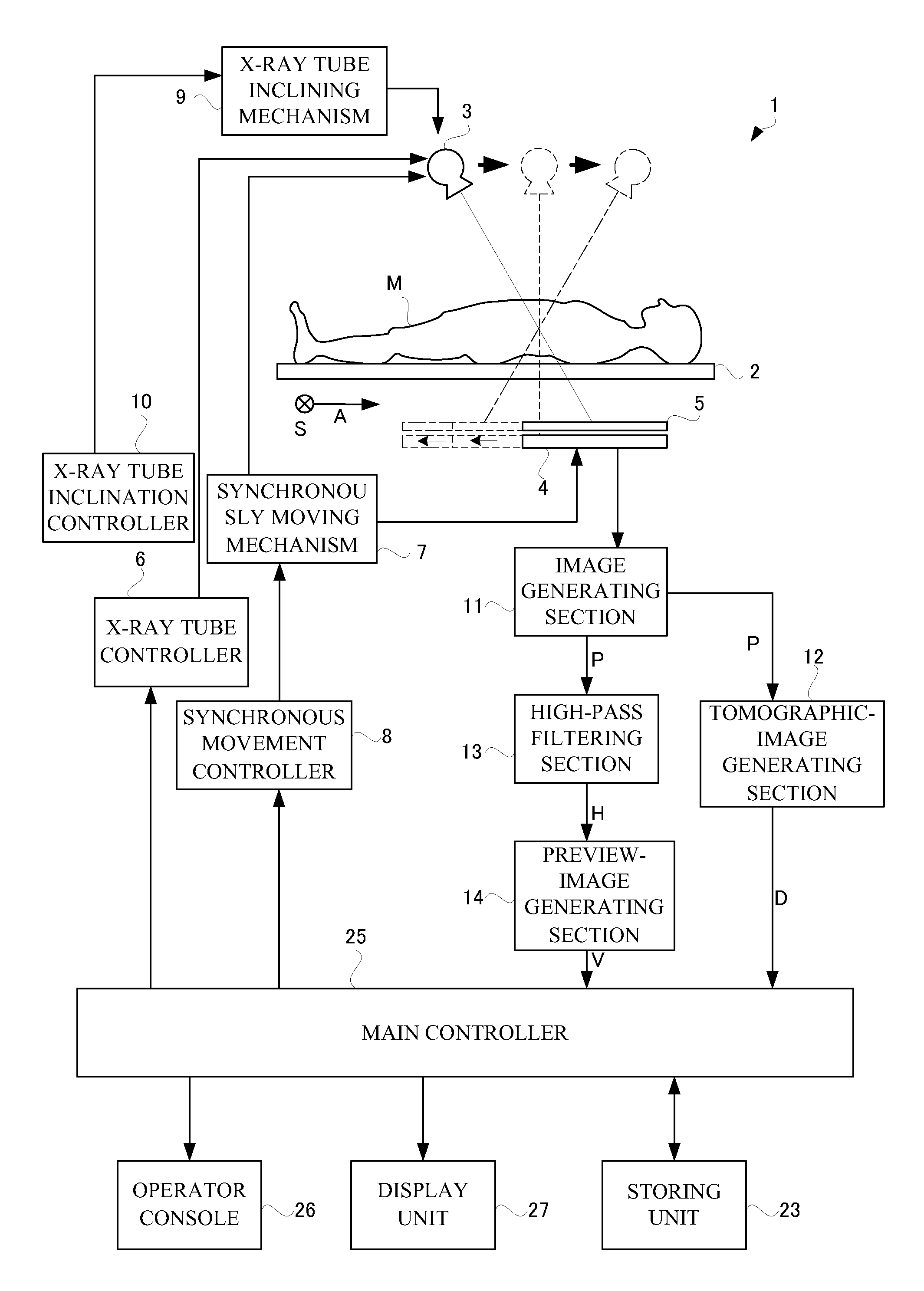
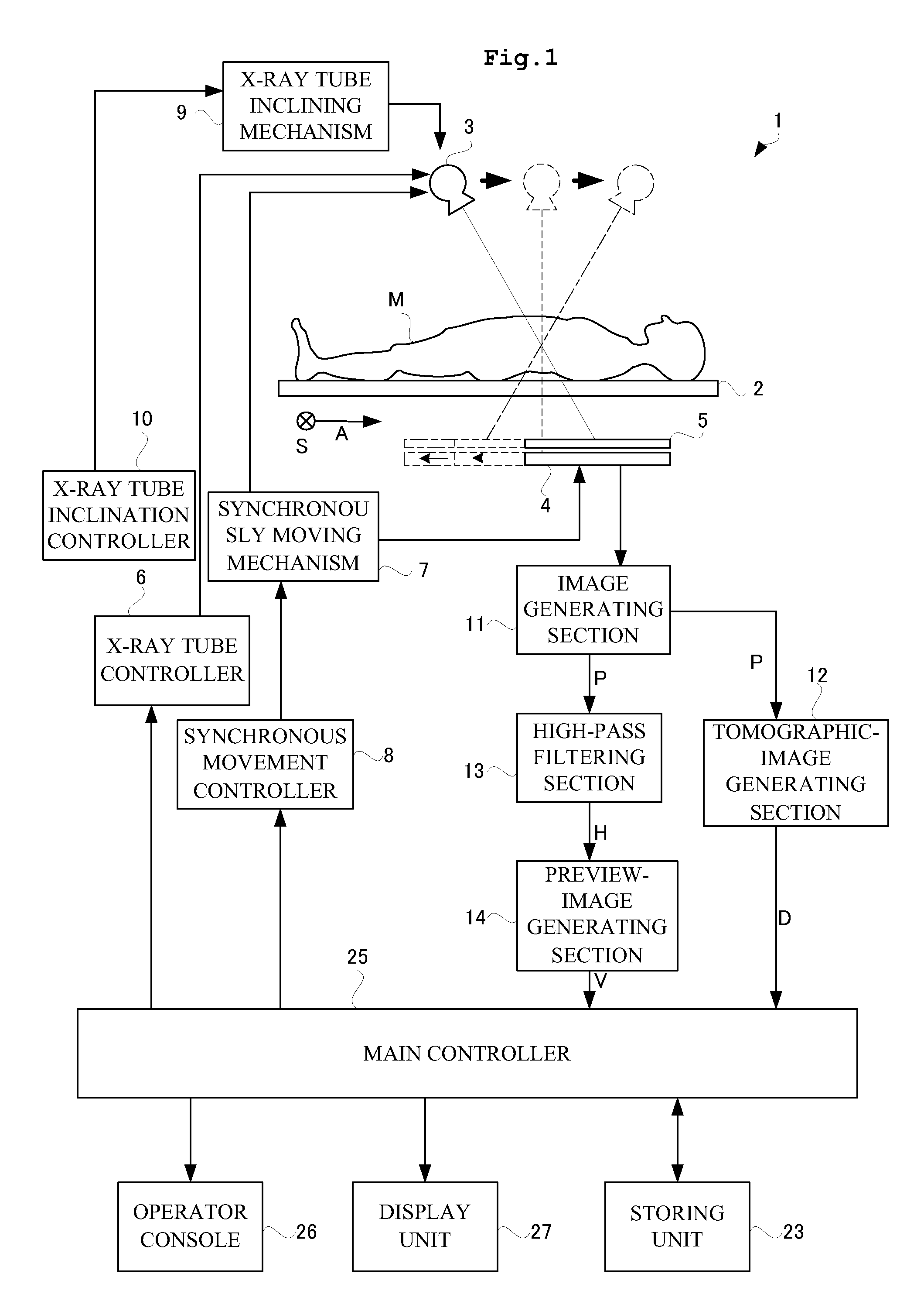
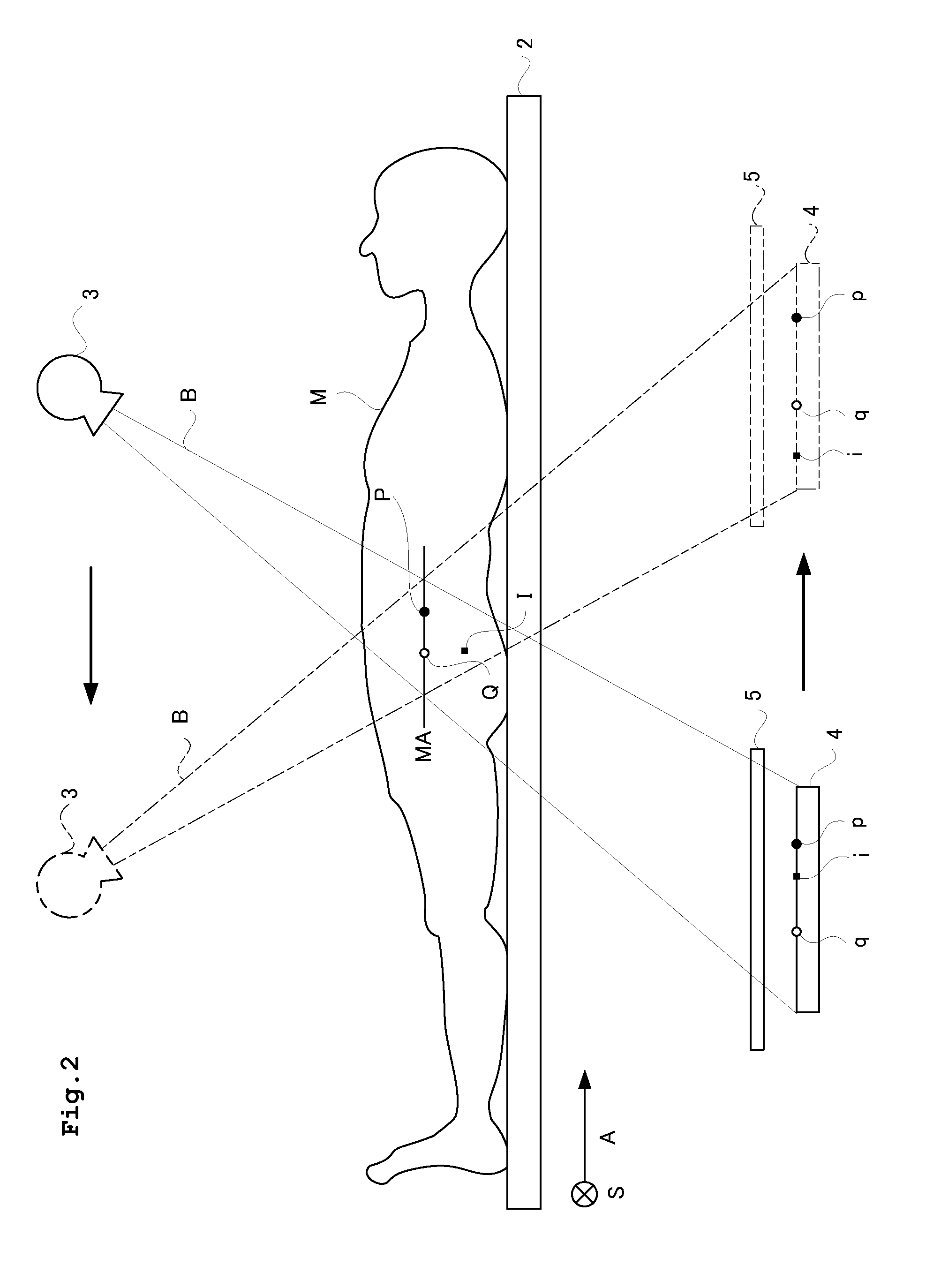
[0078]Description will be given next of a radiation tomography apparatus according to Example 2. FIG. 9 is a functional block diagram illustrating an X-ray apparatus according to Example 1 of this invention. As shown in FIG. 9, an X-ray apparatus 1 in Example 1 includes a top board 2 for supporting a subject M placed thereon as a target for X-ray tomography, an X-ray tube 3 disposed above the top board 2 (on one face side of the top board 2) for irradiating the subject with a cone-shaped X-ray beam, an FPD 4 below the top board 2 (on the other face side of the top board) for detecting X-rays transmitting through the subject M, a top-board moving mechanism 7a for moving the top board 2 relative to the X-ray tube 3 and the FPD 4 while a relationship between the X-ray tube 3 and the FPD 4 in which a center axis of the cone-shaped X-ray beam conforms to a center of the FPD 4 is maintained, a top-board movement controller 8a for controlling the top-board moving mechanism 7a, and an X-ray...


PUM


Abstract

Provided is a radiation tomography apparatus that performs radiography for a series of fluoroscopic images while moving a radiation source and a radiation detecting device synchronously in opposite directions to each other and obtains a tomographic image of a subject based on the fluoroscopic images. The radiation tomography apparatus allows to preview the tomographic image obtained through the radiography immediately after completion of the radiography. A preview image for previewing a sectional image of a subject is generated besides a tomographic image for diagnosis. The preview image can be obtained through simpler computing processing. Consequently, an operator sees once the preview image displayed on a display unit prior to the tomographic image for diagnosis, thereby determining to conduct radiography again quickly.

Description

BACKGROUND[0001]1. Field of the Invention[0002]This invention relates to a radiation tomography apparatus provided with a radiation source and an FPD. In particular, this invention relates to a radiation tomography apparatus in which a series of fluoroscopic images are taken while the radiation source and the FPD move synchronously in opposite directions to each other to obtain a tomographic image of a subject based on the fluoroscopic images.[0003]2. Description of the Related Art[0004]Medical institutions are equipped with a radiation tomography apparatus 51 for obtaining a tomographic image of a subject M. Such radiation tomography apparatus 51 includes a configuration in which a radiation source 53 that emits radiation and an FPD 54 that detects radiation move synchronously to take a series of fluoroscopic images successively, and then the series of fluoroscopic images are superimposed to obtain a tomographic image (see FIG. 14.) In such radiation tomography apparatus 51, during...

Claims


Application Information

Patent Timeline
Patent Type & Authority Applications(United States)
IPC IPC(8): A61B6/02
CPCA61B6/025A61B6/4085A61B6/4233G06T2211/436A61B6/5241G06T11/006G06T2211/421A61B6/5205
Inventor NOTOHARA, DAISUKENISHINO, KAZUYOSHI
Owner SHIMADZU CORP
Why Eureka
Try Eureka