Radiation imaging apparatus and operation method thereof

a technology of radiation imaging apparatus and operation method, which is applied in the field of radiation imaging apparatus, can solve problems such as patient useless radiation exposur

Inactive Publication Date: 2013-06-13
FUJIFILM CORP
View PDF0 Cites 34 Cited by
  • Summary
  • Abstract
  • Description
  • Claims
  • Application Information

AI Technical Summary

Benefits of technology

[0011]An object of the present invention is to provide a radiation imaging apparatus and an operation method thereof in which a radiation dose applied to a patient during pre-exposure is reflected without waste in a radiographic image for use in diagnosis.

Problems solved by technology

The pre-exposure is useful for determining the appropriate main exposure condition, but causes useless radiation exposure to the patient because the X-ray dose applied during the pre-exposure is not reflected in the X-ray image.

Method used


Image

  • Radiation imaging apparatus and operation method thereof
  • Radiation imaging apparatus and operation method thereof
  • Radiation imaging apparatus and operation method thereof

Examples


Embodiment Construction

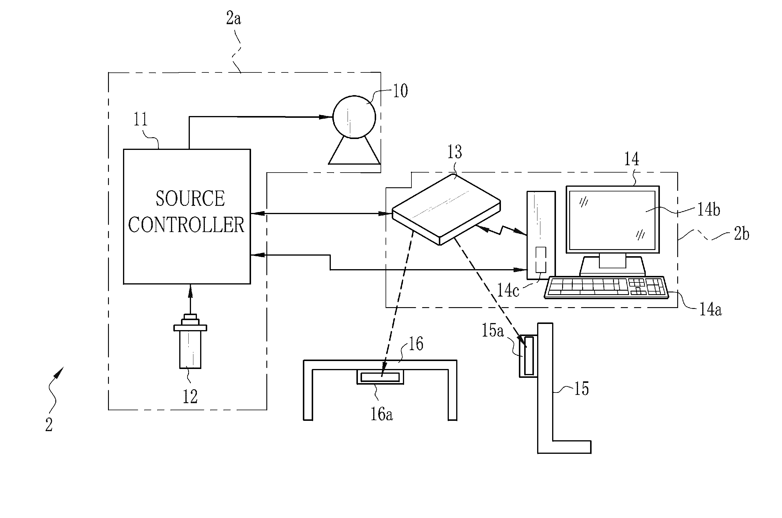
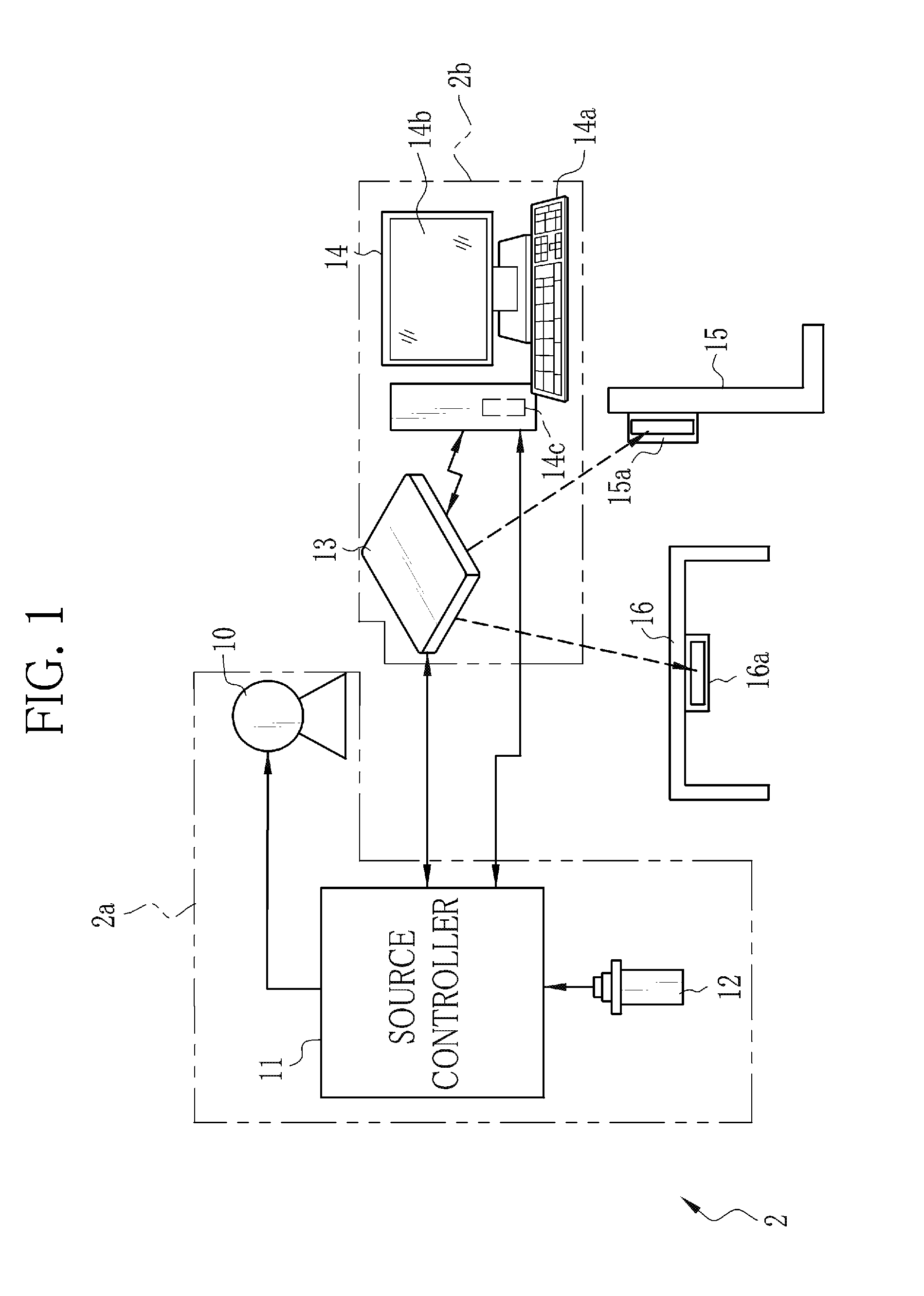
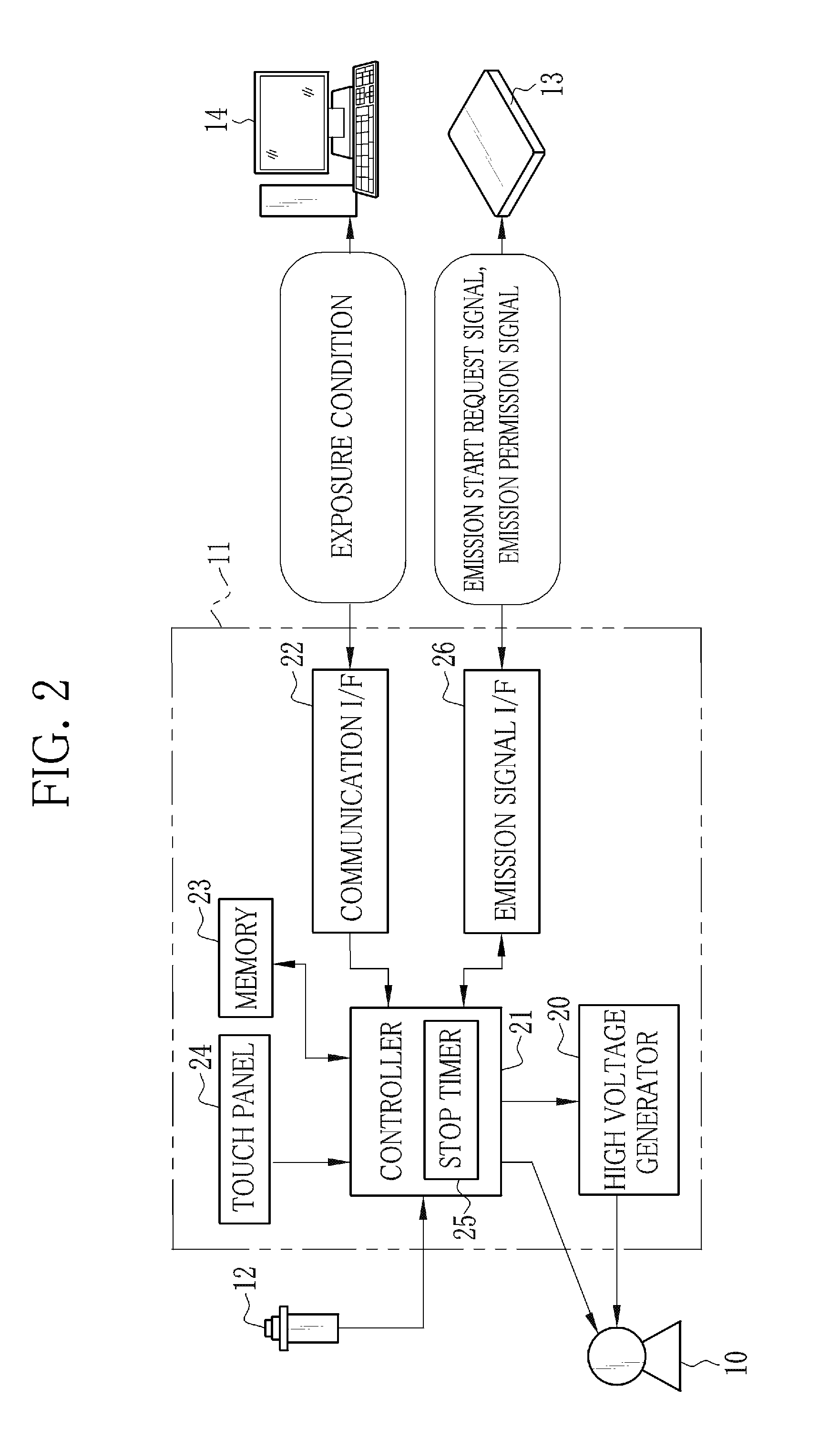
[0053]As shown in FIG. 1, an X-ray imaging system 2 is constituted of an X-ray source 10, a source controller 11, an emission switch 12, an electronic cassette 13, a console 14, and an imaging stand 15, and an imaging table 16. The X-ray source 10 contains an X-ray tube for emitting X-rays. The source controller 11 controls the operation of the X-ray source 10. The emission switch 12 commands the start of warming-up and the start of X-ray emission to the X-ray source 10. The electronic cassette 13 detects the X-rays that have passed through a patient's body (object), and outputs an X-ray image. The console 14 performs the operation control of the electronic cassette 13 and the display process of the X-ray image. The imaging stand 15 and the imaging table 16 are used in X-ray imaging of the patient who is in a standing position and a lying position, respectively. The X-ray source 10, the source controller 11, and the emission switch 12 compose an X-ray generating apparatus 2a. The el...


PUM


Abstract

An X-ray imaging system performs pre-exposure and main exposure to take a single X-ray image. In the pre-exposure, an AEC circuit reads out a dose detection signal outputted from a detection pixel, and compares an integral value of the dose detection signal with a threshold value. When the integral value has reached the threshold value, the AEC circuit stops X-ray emission. A main exposure condition determination unit determines main irradiation time, being one item of a main exposure condition, based on irradiation time in the pre-exposure, an integral dose in the pre-exposure, and a necessary dose required for production of the X-ray image. After that, the main exposure is performed immediately with the determined main irradiation time. Normal pixels perform a charge accumulation operation continuously from the start of the pre-exposure to the end of the main exposure, and the X-ray image is produced from accumulated electric charge.

Description

BACKGROUND OF THE INVENTION[0001]1. Field of the Invention[0002]The present invention relates to a radiation imaging apparatus and an operation method of the radiation imaging apparatus.[0003]2. Description Related to the Prior Art[0004]In a medical field, a radiation imaging system, for example, an X-ray imaging system using X-rays is widely known. The X-ray imaging system is constituted of an X-ray generating apparatus for generating the X-rays, and an X-ray imaging apparatus that takes an X-ray image by reception of the X-rays passed through a patient's body. The X-ray generating apparatus includes an X-ray source for emitting the X-rays to the patient's body, a source controller for controlling the operation of the X-ray source, and an emission switch for issuing an emission start signal of the X-rays to the source controller. The X-ray imaging apparatus includes an X-ray image detecting device that detects the X-ray image by conversion of the X-rays passed through the patient's...

Claims


Application Information

Patent Timeline
Patent Type & Authority Applications(United States)
IPC IPC(8): A61B6/00
CPCA61B6/542A61B6/585H04N5/235A61B6/548A61B6/4233A61B6/527A61B6/545H04N5/32H04N23/70
Inventor TAJIMA, TAKASHI
Owner FUJIFILM CORP
Why Eureka
Try Eureka