Intelligent Atlas for Automatic Image Analysis of Magnetic Resonance Imaging

Inactive Publication Date: 2013-07-04
THE JOHN HOPKINS UNIV SCHOOL OF MEDICINE
View PDF3 Cites 21 Cited by
  • Summary
  • Abstract
  • Description
  • Claims
  • Application Information

AI Technical Summary

Problems solved by technology

Even though quantitative analyses may significantly improve our ability to detect and characterize abnormalities, currently almost no quantitative techniques can be accepted as a part of routine radiological diagnosis.
This is partly due to difficulties in analyzing tissue based on radiological, such as Magnetic Resonance (MR) images.
For example, high quality segmentation of the brain boundary requires a considerable amount of manual labor, whic

Method used


Image

  • Intelligent Atlas for Automatic Image Analysis of Magnetic Resonance Imaging
  • Intelligent Atlas for Automatic Image Analysis of Magnetic Resonance Imaging
  • Intelligent Atlas for Automatic Image Analysis of Magnetic Resonance Imaging

Examples


Embodiment Construction

[0020]Some embodiments of the current invention are discussed in detail below. In describing the embodiments, specific terminology is employed for the sake of clarity. However, the invention is not intended to be limited to the specific terminology so selected. A person skilled in the relevant art will recognize that other equivalent components can be employed and other methods developed without departing from the broad concepts of the current invention. All references cited herein are incorporated by reference as if each had been individually incorporated.

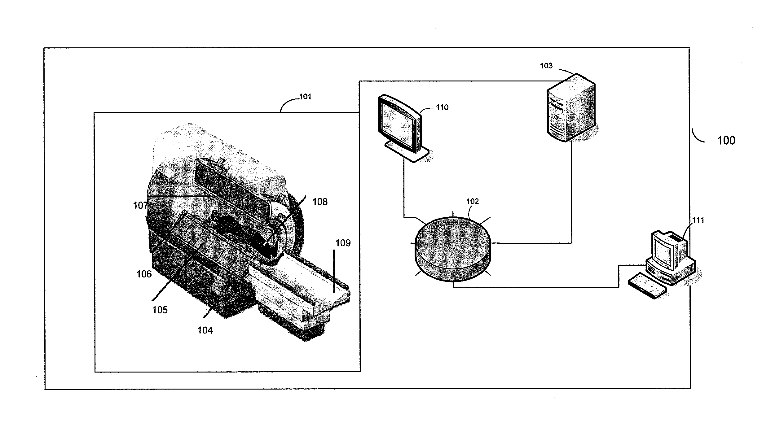
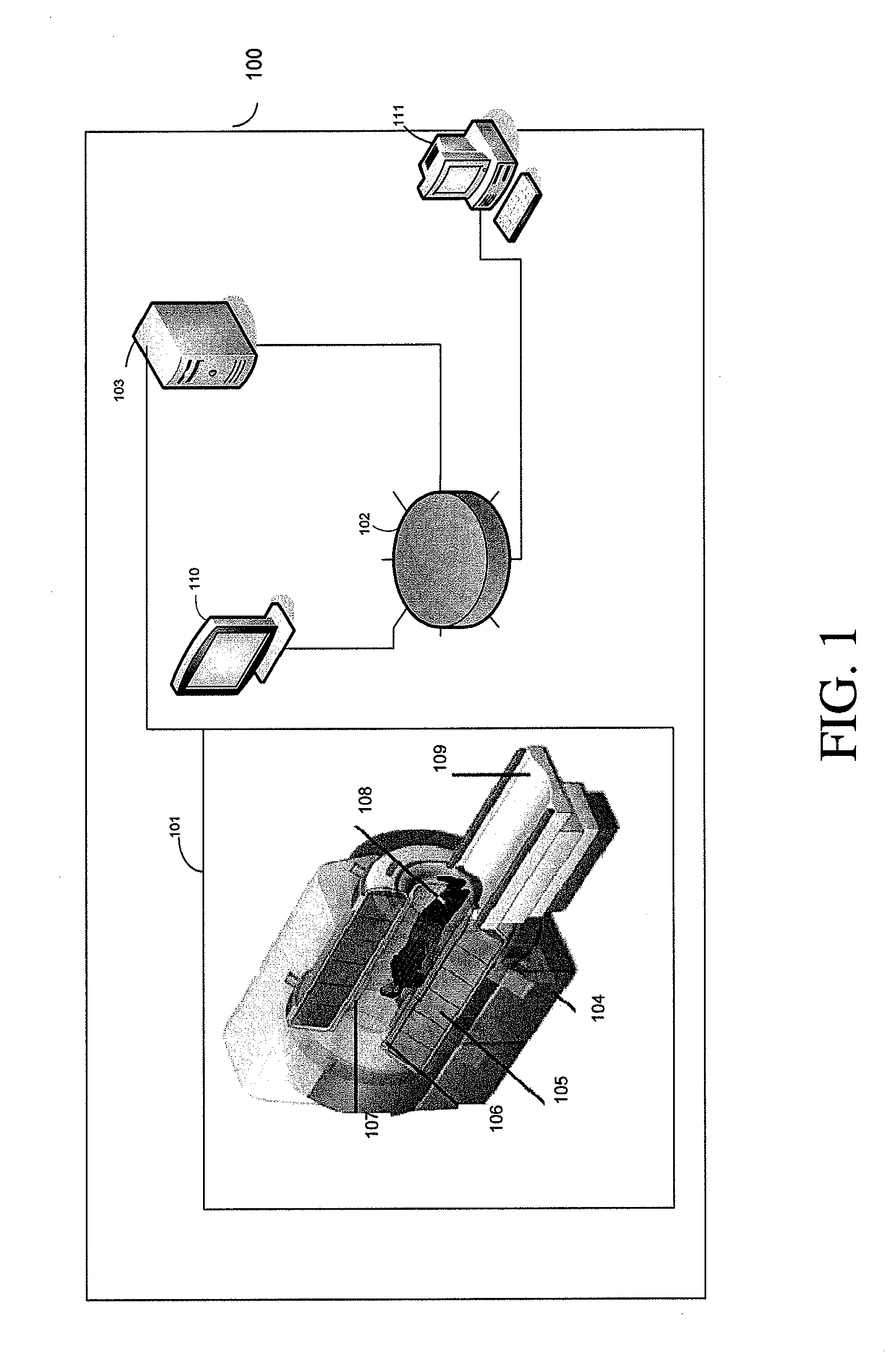
[0021]FIG. 1 is a schematic illustration of a non-invasive imaging system 100 according to some embodiments of the current invention. The non-invasive imaging system 100 includes an imaging scanner 101, a data storage unit 102, and a signal processing system 103. Imaging scanner 101 may be, but is not limited to, a magnetic resonance imaging (MRI) scanner, a computed tomography (CT) scanner, a positron emission tomography (PET) sc...


PUM


Abstract

A non-invasive imaging system, including an imaging scanner suitable to generate an imaging signal from a tissue region of a subject under observation, the tissue region having at least one substructure; a signal processing system in communication with the imaging scanner to receive the imaging signal from the imaging scanner; and a data storage unit in communication with the signal processing system, wherein the data storage unit stores an anatomical atlas comprising data encoding spatial information of the at least one substructure in the tissue region, and a pathological atlas corresponding to an abnormality of the tissue region, wherein the signal processing system is adapted to automatically identify, using the imaging signal, the anatomical atlas, and the pathological atlas, a presence of the abnormality or a precursor abnormality in the subject under observation.

Description

CROSS-REFERENCE OF RELATED APPLICATION[0001]This application claims priority to U.S. Provisional Application No. 61 / 329,857 filed on Apr. 30, 2010, the entire contents of which are hereby incorporated by reference.[0002]The U.S. Government has a paid-up license in this invention and the right in limited circumstances to require the patent owner to license others on reasonable terms as provided for by the terms of Grant No.: R01AG20012 and P41RR15241, awarded by the National Institutes of Health.BACKGROUND[0003]1. Field of Invention[0004]The current invention relates to non-invasive imaging systems and methods, and more particularly to imaging systems and methods that provide computer assisted diagnosis of tissue abnormalities in human and animal subjects.[0005]2. Discussion of Related Art[0006]Conventional radiological diagnosis can be qualitative or subjective. Even though quantitative analyses may significantly improve our ability to detect and characterize abnormalities, currentl...

Claims


Application Information

Patent Timeline
IPC IPC(8): G06T7/00A61B5/00
CPCA61B5/055A61B6/032A61B6/037G06T7/0012A61B6/501A61B6/5217A61B5/72A61B6/50G16H50/30A61B5/00G06T7/0014G06T2207/20221A61B5/004G06T7/143G06T7/174G06T7/248G06T7/149G01R33/56341G06T2207/10092A61B5/4064A61B2576/026A61B2576/00A61B2576/02G06T15/00G01R33/20G01R33/5608A61B6/52G01T1/2985G01T1/161G16H50/20G16H50/50G06F18/2132G06F18/2321A61B6/461G06T2207/10081G06T2207/10088G06T2207/10104G06T2207/10108
Inventor MORI, SUSUMUMILLER, MICHAEL IOISHI, KENICHI
Owner THE JOHN HOPKINS UNIV SCHOOL OF MEDICINE
Why Eureka
Try Eureka