Cassette for facilitating optical sectioning of a retained tissue specimen

a tissue specimen and optical sectioning technology, applied in the field of cassettes for retaining tissue specimens, can solve the problems of inconvenient optical sectioning, inconvenient operation, and inability to use optical imaging techniques, and achieve the effect of facilitating optical examination of tissue specimens

Inactive Publication Date: 2013-07-18
CALIBER IMAGING & DIAGNOSTICS
View PDF4 Cites 22 Cited by
  • Summary
  • Abstract
  • Description
  • Claims
  • Application Information

AI Technical Summary

Benefits of technology

[0012]Accordingly, it is the principal object of the present invention to provide a cassette for retaining a specimen of surgically exposed tissue from a patient which facilitates optical examination of the tissue by a confocal microscopic or other optical imaging microscope.
[0015]It is a further object of the present invention to provide a cassette for retaining a tissue specimen which enables Mohs surgery by optical sectioning the specimen with a confocal microscopic or other optical imaging microscope.
[0016]It is a still another object of the present invention to provide a cassette for a tissue specimen which can be used to archive and identify the tissue specimen more easily than the prior art tissue specimens from Mohs surgery which are physically sectioned and prepared on multiple slides.
[0018]A still further object of the present invention is to provide a cassette for retaining a tissue specimen which allows the entire sample to be observable in the cassette through the top and bottom of the cassette.
[0020]The base member may has at least one injection port through which fluids, via a syringe needle, may be inserted and removed from the tissue specimen retained in the cavity of the cassette. Such fluids can facilitate imaging of the specimen by the optical imaging microscope, or can be used to place the specimen in a preservative for archiving purposes. A label with indicia identifying the tissue specimen may be applied to the cassette.

Problems solved by technology

However, since numerous sections must be made from each tissue specimen and then histologically prepared on slides, this procedure is both tedious and time consuming.
This procedure is adequate for obtaining a section which can be placed on a slide for review under a microscope, but is not useful with optical imaging techniques, such as provided by confocal microscopes, which can examine a surgically exposed tissue specimen without the need for traditional microtome sectioning or slide preparation.
One problem with optical imaging of a tissue specimen for Mohs surgery is that the tissue specimen is generally too thick, for example 2-3 mm, to enable optically imaging of the edges of the specimen to determine if the specimen contains all of the tumor.
Typically, a confocal microscope is limited to producing adequate images of tissue sections at 100-200 microns.
This necessitates that the tissue specimen be wetted or immersed in a fluid having an optical index suitable for the objective lens, otherwise, the imaging performance of the system is severely degraded.
Further, prior to mechanical sectioning, such a specimen is incompatible with optical imaging techniques since fluid cannot be present with a tissue specimen in a vacuum, and the platform does not provide a surface through which optical imaging can be performed.
This requires storage of the slides for many years which is cumbersome for large volumes of sectioned tissue specimens.
Such a device is limited to dialysis and provides no mechanism for retaining a tissue specimen in a planar orientation for optical sectioning.

Method used


Image

  • Cassette for facilitating optical sectioning of a retained tissue specimen
  • Cassette for facilitating optical sectioning of a retained tissue specimen
  • Cassette for facilitating optical sectioning of a retained tissue specimen

Examples


Embodiment Construction

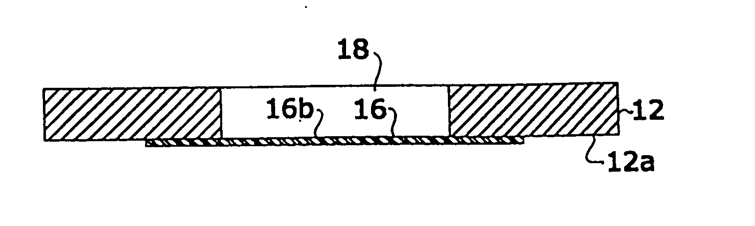
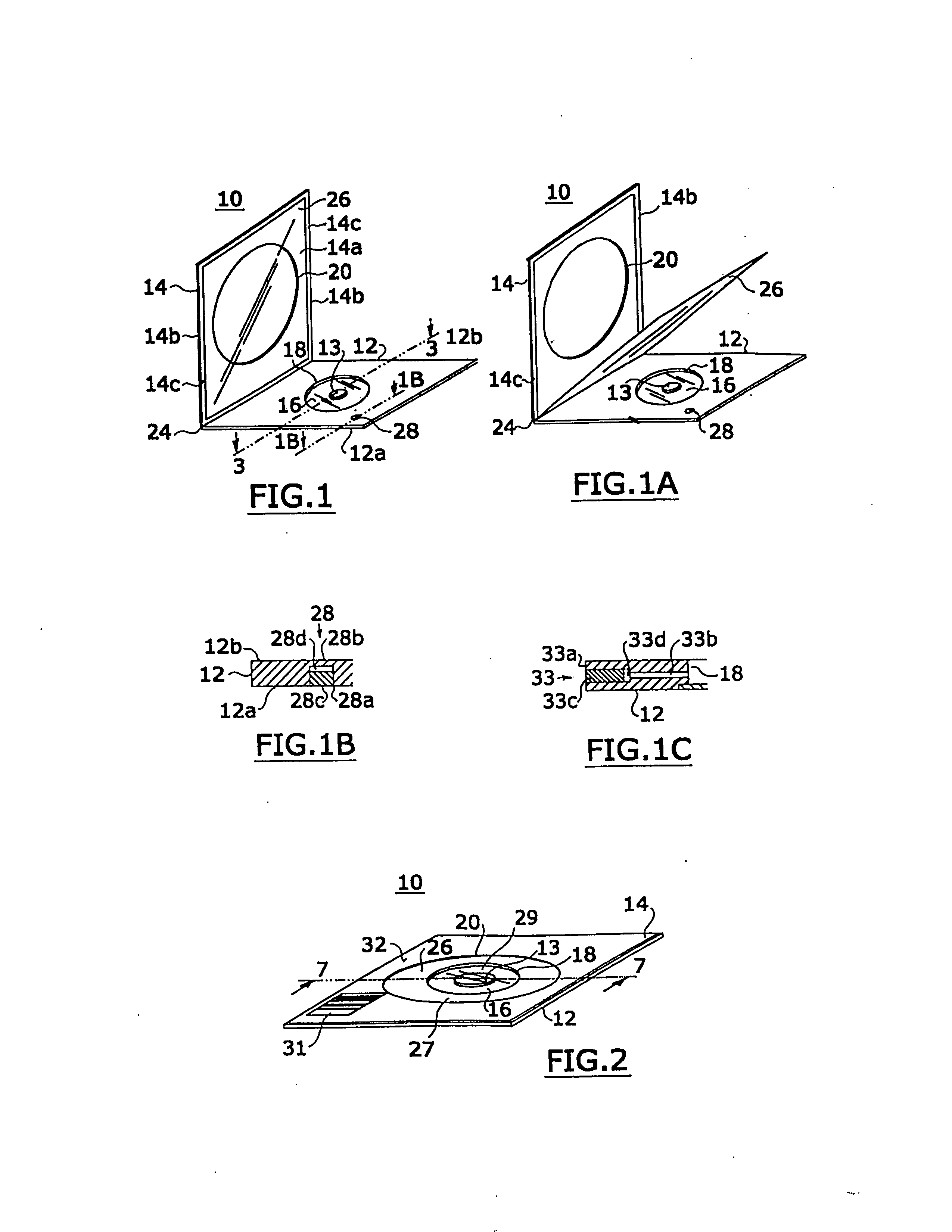
[0048]Referring to FIGS. 1-3, a cassette 10 of the present invention is shown having a base member 12 and an upper member 14. Base member 12 has an aperture 18 and a rigid optically transparent window 16 situated in aperture 18. The upper member 14 has an aperture 20 preferably substantially greater in size than the aperture 18 of base member 12. Base and upper members 12 and 14 are hinged at a hinge 24 which allows cassette 10 to have an open state, as shown in FIG. 1, and a closed state, as shown in FIG. 2, where upper member 14 covers base member 12. Although preferably base and upper members 12 and 14 are attached by a hinge 24, they may, in the alternative, be separate and unattached to each other when cassette 10 is in an open state.

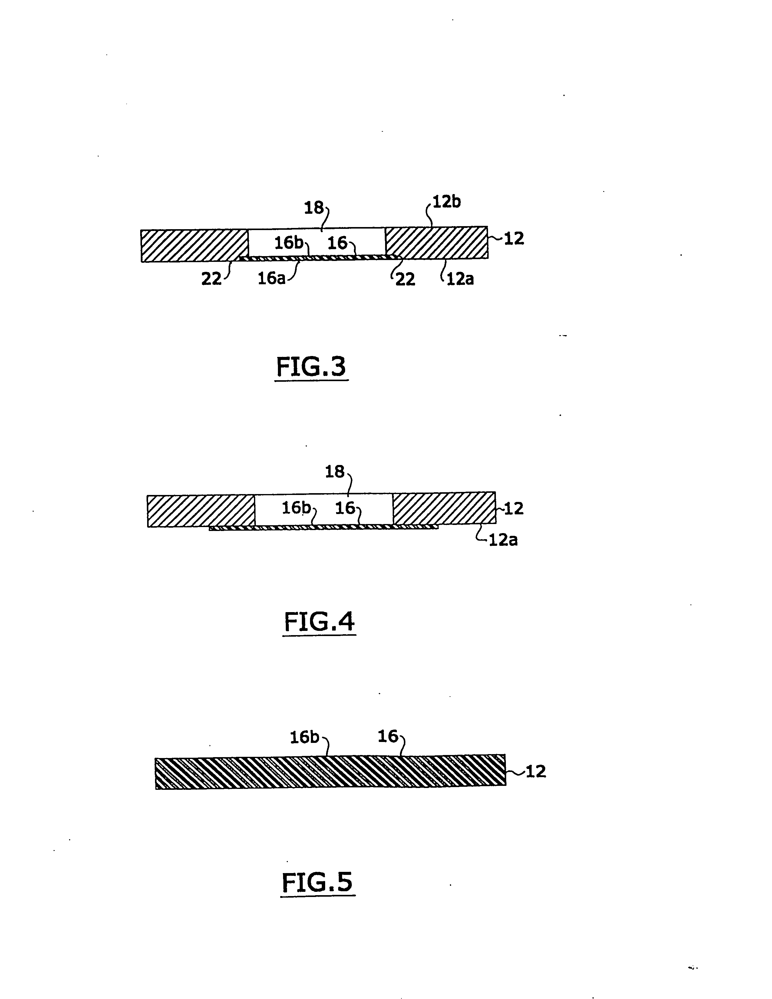
[0049]As best shown in FIG. 3, at the bottom surface 12a of base member 12, the base member has an annular shelf 22 in communication with aperture 18. Window 16 is inset on shelf 22, such that bottom surface 12a of the base member is planar with th...


PUM

PropertyMeasurementUnit
thickaaaaaaaaaa
thickaaaaaaaaaa
heightaaaaaaaaaa

Abstract

A cassette for retaining a specimen of surgically exposed tissue from a patient in an orientation that facilitates optical sectioning of the tissue by a confocal microscopic or other optical imaging microscope. The cassette includes a base member having a rigid optically transparent window upon which a tissue specimen is situated, a pliable membrane locatable over a substantial portion of the base member including the window, and an upper member, having an aperture therethrough, which can cover the base member to provide an enclosed cavity between the membrane and the window sealing the tissue specimen therein. The edges of the tissue specimen may be positioned planar against the window and retained in that position by bonds formed between the membrane and window at multiple points or locations around the tissue specimen. The specimen retained in the cavity is imagible a microscope through the window of the base member.

Description

[0001]This application claims the benefit of priority to U.S. Provisional Application No. 60 / 120,534, filed Feb. 17, 1999, which in herein incorporated by reference.FIELD OF THE INVENTION[0002]The present invention relates to a cassette for retaining a tissue specimen, and relates particularly to a cassette for retaining a specimen of surgically exposed tissue from a patient in an orientation that facilitates optical sectioning of the tissue by a confocal microscope, or other optical imaging microscope, for Mohs micrographic surgery. The invention further relates to a method of using the cassette for preparing a tissue specimen for examination by a confocal microscope or other optical imaging microscope, and a system for optically sectioning a tissue specimen retained in a cassette.BACKGROUND OF THE INVENTION[0003]In Mohs micrographic surgery, tissue having a tumor, typically a carcinoma on the skin of the head or neck, is excised from a patient under microscopic control. The excise...

Claims


Application Information

Patent Timeline
Patent Type & Authority Applications(United States)
IPC IPC(8): G02B21/34B01L3/00G01N1/31
CPCB01L3/508B01L2200/0642B01L2300/021B01L2300/043G02B21/34B01L2300/0609B01L2300/0822B01L2300/123G01N2001/315B01L2300/044G02B21/0012G02B21/26G02B21/33G02B21/368
Inventor EASTMAN, JAY M.FOX, WILLIAM J.GREENWALD, ROGER J.ROESSER, KEVIN P.ZAVISLAN, JAMES M.
Owner CALIBER IMAGING & DIAGNOSTICS
Why Eureka
Try Eureka