Perfusion scanning detects angiogenesis from similarity in evolution of local concentrations of contrast agent

a contrast agent and scanning technology, applied in the field of perfusion scanning detection angiogenesis, can solve the problem that the known applications of perfusion imaging techniques cannot readily characterize the perfusable structure, and achieve the effect of improving image quality and similar noise characteristics

Active Publication Date: 2013-07-18
ANGIOGENESIS ANALYTICS BV
View PDF4 Cites 0 Cited by
  • Summary
  • Abstract
  • Description
  • Claims
  • Application Information

AI Technical Summary

Benefits of technology

[0028]An advantage of this approach is the following. Noise in the evolution as determined at the specific location (e.g., the specific pixel) and noise in the evolution as determined at an adjacent location (e.g., a pixel adjacent to the specific pixel) are typically correlated. For example, the resolution of an image obtained through conventional medical imaging techniques turns out, in practice, to be higher than the physical resolution of the scanner used in this imaging technique. This results from some interpolation algorithm being applied in the scanner proper. Noise correlation in neighboring pixels can also be due to pre-processing techniques implemented by the scanner in or

Problems solved by technology

A problem recognized by the inventor is that the known applications of the perfusion imaging techniques cannot readily charact

Method used


Image

  • Perfusion scanning detects angiogenesis from similarity in evolution of local concentrations of contrast agent
  • Perfusion scanning detects angiogenesis from similarity in evolution of local concentrations of contrast agent
  • Perfusion scanning detects angiogenesis from similarity in evolution of local concentrations of contrast agent

Examples


Embodiment Construction

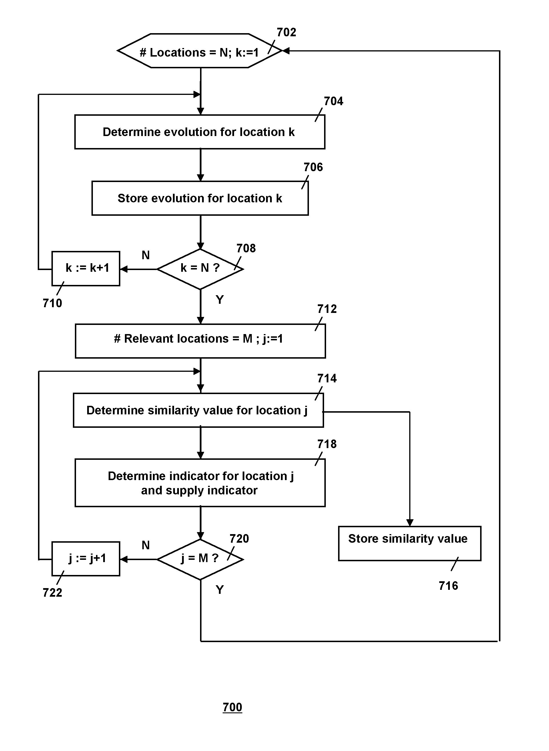
[0054]The invention relates to using a perfusion scanning medical imaging technique to generate an image of a perfusable structure of an organism. A fluid is flowing through the structure, and a dose of a traceable agent has been introduced into the fluid, or generated within the fluid. The evolution of the spatial concentration of the agent, e.g., a set of values of the magnitude of the concentration assumed at various moments over a period of time, is determined for a plurality of locations within the structure. The spatial pattern of the evolutions is analyzed and an image is generated on the basis of this analysis in order to enable the medical practitioner to draw conclusions about the dispersion characteristics of the perfusable structure.

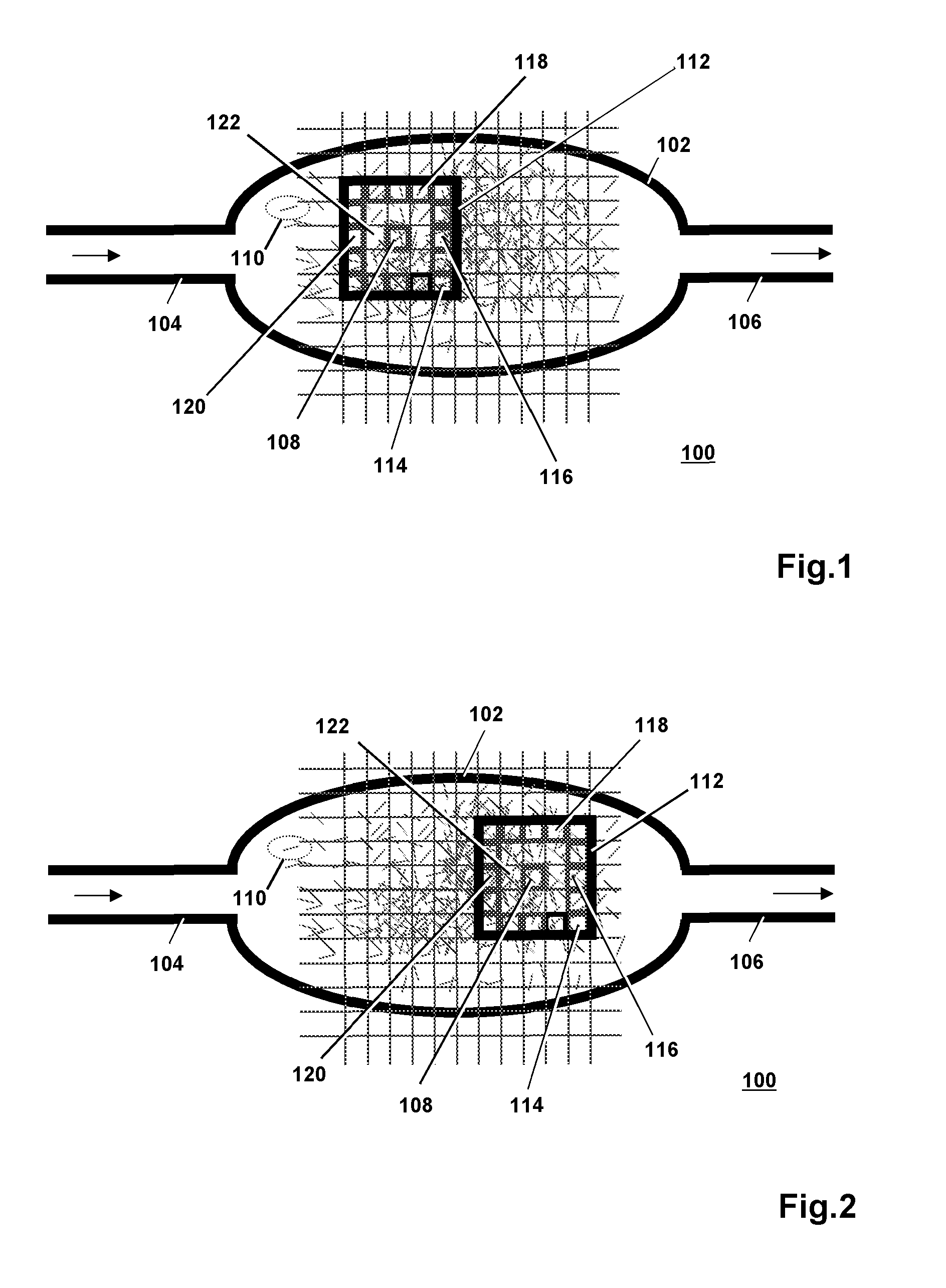
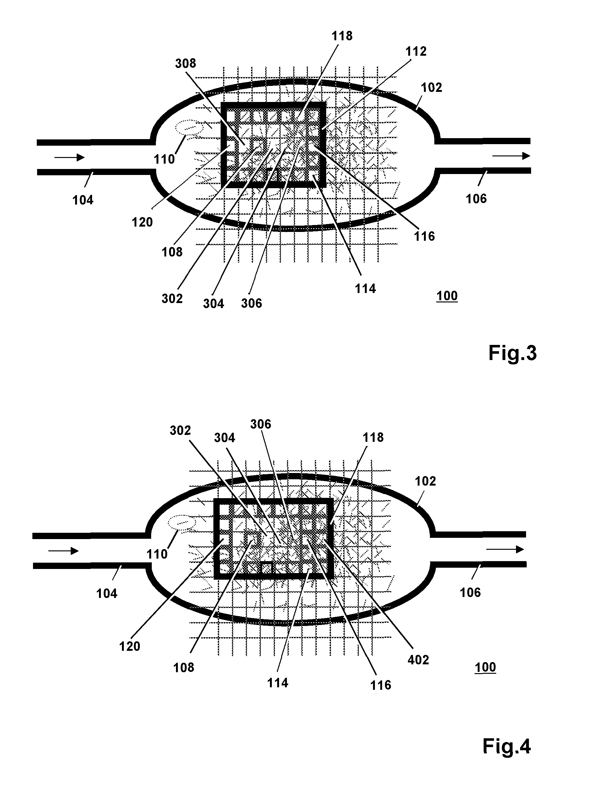
[0055]FIG. 1 is a schematic image 100 of a perfusable structure 102 of an organism, e.g., a gland such as the prostate in a male mammal. The main blood supply to the prostate is provided by the internal iliac arteries. These internal iliac ar...


PUM


Abstract

The invention relates to using a perfusion scanning medical imaging technique to generate an image of a perfusable structure of an organism. A fluid is flowing through the structure, and a dose of a traceable agent is present in the fluid. The evolution of the spatial concentration of the agent, e.g., a set of values of the magnitude of the concentration assumed at various moments over a period of time, is determined for a plurality of locations within the structure. The spatial pattern of the evolutions is analyzed and an image is generated on the basis of this analysis in order to enable the medical practitioner to draw conclusions about the dispersion characteristics of the perfusable structure.

Description

FIELD OF THE INVENTION[0001]The invention relates to a method of controlling generating an image of a perfusable structure of an organism, while a fluid is flowing through the perfusable structure. The invention further relates to a system for controlling generating such image and to control software on a computer-readable medium.BACKGROUND ART[0002]A perfusable structure is a structure that enables the passage of a fluid (e.g., a liquid or a gas) through the structure. An example of a perfusable structure is the vascular structure of an organ of an organism (plant, animal, human), such as the vascular structure of the brains or heart of a human or of an animal, or the vascular structure of a gland, e.g., the prostate of a male mammal. Medical imaging techniques based on monitoring the perfusion of a fluid (lymph) through the vessels of a lymphatic system or blood through the blood vessels to an organ or to a tissue are commonly referred to as “perfusion scanning techniques”.[0003]E...

Claims


Application Information

Patent Timeline
IPC IPC(8): G06F19/00
CPCG06F19/321G06F19/3468G06F19/36G16H30/20G16H20/17
Inventor MISCHI, MASSIMO
Owner ANGIOGENESIS ANALYTICS BV
Why Eureka
Try Eureka