
System for Suppressing Vascular Structure in Medical Images

Active Publication Date: 2013-08-22
SIEMENS HEALTHCARE GMBH
View PDF14 Cites 14 Cited by
  • Summary
  • Abstract
  • Description
  • Claims
  • Application Information

AI Technical Summary

Benefits of technology

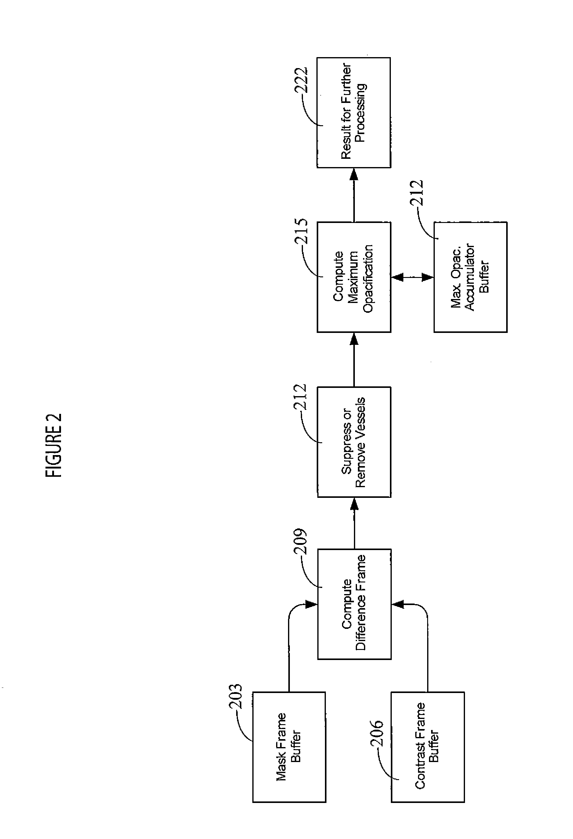
The patent describes a system that improves the visibility of blood vessels in X-ray images by reducing the impact of other structures, such as bones and muscles. This is particularly useful for capturing the initial phase of a contrast agent injection, where the capillaries are the most visible. The system uses a series of images taken before and after the injection to create a detailed map of the vessels in the area. By identifying and removing larger vessels with a system of algorithms, the system can generate an enhanced image that is easier to visualize. This technology can help healthcare professionals better understand and analyze the blood flow in a patient's body.

Problems solved by technology

Unfortunately, such capillary perfusion is not easily visualized due to both faint appearance of the capillaries relative to larger vessels and the capillaries being obscured by the larger vessels in an image.

Method used


Image

  • System for Suppressing Vascular Structure in Medical Images
  • System for Suppressing Vascular Structure in Medical Images
  • System for Suppressing Vascular Structure in Medical Images

Examples


Embodiment Construction

[0011]A system generates a maximally perfused image in which individual image picture elements (pixels) represent peak (substantially maximally opacified) contrast agent at their respective image locations in multiple images acquired over the duration of contrast agent flow. In the case of a maximally perfused image however, the vessel structure is advantageously removed from each image prior to processing for inclusion into a substantially maximally opacified resultant image. The resultant image advantageously provides enhanced visualization of capillary (or parenchymal) perfusion.

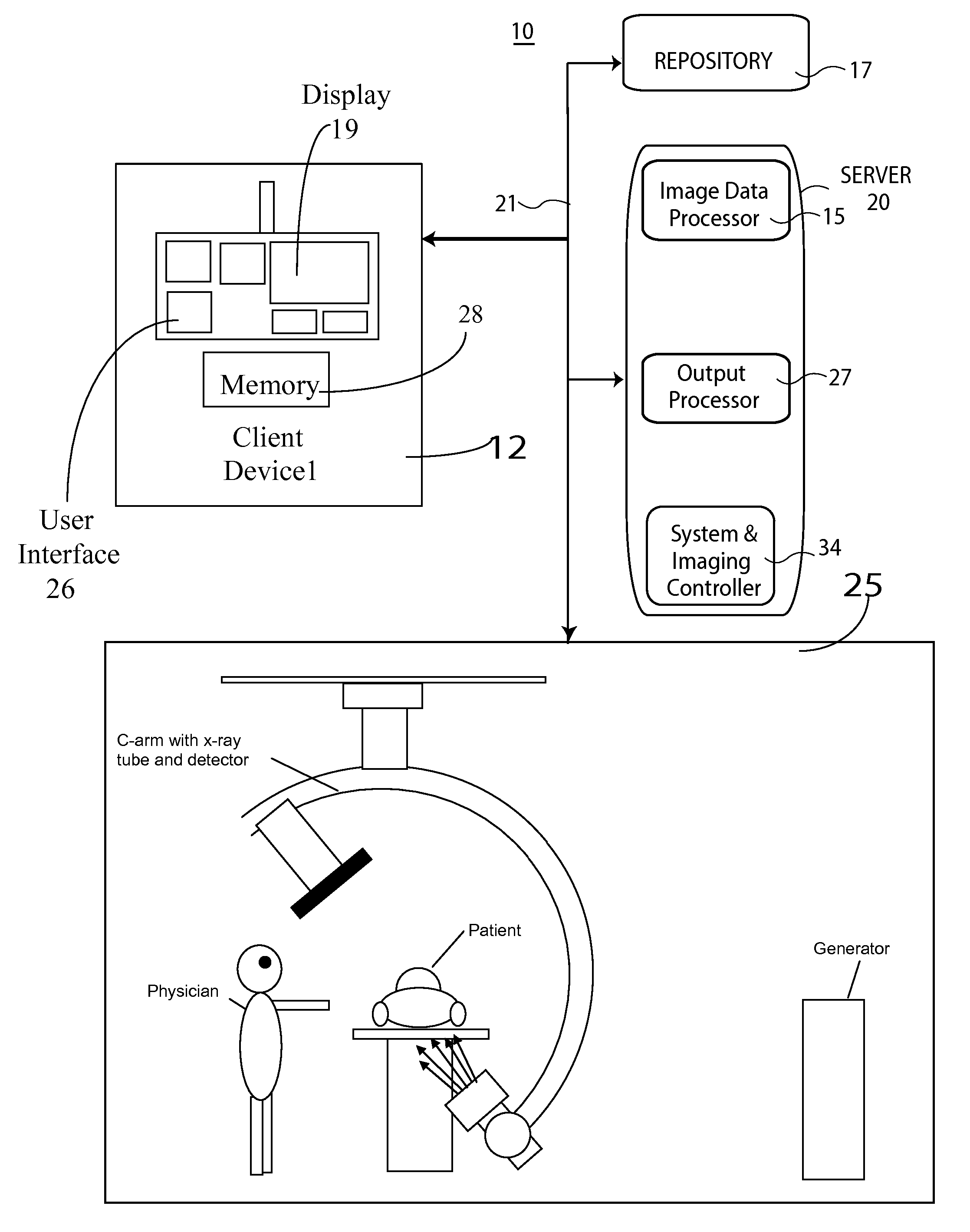
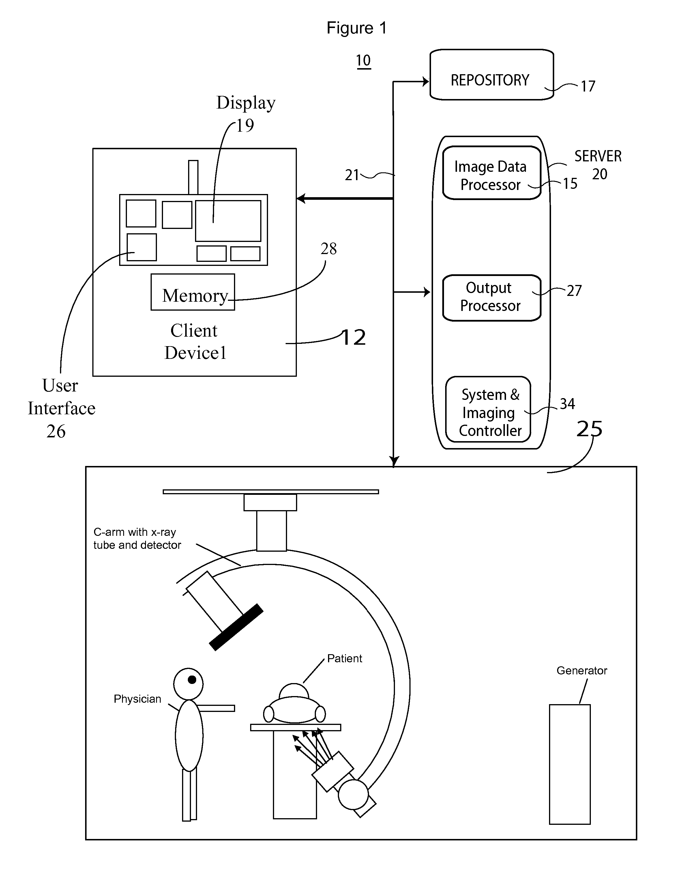
[0012]FIG. 1 shows system 10 for generating medical image data representing smaller vessels including capillaries of a region of patient anatomy. System 10 includes one or more processing devices (e.g., workstations, computers or portable devices such as notebooks, Personal Digital Assistants, phones) 12 that individually include memory 28, a user interface 26 enabling user interaction with a Graphical Us...


PUM


Abstract

A system generates medical image data representing smaller vessels including capillaries of a region of patient anatomy. An image data processor identifies pixels of larger vessels in individual images of difference images where the larger vessels have a size exceeding a predetermined threshold size. The image data processor generates an enhanced visualization small vessel image comprising substantially peak luminance values of individual pixels exclusive of pixels of the identified larger vessels. A peak luminance value of an individual pixel is generated in response to a peak luminance value of luminance values of pixels, spatially corresponding to the individual pixel and present in images comprising the plurality of temporally sequential individual difference images. An output processor outputs substantially peak luminance values as a vessel image.

Description

[0001]This is a non-provisional application of provisional application Ser. No. 61 / 599,967 filed Feb. 17, 2012, by J. Baumgart.FIELD OF THE INVENTION[0002]This invention concerns a system for generating an enhanced visualization small vessel medical image representing smaller vessels including capillaries of a region of patient anatomy using substantially peak pixel luminance values.BACKGROUND OF THE INVENTION[0003]In known subtracted angiographic x-ray image acquisition, an object of interest is typically patient vasculature. There are instances, however, where perfusion of contrast agent through capillaries is of interest. Unfortunately, such capillary perfusion is not easily visualized due to both faint appearance of the capillaries relative to larger vessels and the capillaries being obscured by the larger vessels in an image. A system according to invention principles addresses these deficiencies and related problems.SUMMARY OF THE INVENTION[0004]A system suppresses vascular st...

Claims


Application Information

Patent Timeline
IPC IPC(8): A61B5/145
CPCA61B5/14G06T5/50G06T2207/10116G06T2207/20148G06T2207/30101Y10S128/922A61B6/4441A61B6/481A61B6/504A61B6/5235A61B6/5258G06T2207/20224A61B5/150748G06T7/136
Inventor BAUMGART, JOHN
Owner SIEMENS HEALTHCARE GMBH
Learn More