Corneal graft evaluation based on optical coherence tomography image

a tomography and corneal graft technology, applied in image enhancement, instruments, applications, etc., can solve the problems of reducing vision clarity, affecting so as to achieve the effect of improving the quality of corneal grafts and reducing the risk of ruptur

Inactive Publication Date: 2013-09-26
SINGAPORE HEALTH SERVICES PTE +1
View PDF0 Cites 9 Cited by
  • Summary
  • Abstract
  • Description
  • Claims
  • Application Information

AI Technical Summary

Benefits of technology

[0007]The present invention aims to provide new and useful methods and computer systems for investigating a corneal transplant using a two-

Problems solved by technology

When cornea tissues are damaged or diseased, the cornea becomes cloudy and vision clarity is reduced.
Since the cornea transplant wound (i.e. the intersection between the replacement cornea and the remaining portion of the eye) extends throughout the thickness of the replacement cornea, and along its entire circular periphery,

Method used


Image

  • Corneal graft evaluation based on optical coherence tomography image
  • Corneal graft evaluation based on optical coherence tomography image
  • Corneal graft evaluation based on optical coherence tomography image

Examples


Embodiment Construction

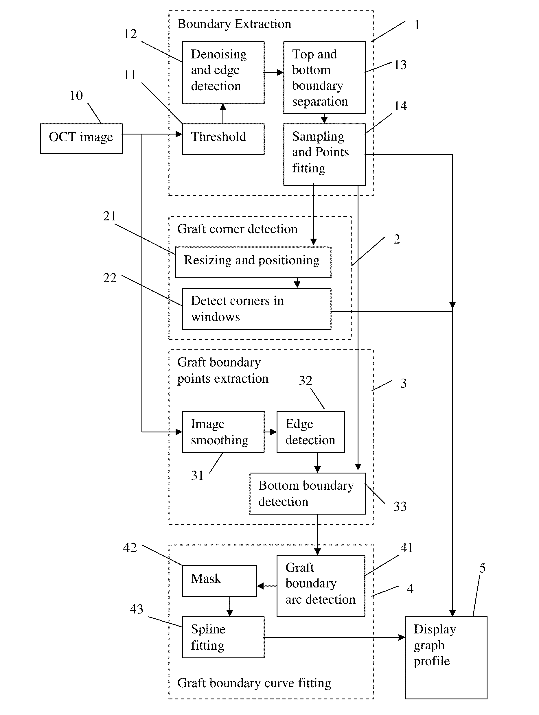
[0030]Referring to FIG. 4, a flow chart is given of an embodiment of the invention, known as the COLGATE (COrneaL GrAft Thickness Evaluation) system. The input to the image is a 2-dimensional OCT image 10 such as the image shown in FIG. 8(a). The image may be one slice from a 3-dimensional OCT image containing a large number of 2-d slices, e.g. scanned in a star-shape formation. The 2-dimensional OCT image is selected (e.g. manually) to contain the graft. Optionally, COLGATE can be run on each of a number of 2-dimensional OCT images sequentially.

[0031]In step 1, the embodiment extracts the boundary of the body formed by the combination of the graft and the remaining portion of the cornea. The top surface of this body is the original cornea (which is unchanged by the DSAEK), and the bottom surface of the body includes the lower surface of the graft and a portion of the lower surface of the original cornea. In step 2, the embodiment detects the corners of the transplanted graft. In st...


PUM


Abstract

An OCT image of an eye which has been subject to a DSAEK corneal transplant, in which a Descement's membrane in the cornea has been replaced by a graft, is processed to identify the outline of the graft. The process includes the steps of: computationally extracting the boundary of the cornea including the graft; computationally detecting the corners of the graft; computationally extracting points on the boundary between the graft and the original cornea; and computationally fitting the points on the boundary between the graft and the original cornea smoothly into a curve. The outline of the graft is then displayed. A graft profile may be generated, indicating the thickness of the graft at each point along its length.

Description

FIELD OF THE INVENTION[0001]The present invention relates to a method for processing an optical coherence tomography (OCT) image of an eye which has been subject to a corneal graft, to obtain data characterizing the image, such as data identifying the portion of the image which shows the graft.BACKGROUND OF THE INVENTION[0002]The cornea is a clear, transparent window of the eye. It contributes two thirds of the eye's focusing power. A healthy cornea should be clear and free of impurities. When cornea tissues are damaged or diseased, the cornea becomes cloudy and vision clarity is reduced. Cornea transplantation is a surgical procedure to replace a damaged or diseased cornea with a healthy one from a donor.[0003]Penetrating Keratoplasty (PKP) or full thickness cornea transplantation is a procedure in which the entire cornea is completely replaced by that of a donor. The original cornea is cut away from the rest of the eye, and the replacement cornea (which has a dome shape) is attach...

Claims


Application Information

Patent Timeline
IPC IPC(8): G06T7/00
CPCA61B3/102A61F2/142G06T7/0012G06T2207/30041G06T7/0089G06T2207/10101G06T7/0083G06T7/12G06T7/149
Inventor LEE, BENG HAICHENG, JUNLIU, JIANGWONG, WING KEE DAMONTAN, NGAN MENGMEHTA, JODHBIR SINGHTAN, TIANG HWEE DONALDWONG, TIEN YIN
Owner SINGAPORE HEALTH SERVICES PTE
Why Eureka
Try Eureka