Medium, corneal stromal slice prepared by medium and preparation method

A technology of corneal stroma and culture medium, applied in biochemical equipment and methods, culture process, tissue culture, etc., which can solve the problems of complex production process, difficulty in wide application, and shortage of tissue sources, and achieve good growth and good biocompatibility Effects of stability, good biocompatibility and cell affinity

Active Publication Date: 2020-06-05
AIER EYE HOSPITAL GRP CO LTD
View PDF8 Cites 2 Cited by
  • Summary
  • Abstract
  • Description
  • Claims
  • Application Information

AI Technical Summary

Problems solved by technology

However, some of these cytokines have been commercialized, while others need to be extracted from human tissues. The

Method used


Image

  • Medium, corneal stromal slice prepared by medium and preparation method
  • Medium, corneal stromal slice prepared by medium and preparation method
  • Medium, corneal stromal slice prepared by medium and preparation method

Examples


Embodiment 1

[0061] Example 1 Preparation of Decellularized Corneal Lens

[0062] (1) Disinfection: Soak the corneal stromal lens with the accurate thickness of the cells in physiological saline or phosphate buffer solution containing penicillin at a concentration of 0.01-0.1 mg / ml and streptomycin at a concentration of 0.05-0.5 mg / ml for 1 -5h, then rinse with 0.9% normal saline or phosphate buffer;

[0063] (2) Decellularization treatment: rinsing with phosphate buffer for 3 times, soaking in sodium chloride solution with a concentration of 1.5mol / L for 48 hours, changing the solution every 24 hours, and then using 0.9 Rinse with % normal saline or phosphate buffer for 48 hours, then soak in phosphate buffer for 72 hours, and change the solution every 24 hours;

[0064] (3) Sterilization: irradiate with gamma rays, and the irradiation dose is 20-30 kGy.

Embodiment 2

[0065] The preparation of embodiment 2 cornea extract

[0066] 1. Take some fresh porcine corneas and remove the epithelium and endothelium.

[0067] 2. After washing with PBS, soak in a mixture containing 50% glycerol and 50% DMEM, and store at -80°C to inactivate the corneal tissue.

[0068] 3. After thawing, wash with PBS, dry and weigh.

[0069] 4. Put some corneal stroma with known total weight into a mortar, pour liquid nitrogen into it, and grind until it becomes a fine powder.

[0070] 5. Add sterile, 4°C PBS in an amount of 5-10ml / g, and homogenate.

[0071] 6. Shake the suspension at 300rpm at 4°C for 48h, then centrifuge at 20000-25000g for 20-30min to remove undissolved debris.

[0072] 7. Aspirate the supernatant, filter it with a 0.22 μm filter head, measure the concentration of the extract by the BCA method, and store it at -80°C after aliquoting.

Embodiment 3

[0073] The preparation of embodiment 3 corneal stroma sheet

[0074] The corneal lens used in this example is prepared from Example 1.

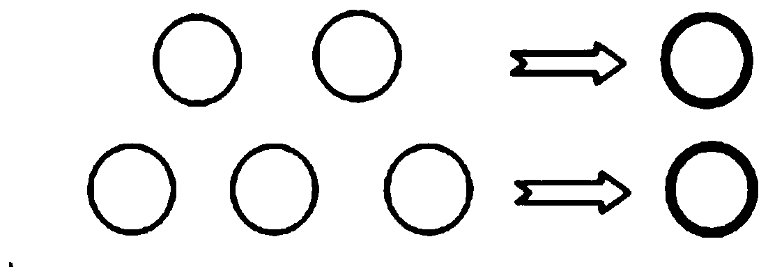
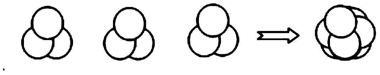
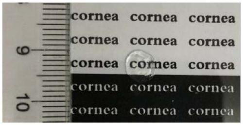
[0075] Take a piece of corneal lens, and coat the surface with a mixed solution of 2 thrombin with a concentration of 50U / ml and calcium chloride with a concentration of 40mmol / L, and then coat with 2ul of fibrinogen with a concentration of 20mg / ml. Then quickly put another lens on top of the first lens, repeat the above steps, according to figure 1 3 corneal lenses are laminated to form the corneal lens layer of the present invention. The prepared corneal lens layer was prepared with DMEM / F12 medium containing 7.5 μg / ml corneal extract, 15 μmol / L Y-27632, 15ng / ml ITS, 20ng / ml FGF, 2mmol / L ascorbic acid, and 0.5vol% fetal calf serum Cultivate for 24h. The appearance of the prepared corneal stroma sheet sample is shown in image 3 .


PUM


Abstract

The invention relates to the technical field of medical biomaterials, in particular to a culture medium, a corneal stromal slice prepared by the culture medium and a preparation method. The medium provided by the invention is a Dulbecco's modified eagle medium/F12 (DMEM/F12) medium containing a porcine corneal extract, Y-27632, insulin-transferrin-selenium (ITS), fibroblast growth factors (FGF), ascorbic acid and fetal bovine serum. According to the medium, by selecting suitable active components and proportion, a corneal stromal cell and corneal stromal slice prepared by employing the mediumhave the functions of maintaining the corneal stromal cell phenotype, promoting proliferation, improving vitality, resisting fibrosis and resisting apoptosis, and the like in comparison with a cornealstromal cell and corneal stromal slice prepared by a traditional medium. The corneal stromal slice disclosed by the invention is prepared by taking a corneal lens as a raw material and culturing thecorneal lens by employing the medium; and compared with animal-derived corneal repair material, the corneal lens has good biocompatibility and no antigenicity, can be used as substitutes of various donor materials for corneal transplantation, and can be accepted by the majority of patients and is clinically applied for long.

Description

technical field [0001] The invention relates to the technical field of medical biomaterials, in particular to a culture medium, a corneal stroma sheet prepared by it and a preparation method. Background technique [0002] In ophthalmic diseases, more than 3 million patients are blinded by corneal diseases, and about 80% of them can be relieved of blindness and restore their vision through traditional keratoplasty. At present, corneal transplantation is the only hope for blind patients with corneal disease to restore vision and sight. However, due to the extremely limited number of corneal donations in my country, most patients cannot see again because they cannot obtain available donor corneas. At the same time, some corneal diseases can be replaced by dead or sick corneal stromal cells through cell therapy, so as to avoid the development of corneal blindness, so as to achieve the effect of curing diseases and restoring corneal function, and become another treatment for corn...

Claims


Application Information

Patent Timeline
IPC IPC(8): C12N5/079A61F2/14
CPCC12N5/0621A61F2/142C12N2500/84C12N2500/25C12N2501/113C12N2501/115C12N2501/119C12N2501/117C12N2500/38C12N2501/999A61F2240/001
Inventor 陈建苏李申阳崔泽凯
Owner AIER EYE HOSPITAL GRP CO LTD
Why Eureka
Try Eureka