
Multimodality correlation of optical coherence tomography using secondary reference images

a coherence tomography and secondary reference image technology, applied in the field of optical coherence tomography, can solve the problems of limiting the amount of data one can process in one exam, unable to look at all of the different areas at one time, and taking a long time, so as to achieve accurate oct data registration, improve quality, and improve the effect of quality

Inactive Publication Date: 2013-11-14
SONOMED IP HLDG
View PDF4 Cites 18 Cited by
  • Summary
  • Abstract
  • Description
  • Claims
  • Application Information

AI Technical Summary

Benefits of technology

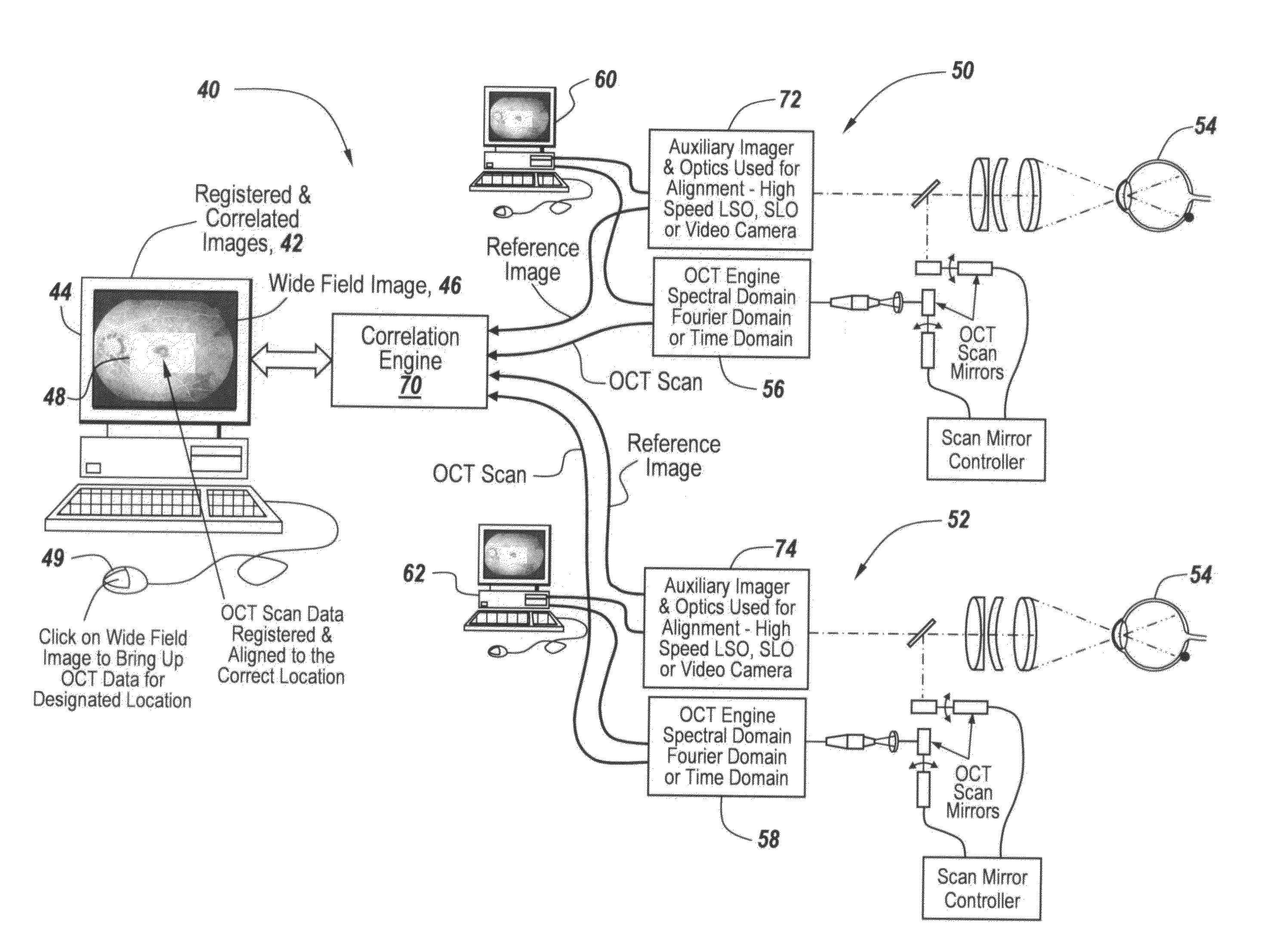
The patent describes a way to compare optical coherence tomography (OCT) images of the eye taken from different machines or at different times. By using a reference image created by all OCT machines, the images can be correlated and registered, allowing for easier comparison and interpretation. The OCT machine can also use an active tracking mechanism to ensure accurate registration of the OCT data in relation to the reference image. The technical effect is that this approach enhances the accuracy and reliability of OCT imaging for diagnostic and therapeutic purposes.

Problems solved by technology

Thus, typically what happens is that during an OCT exam one can image a small area very densely but it takes quite a bit of time.
Thus, there are limits on how much data one can process in one exam.
The trouble is that one is currently unable to look at all of the different areas together at one time.
This means one uses the B scans and provides a calculated summation to create an image, namely a forward facing image from that data which looks like a photograph or an image one would see looking at the eye, but the resolution is very low and of very low quality.
A lot of times it can have motion induced artifacts as the patient moves his eyes during the examination with the result that one does not obtain good resolution.
As a result, The OCT can have a gap in it and the data can be misaligned so when one creates that enface image it's not real.
It has errors in it and it presents an issue when one tries to register that feature or data against the other image of the eye to try to find the location where it fits.
However, when one has such errors, it makes it very difficult to align the blood vessel patterns.
If the blood vessel patterns do not match, automatic alignment will fail.
Also just being an enface image, the resolution of the OCT scan is always lower than the actual resolution of the reference image so it is difficult to get a good alignment, especially if there are no very clear structures that are visible to the eye.

Method used


Image

  • Multimodality correlation of optical coherence tomography using secondary reference images
  • Multimodality correlation of optical coherence tomography using secondary reference images
  • Multimodality correlation of optical coherence tomography using secondary reference images

Examples


Embodiment Construction

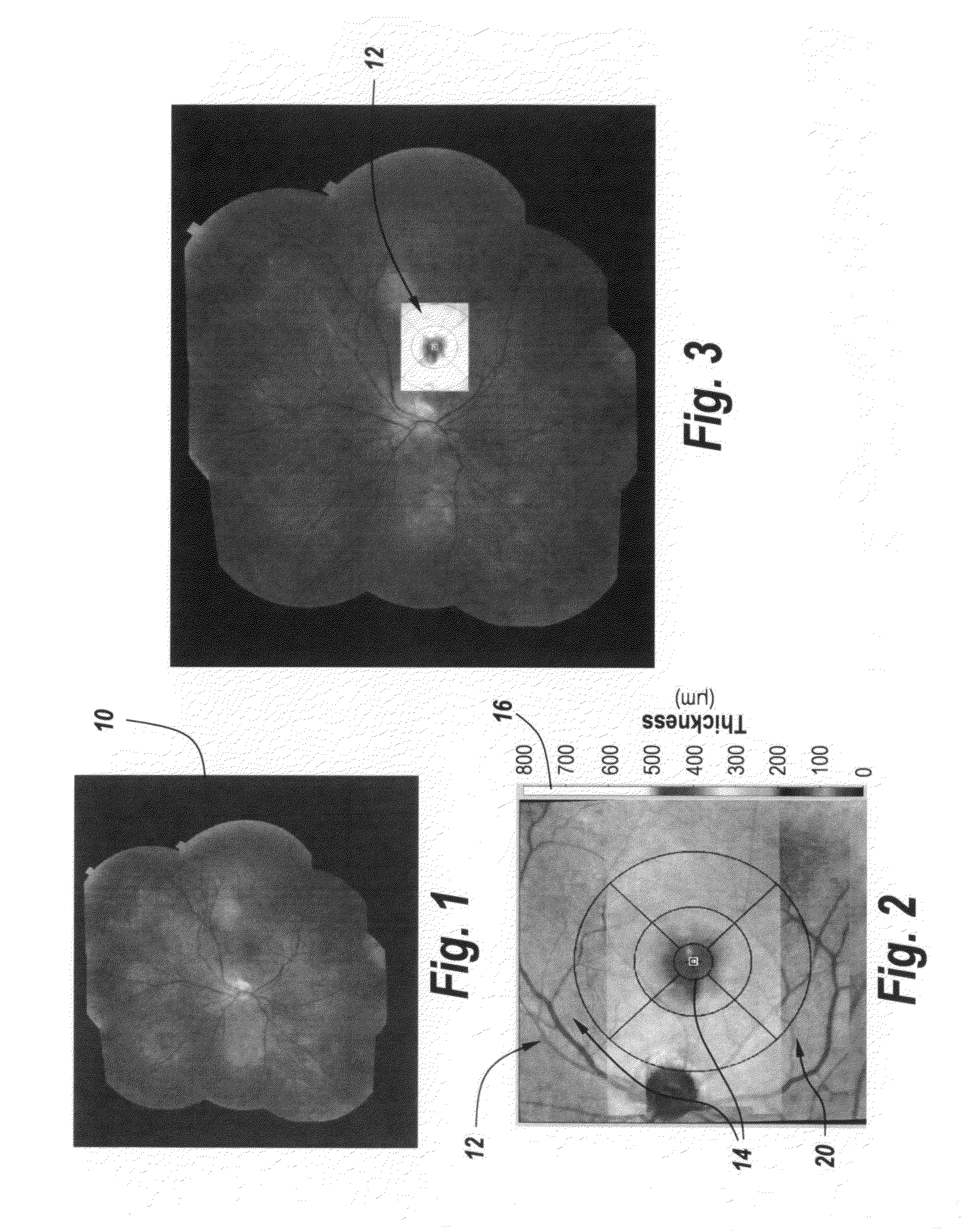
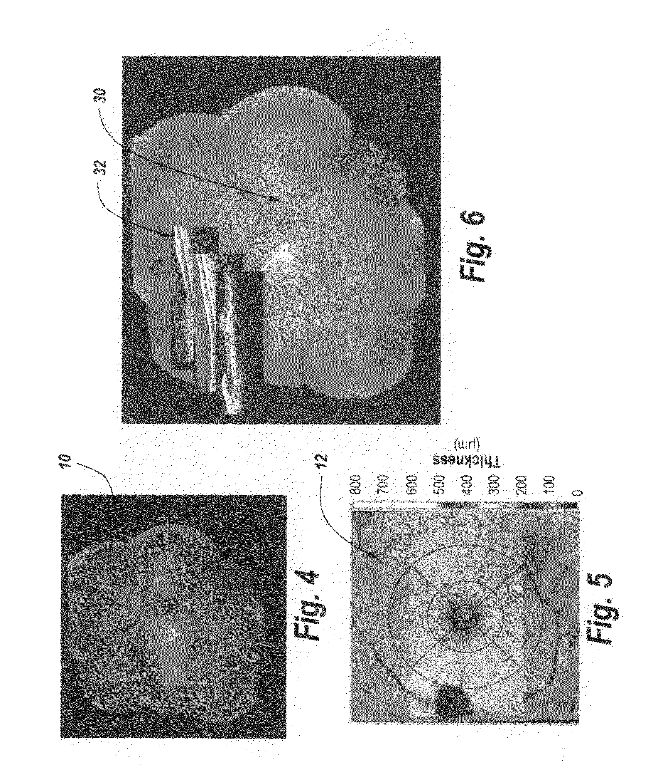
[0037]Referring now to FIG. 1, the purpose of this Figure along with FIGS. 2 and 3 is to show how the subject system registers OCT scan data to a wide field photography mosaic of the eye of the patient undergoing an OCT scan. It will be noted that there are a number of ways to generate a mosaic of the eye such as that shown in FIG. 1 by reference character 10. In one example this is done by a series of photographs of smaller regions of the eye and by controlling the patient's gaze one can image different areas. Thereafter these areas are all automatically stitched together by an algorithm to create a wide field image. There are also some devices such as manufactured by Optos which create an Optomap to generate an image like the one shown in FIG. 1 with one shot that creates the wide field image. It should also be noted that although the drawings depict images of the retina, the same methods can also be applied to OCT scans and images of the anterior segment of the eye.

[0038]Referrin...


PUM


Abstract

Reference images from one or more OCT scanners are correlated with associated OCT scan data, which is in turn registered and correlated to a wide field image so as to present the OCT scan data registered and aligned to the correct location on the wide field image so as to permit displaying OCT scan data taken at different times or on different machines on a single screen all registered to the wide field image.

Description

FIELD OF THE INVENTION[0001]This invention relates to optical coherence tomography (OCT) and more particularly to displaying the tomography scans from a number of tomographic sources.BACKGROUND OF THE INVENTION[0002]Typically one has OCT scans being done in a localized region of the eye with the result of the localized scan being displayed. In addition to this data from a localized area there are also other modalities that image a larger area of the eye or that image the eye in a different way, for example monochrome and color photography or laser scanning. It would be useful for the clinician to be able to look at those other modality images, and be able to see the OCT scans captured from another machine in alignment with the images.[0003]The purpose is to allow the clinician can look at multiple exams of the patient's eye integrated into one screen. One of course one could display the images from different machines on different screens, but it is more helpful to the clinician to a...

Claims


Application Information

Patent Timeline
IPC IPC(8): G06T11/60A61B3/10
CPCG06T11/60A61B3/102G06Q40/00G06Q40/08
Inventor CARNEVALE, MATTHEW
Owner SONOMED IP HLDG
Learn More