Non-coding transcripts for determination of cellular states
a technology of cellular state and non-coding transcripts, which is applied in the direction of biochemical apparatus and processes, biomass after-treatment, specific use bioreactors/fermenters, etc., can solve the problems of poor technical skills and/or pathologists' experience, biased interpretation of pathology test results, and cancer as a leading cause of death worldwid
- Summary
- Abstract
- Description
- Claims
- Application Information
AI Technical Summary
Benefits of technology
Problems solved by technology
Method used
Image

Examples
example 1
Exemplary Methods for Identification of Genes that Produce Short RNAs Out of their Exons
[0272]Human samples from breast and pancreas (both normal and diseased / abnormal) were obtained for deep sequencing, e.g., next generation sequencing. The NGS focused on generating a profile of the short RNAs contained in those samples. The NGS was carried out on a Life Technologies SOLiD 3+ platform. For each sample, a large dataset that contained the sequenced reads in Life Technologies' “colorspace” format was obtained. Using a read mapping program, e.g., Burrows-Wheeler Alignment tool as described in Li and Durbin (2009) Bioinformatics 25(14): 1754-1760, the sequenced reads were mapped on the assembly of the human genome (e.g., using hg19 which can be assessed at hgdownload.cse.ucsc.edu / downloads.html#human. If a sequenced read mapped at multiple locations of the genome, then all instances of the read were discarded. This ensured that the genomic locations that gave rise to sequenced RNA reads...
example 2
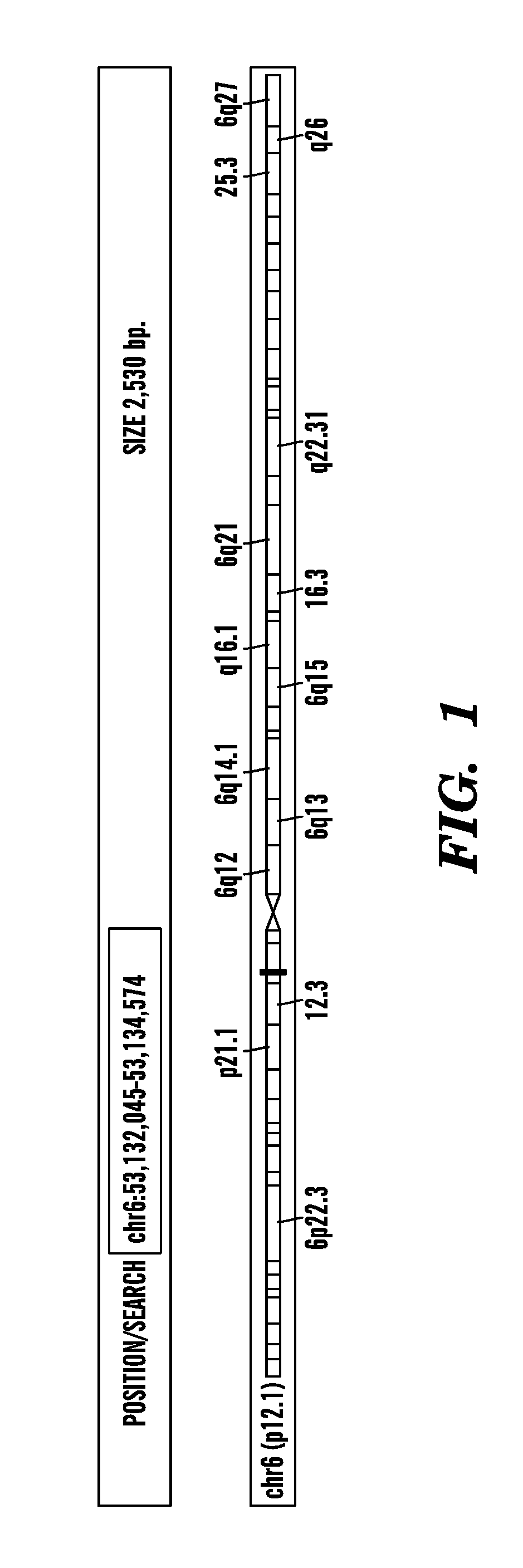
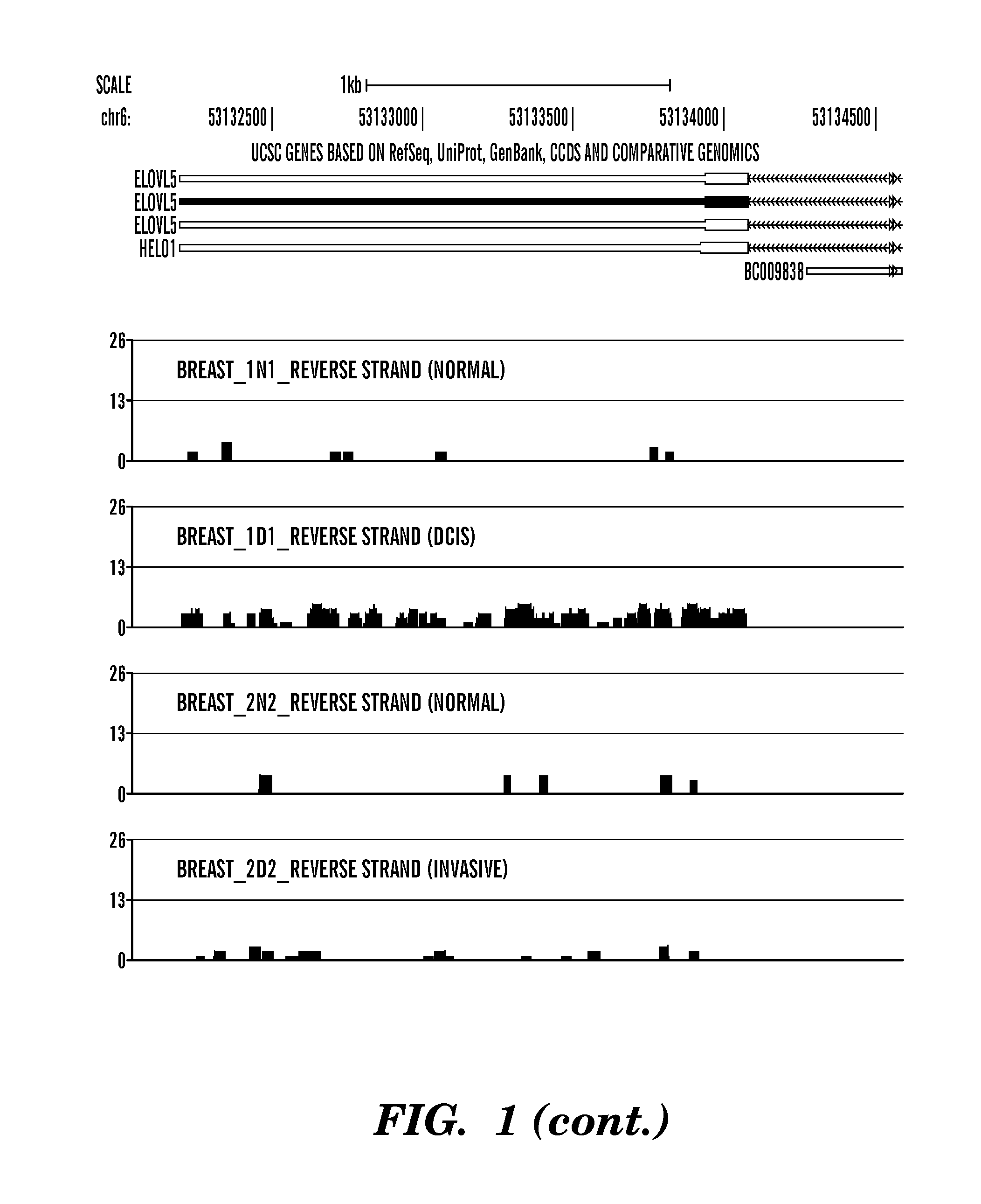
Determination of a State / Condition of a Breast Tissue Sample Based on Detecting Short RNAs Produced from One or More Exons of ELOVL5
[0284]This example shows the amount of short RNAs present in the last exon of the gene known as ELOVL5 (elongation of very long chain fatty acids protein 5) from different breast samples, including 2 normal (Breast—1N1 and Breast—2N2), 1 ductal in situ carcinoma (Breast—1D1) and 1 invasive (Breast—2D2). The last exon of the ELOVL5 gene includes the gene's 3′UTR. ELOVL5 is located on the reverse strand (going from 3′ to 5′) of the human genome, as indicated in FIG. 1 by left-pointing arrowheads (i.e. ). Sequenced reads that map to the same genomic location contribute independently to that location: the height of the red bar at a given genomic location represents the number of overlapping sequenced reads that map there. Note that the Y-axis is logarithmic (base 2) and ranges from 0 (0 reads) to 26 (226 reads). As shown in FIG. 1, for both normal samples (...
example 3
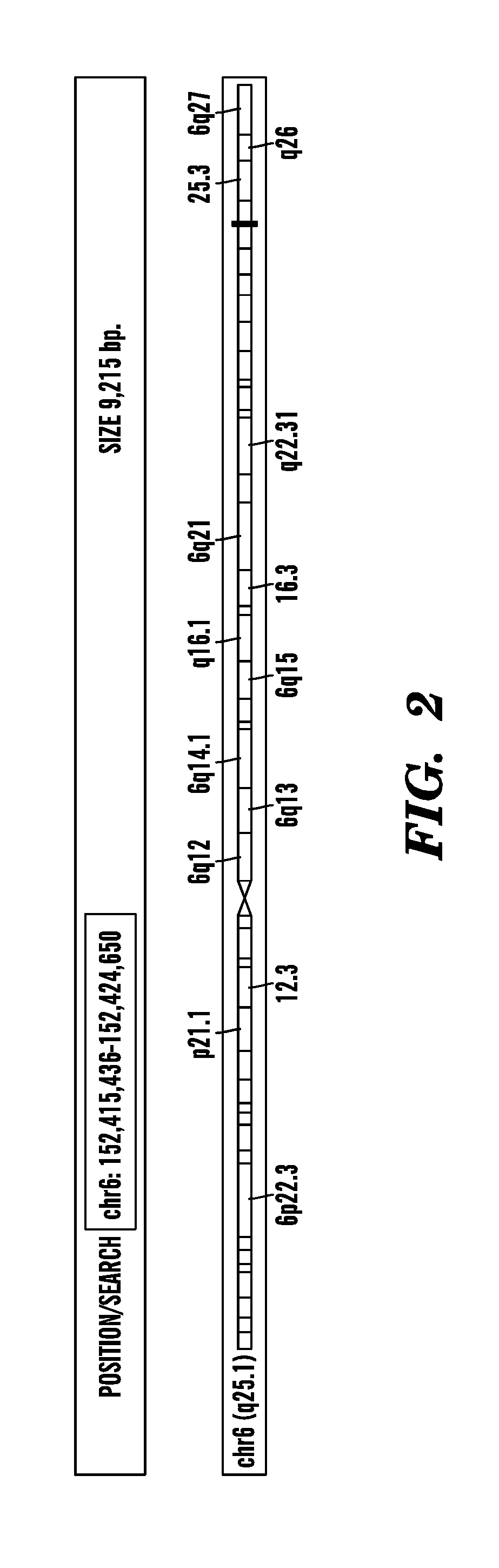
Determination of a State / Condition of a Breast Tissue Sample Based on Detecting Short RNAs Produced from One or More Exons of ESR1
[0285]This example shows the amount of short RNAs present in the last two exons of the gene known as ESR1 (estrogen receptor 1) from different breast samples, including 2 normal (Breast—1N1 and Breast—2N2), 1 ductal in situ carcinoma (Breast—1D1) and 1 invasive (Breast—2D2). The last exon includes the gene's 3′UTR. ESR1 is located on the forward strand (going from 5′ to 3′) of the human genome, as indicated by right-pointing arrowheads (i.e. >>>>>>>>) at the top left of FIG. 2 and the use of blue bars to mark the location where the sequenced reads (corresponding to short RNAs) have mapped. In the top part of FIG. 2, exons are indicated by solid color rectangles separated by intronic regions that are indicated by long lines with arrowheads (i.e., ). Sequenced reads that map to the same genomic location contribute independently to that location: the height ...
PUM
| Property | Measurement | Unit |
|---|---|---|
| diameter | aaaaa | aaaaa |
| diameter | aaaaa | aaaaa |
| length | aaaaa | aaaaa |
Abstract
Description
Claims
Application Information