Diffusion tensor magnetic resonance imaging method

a technology of tensor magnetic resonance imaging and diffusion tensor, which is applied in the field of magnetic resonance imaging, can solve the problems of prolonging scanning time and ineffective elimination of the influence of respiratory movement by the patient, and achieves the effect of greatly reducing the influence of respiratory movement and substantially reducing the scanning tim

Inactive Publication Date: 2013-12-19
SIEMENS AG
View PDF5 Cites 6 Cited by
  • Summary
  • Abstract
  • Description
  • Claims
  • Application Information

AI Technical Summary

Benefits of technology

[0015]In the embodiments of the present invention the cardiac DTI image data can be obtained when the subject respirates freely by combining the technology of the echo planar imaging (EPI) of STEAM having two electrocardiogram (ECG) triggers with the technology of the two-dimensional (2D) Prospective Acquisition Correction (PACE). Experimental data indicates that the influence of respiratory movement is greatly reduced and the scanning time is substantially shortened, so as to solve the aforementioned problems in the existing technology.
[0016]At the same time, the technical solution of the present invention does not bring further complexity and limitation into the sequence, and the DTI image data can be accomplished through the existing image reconstruction algorithm. Moreover, the desired 3D myocardial fiber image can be finally reconstructed from the initial data. The entire scanning process can be completed within 5 minutes. The experimental results show that the basic ventricular fiber helical structure can be acquired from the final 3D myocardial fiber image.

Problems solved by technology

However, the problem with the above-mentioned STEAM EPI technology having two electrocardiogram triggers is that the influence of respiratory movement by the patient cannot be effectively eliminated, therefore patients are required to cooperate by strictly holding their breath during signal acquisition, i.e., to hold their breath intermittently several times, even though breathholding is a significant challenge for certain patients.
In addition, since intermittently holding the breath several times often results in the prolonging of the scanning time, generally speaking, approximately 30 minutes are needed to acquire the cardiac DTI data using the existing technology.

Method used


Image

  • Diffusion tensor magnetic resonance imaging method
  • Diffusion tensor magnetic resonance imaging method
  • Diffusion tensor magnetic resonance imaging method

Examples


Embodiment Construction

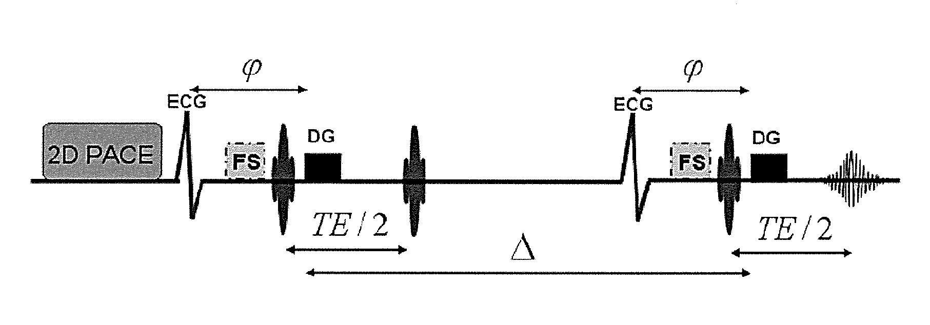
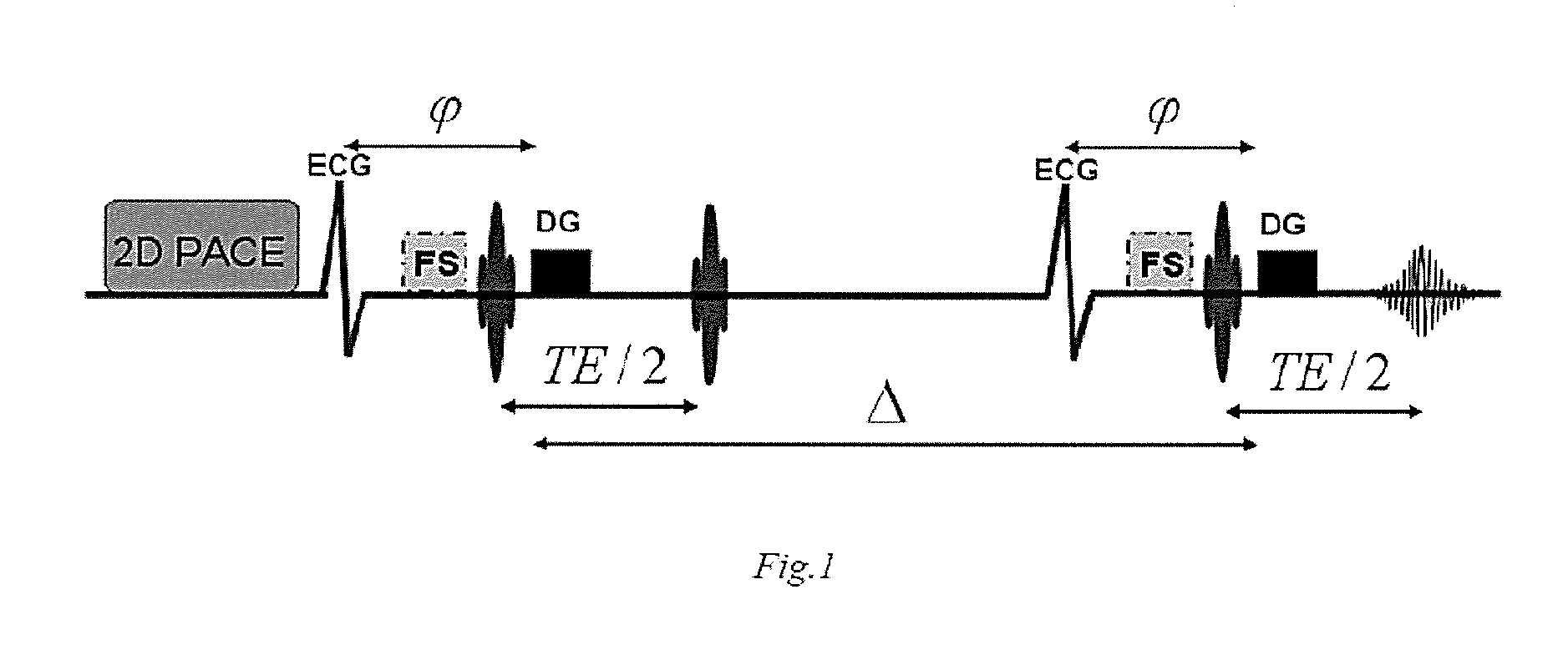
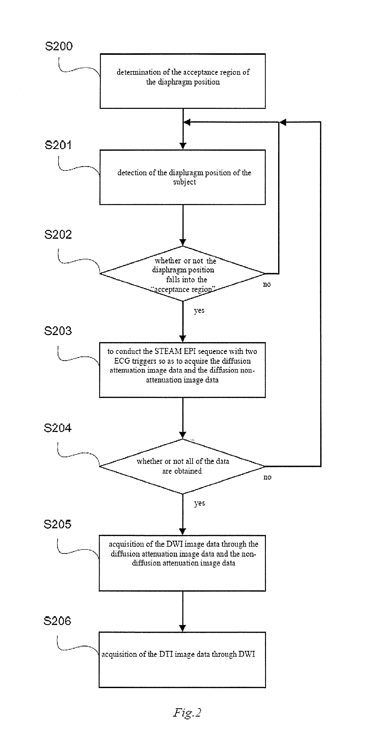
[0023]As mentioned above, respiratory movement has great influence on acquiring the cardiac DTI and the subject is usually forced to hold his / her breath intermittently several times in existing cardiac DTI to attenuate the influence of the respiratory movement. In order to solve this problem, FIG. 1 shows a schematic diagram of the STEAM echo planar imaging sequence of cardiac DTI according to the particular embodiment of the present invention. In this particular embodiment, as shown in FIG. 1, two-dimensional (2D) Prospective Acquisition Correction (PACE) technology is employed to correct the respiratory movement during the acquisition of the cardiac DTI data, so that the subject can respire freely during the measurement.

[0024]In the applied two-dimensional PACE technology, the diaphragm position of a subject is detected using two-dimensional gradient echo sequence with low resolution. First, a brief “learning time” is used to analyze the respiratory state of the subject and the mi...


PUM


Abstract

In a diffusion tensor magnetic resonance imaging method for imaging a myocardial fiber structure, the diaphragm position of a subject is detected and a determination is made as to whether the diaphragm position of the subject falls into the acceptance region or not. If it does not, continue the diaphragm position of the examination subject is continued to be detected. If and when the diaphragm position is in the acceptance region, an echo planar imaging sequence with stimulated echo is executed with two electrocardiogram triggers, so as to acquire diffusion tensor image data of the myocardial fiber structure. The cardiac DTI image data thus can be obtained under free respiration of the subject, and the influence of respiratory movement is reduced and the scanning time is shortened.

Description

BACKGROUND OF THE INVENTION[0001]1. Field of the Invention[0002]The present invention relates to the technical field of magnetic resonance imaging, in particular to a diffusion tensor magnetic resonance imaging method, especially a diffusion tensor magnetic resonance imaging method for imaging a myocardial fiber structure.[0003]2. Description of the Prior Art[0004]Cardiac Diffusion Tensor Imaging (DTI) provides an effective and noninvasive detection means for the reconstruction of an image of a myocardial fiber structure, and is capable of being used for measuring the abnormal deformation of myocardial structure in some specific heart diseases.[0005]In existing technology, the echo planar imaging (EPI) technology of stimulated echo acquisition mode (STEAM) having two ECG triggers (electrocardiogram triggers) is a common mode for acquiring the cardiac DTI. This technology uses two electrocardiogram triggers that are added in the STEAM EPI sequence, with identical diffusion encoding g...

Claims


Application Information

Patent Timeline
Patent Type & Authority Applications(United States)
IPC IPC(8): G01R33/563
CPCG01R33/56341G01R33/5676
Inventor HUANG, YU QING
Owner SIEMENS AG
Why Eureka
Try Eureka