Optical tomographic imaging otoscope with integrated display and diagnosis

a tomographic imaging and integrated technology, applied in the field of otoscopes that examine only the condition of the eardrum, can solve the problems of difficult to differentiate between bacterial and general infections, difficult to diagnose the causes of otitis media defection, so as to simplify the disease-diagnosis procedure and improve resolution. , the effect of reducing the error in related diagnoses

Inactive Publication Date: 2014-01-09
KYUNGPOOK NAT UNIV IND ACADEMIC COOP FOUND
View PDF3 Cites 22 Cited by
  • Summary
  • Abstract
  • Description
  • Claims
  • Application Information

AI Technical Summary

Benefits of technology

[0026]According to the present invention, it is possible to concurrently examine the condition of the section of the ear drum and the condition of the middle ear, provide better resolution than the existing CT or MRI apparatus, simplify the disease-diagnosing procedures, diagnose causes for the defection of the otitis media at an early stage so that prescriptions according to the causes can be suitably prepared, and reliably differentiate between bacterial and general infections so that errors in related diagnoses can be reduced.
[0027]Furthermore, according to the invention, not only the surface condition of the ear drum but also the condition of the section of the ear drum and any disease of the middle ear positioned below the ear drum can be concurrently examined based on the 2D images obtained from the image-photographing part and the section-photographing part, thereby reducing the chance of an erroneous diagnosis. It is possible to examine points in the depth direction into the middle ear so that the thickness of the ear drum can be measured in real time and a diseased region can be accurately determined in real time, thereby obtaining the efficiency and reliability of the diagnosis, and simplify procedures of diagnosis, thereby realizing the convenience of diagnoses and easily obtaining data.

Problems solved by technology

Otorhinolaryngological apparatuses that examine only the condition of the eardrum have limited ability to diagnose otitis media.
However, according to these methods, it is difficult to diagnose causes for the defection of the otitis media at an early stage, differentiate between bacterial and general infections, and examine the condition of the middle ear positioned below the ear drum.
Accordingly, there are many obstacles to address correct diagnosis.
However, it is much difficult to accurately diagnose patients since the resolutions of such apparatuses are limited.
In addition, these methods also act as factors that complicate diagnostic procedures and make it inconvenient in the diagnosis of patients.
Early diagnosis of a disease is difficult.
It is difficult to examine a patient since a separate monitor device is used.
Furthermore, it is difficult to obtain diagnosis data for a patient since it is not easy to store an image.
The related-art lens type otoscope 10 has a drawback in that it is difficult for a doctor to visually examine the ear drum.
It is difficult to correctly examine the condition of the ear drum since the ear drum is small and dark in most cases.
This lens type otoscope 10 has the danger of an erroneous diagnosis since the doctor must make subjective diagnosis by examining only the surface condition of the ear drum, and acts as a factor that can delay the time for diagnosis.
However, since light is radiated through an optical fiber and the image is displayed on a large monitor, the cost of this apparatus increases.
In addition, there are disadvantages in that the resolution of the camera is not satisfactory, colors are not clearly distinguished, and an image that is obtained at the same time as a patient is being diagnose cannot be examined.
However, this related-art endoscope type otoscope 30 has a drawback in that it diagnoses only the surface condition of the ear drum, as in existing otoscopes.
It is therefore impossible to examine the condition of the middle ear behind the ear drum.
In addition, this apparatus is required to illuminate dark regions using an expensive optical fiber, which becomes a factor that increases the unit price of the apparatus.
There are other drawbacks in that a separate monitor is required, it is difficult to examine the image of the ear drum, and the size of the apparatus is increased.
In addition, the size of the apparatus is very large, the price of the apparatus is expensive, and the lens of the apparatus must be adjusted depending on the eyesight of the user, which is inconvenient.

Method used


Image

  • Optical tomographic imaging otoscope with integrated display and diagnosis
  • Optical tomographic imaging otoscope with integrated display and diagnosis
  • Optical tomographic imaging otoscope with integrated display and diagnosis

Examples


Embodiment Construction

[0040]Reference will now be made in detail to various embodiments of the present invention, examples of which are illustrated in the accompanying drawings and described below.

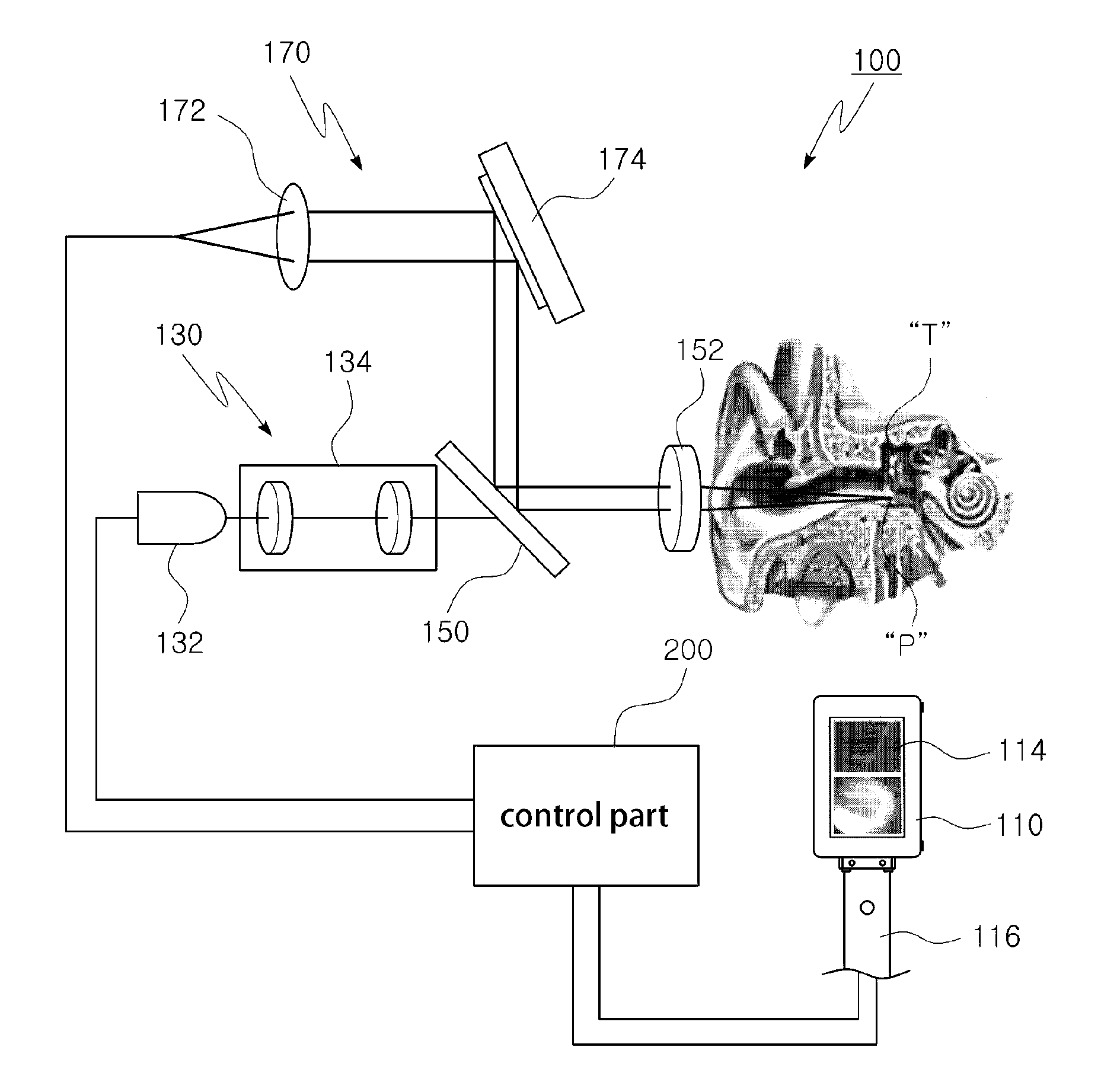
[0041]A diagnosis-and-display integrated optical tomographic imaging otoscope 100 according to the present invention is an apparatus for examining otitis media, and as shown in FIG. 3 to FIG. 6, includes a hollow casing 110 having a certain size.

[0042]The casing 110 is configured, preferably, as a rectangular box. An ear specular 112 is disposed on the front side, a display 114 having a liquid crystal display (LCD) is disposed on the rear side, and a manipulating handle 116 is disposed on the underside.

[0043]In addition, the diagnosis-and-display integrated optical tomographic imaging otoscope 100 according to the present invention also includes a image-photographing part which has a charge-coupled device (CCD) camera 132 inside the casing 110, and takes an image of the ear drum of a patient through the ear spe...


PUM


Abstract

A diagnosis-and-display integrated optical tomographic imaging otoscope for examining otitis media. A hollow casing includes an ear specular disposed on a front surface, a display including an LCD disposed on a rear surface, and a manipulating handle on a lower part. An image-photographing part includes a CCD camera inside the casing, and photographs an ear drum image of a patient through the ear specular. A section-photographing part includes a collimator and a galvanometer mirror inside the casing, and photographs section images of the ear drum and a middle ear of the patient. The ear drum image obtained by the image-photographing part and the section images of the ear drum and the middle ear are obtained in a non-incision method by the section-photographing part, and are displayed in real time on the LCD such that desirable images can be stored.

Description

CROSS-REFERENCE TO RELATED APPLICATIONS[0001]The present application is a continuation of International Application No. PCT / KR2011 / 006599 filed Sep. 7, 2011, which claims priority to Korean Application No. 10-2011-0020524 filed Mar. 8, 2011, which applications are incorporated herein by reference.TECHNICAL FIELD[0002]The present invention relates to an apparatus for examining an otitis media, and more particularly, to a diagnosis-and-display integrated optical tomographic imaging otoscope which can photograph the surface image of the ear drum, the section of the ear drum and condition of the middle ear in a non-incision method and display the photographed images on a liquid crystal display (LCD) of a display. The state of a patient can be promptly examined, thereby significantly increasing the convenience of a diagnosis, diagnosing procedures can be simplified and causes for a disease can be diagnosed at an early stage so that prescriptions according to the causes can be suitably pr...

Claims


Application Information

Patent Timeline
Patent Type & Authority Applications(United States)
IPC IPC(8): A61B1/227
CPCA61B1/227A61B1/00048A61B1/00052A61B5/0033A61B8/13
Inventor KIM, JEE HYUNCHO, NAM HYUNKWON, HYEONG ILJUNG, UN SANGJEONG, HYO SANG
Owner KYUNGPOOK NAT UNIV IND ACADEMIC COOP FOUND
Why Eureka
Try Eureka