
Ultrasound imaging breast tumor detection and diagnostic system and method

a breast tumor and ultrasound imaging technology, applied in the field of ultrasound breast tumor detection and diagnostic system, can solve the problems that the breast ultrasound image analysis program must spend a lot of time, and achieve the effect of reducing the amount of computation and quickly recognizing tumor tissues

Inactive Publication Date: 2014-01-16
NAT TAIWAN UNIV
View PDF12 Cites 24 Cited by
  • Summary
  • Abstract
  • Description
  • Claims
  • Application Information

AI Technical Summary

Benefits of technology

The present invention relates to an ultrasonic imaging breast tumor detection and diagnostic system and method that can quickly and accurately detect tumor tissues in 3D breast ultrasound images. The system and method are designed to cut out multiple regions from the 3D images and analyze them using image processing techniques to rapidly identify tumor tissues. It also includes image smoothing effects to reduce computation and improve image quality. Additionally, the system and method provide a tumor map with different colored marks to make it easier for doctors to identify and determine the severity of the tumor tissue. The system and method use BI-RADS assessment-category scale to classify each tumor tissue region and calculate its position and size for easier recording of breast diagnostic reports.

Problems solved by technology

However, a high-quality breast ultrasound image is often more than a million pixels, and its operation is very impressive, so that the breast ultrasound image analysis program must spend a lot of time.

Method used


Image

  • Ultrasound imaging breast tumor detection and diagnostic system and method
  • Ultrasound imaging breast tumor detection and diagnostic system and method
  • Ultrasound imaging breast tumor detection and diagnostic system and method

Examples


Embodiment Construction

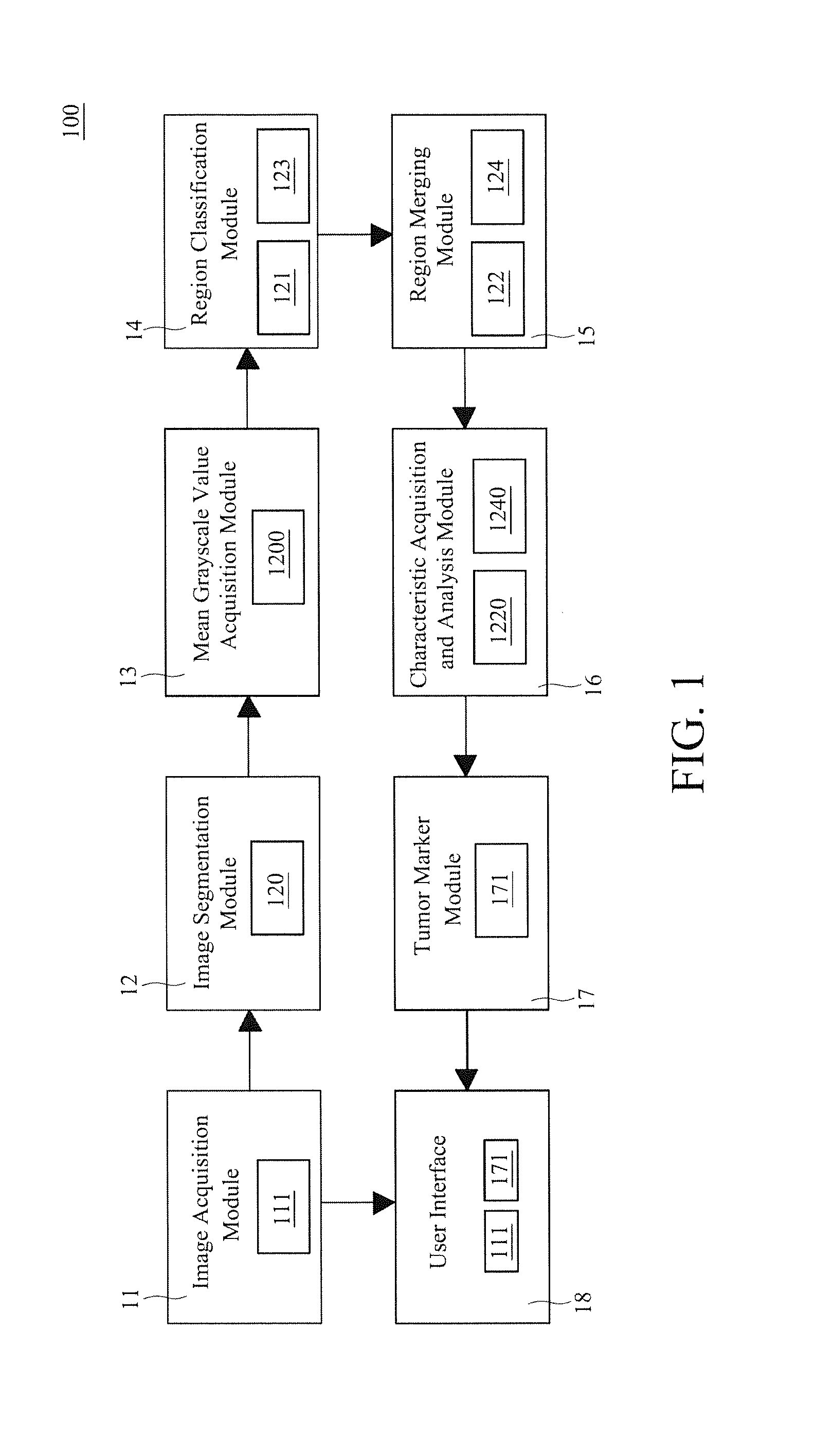
[0038]Please refer to FIG. 1, a system block diagram of an ultrasound imaging breast tumor detection and diagnostic system in accordance with one preferred embodiment of the present invention is shown. As illustrated, the ultrasound imaging breast tumor detection and diagnostic system 100 comprises an image acquisition module 11, an image segmentation module 12, a mean grayscale value acquisition module 13, a region classification module 14, and a region merging module 15.

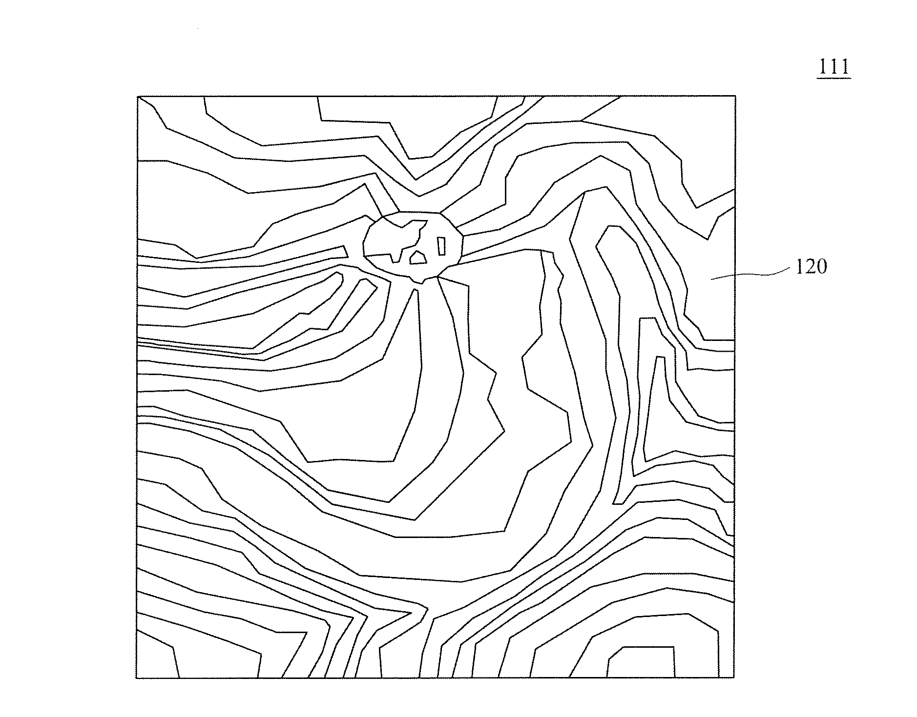
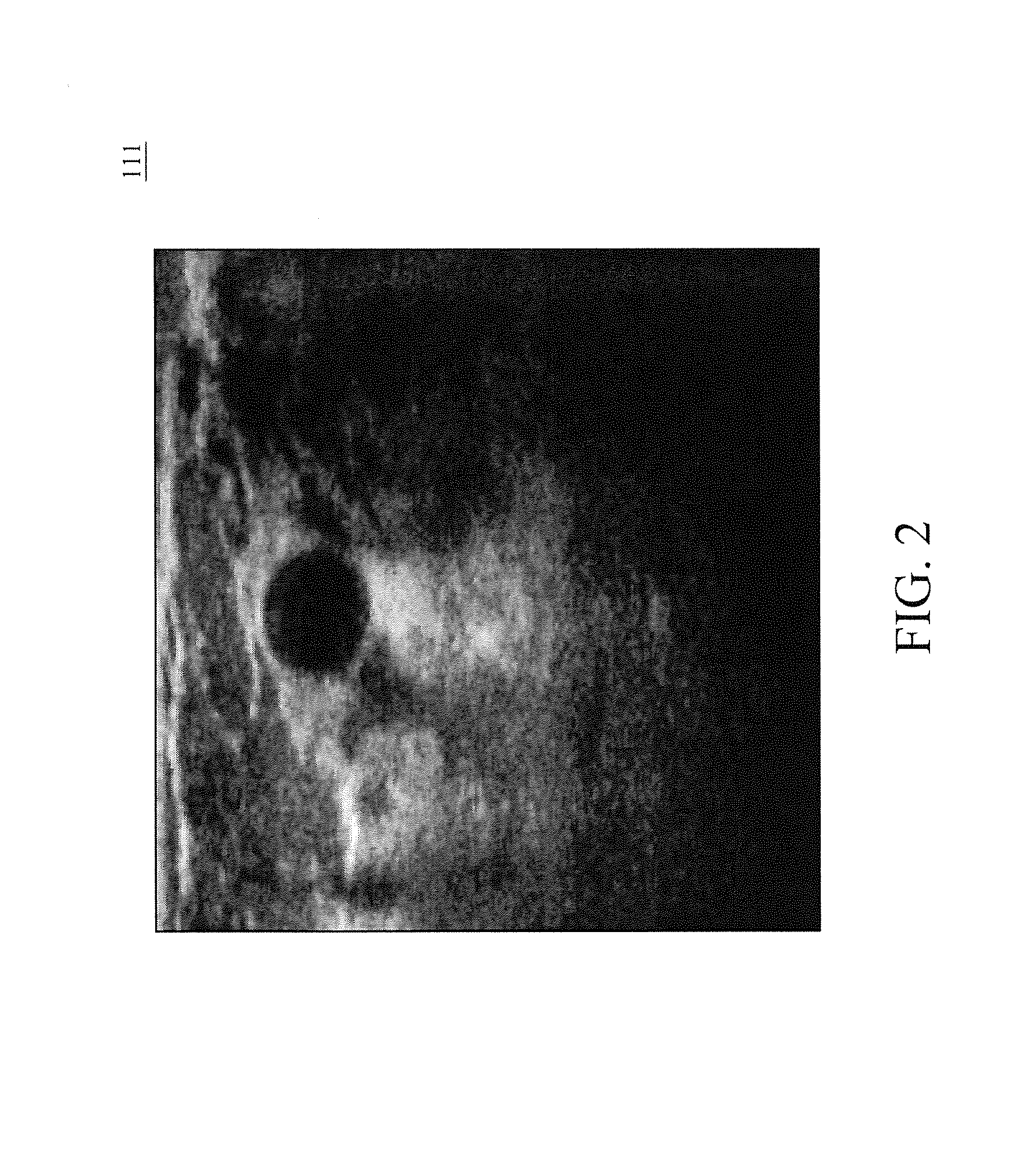
[0039]Firstly, an ultrasound probe performs the breast ultrasound scanning process on the breasts so as to acquire a continuous of 3D breast ultrasound images 111 by the image acquisition module 11, as shown in FIG. 2. The image segmentation module 12 is connected to the image acquisition module 11 to receive the 3D breast ultrasound images 111 and to cluster each of adjacent pixels with a respective similar grayscale value in the 3D breast ultrasound images into the same region by using a 3D means shift algorithm ...


PUM


Abstract

An ultrasound imaging breast tumor detection and diagnostic system and method is disclosed. The method uses the system to acquire a plurality of 3D breast ultrasound images, and then to cut out multiple regions from the 3D breast ultrasound images using a 3D means shift algorithm, and then to acquire the mean grayscale value (MGV) of each region, and then to classify the regions to groups subject to the mean grayscale value (MGV), and to merge each of the regions of the darkest group with adjacent regions of the similar grayscale into a respective suspicious tumor tissue full region, and then to recognize each suspicious tumor tissue full region to be a tumor tissue region or non-tumor tissue region. Thus, using region as the basic computing unit, tumor tissues are quickly recognized from the 3D breast ultrasound images.

Description

1. FIELD OF THE INVENTION[0001]The present invention relative to breast tumor detection and diagnostic technology and more particularly, to an ultrasonic imaging breast tumor detection and diagnostic system for rapidly detecting breast tumor tissues from ultrasound images. The invention relates also to detection and diagnosis of breast tumor using the ultrasonic imaging breast tumor detection and diagnostic system.2. BACKGROUND OF THE INVENTION[0002]Breast cancer has become one of the commonest cancers for women. According to clinical experiences, the cure rate would be rather high if a patient is medically treated right after being diagnosed with breast cancer at an early stage.[0003]Currently, the ultrasound breast examination is one of major techniques for breast lesion checkup because of its advantages such as non-radioactivity, non-invasiveness, and harmlessness to breast tissues, accurate positioning, convenience and ease of use. Most of all, it is less costly compared to CT a...

Claims


Application Information

Patent Timeline
IPC IPC(8): A61B8/08
CPCA61B8/0825A61B8/085A61B8/463A61B8/483A61B8/5223G06T7/0012G06T2207/10136G06T2207/30068G16H50/30
Inventor CHANG, RUEY-FENGHUANG, CHIUN-SHENGCHOU, YI-HONGCHANG, YEUN-CHUNGHSU, WEI-WENSHEN, YI-WEIHUANG, YAN-HAO
Owner NAT TAIWAN UNIV
Learn More