Measurement system for medical images

a measurement system and medical imaging technology, applied in image data processing, diagnostics, applications, etc., can solve the problems of variability in the measurement obtained, the accuracy, reproducibility and optimality of the control and placement of the measurement tool, and the disadvantages of known prior ar

Active Publication Date: 2014-01-30
MIRADA MEDICAL
View PDF7 Cites 27 Cited by
  • Summary
  • Abstract
  • Description
  • Claims
  • Application Information

AI Technical Summary

Benefits of technology

[0036]In such cases, existing tools for automated or semi-automated volumetric techniques either completely fail, or require a significant degree of user intervention to produce a successful

Problems solved by technology

The known prior art has a number of disadvantages regarding the accuracy, reproducibility and optimality of measurement tool control and placement.
The result is variability in the measurements obtained, for example by different users, even when those users are experienced.
(ii) The interface for the interaction required, typically a computer mouse, may be hard to control precisely, leading to inaccurate placement or variability even between the same user making a measurement twice.
However, the selection of optimal image slice, axis direction and control point position placement may be significantly harder.
This may lead to errors from arbitrary choice of placement.
It may also lead to laborious measurements, when repeat measurements are undertaken for opti

Method used


Image

  • Measurement system for medical images
  • Measurement system for medical images
  • Measurement system for medical images

Examples


Embodiment Construction

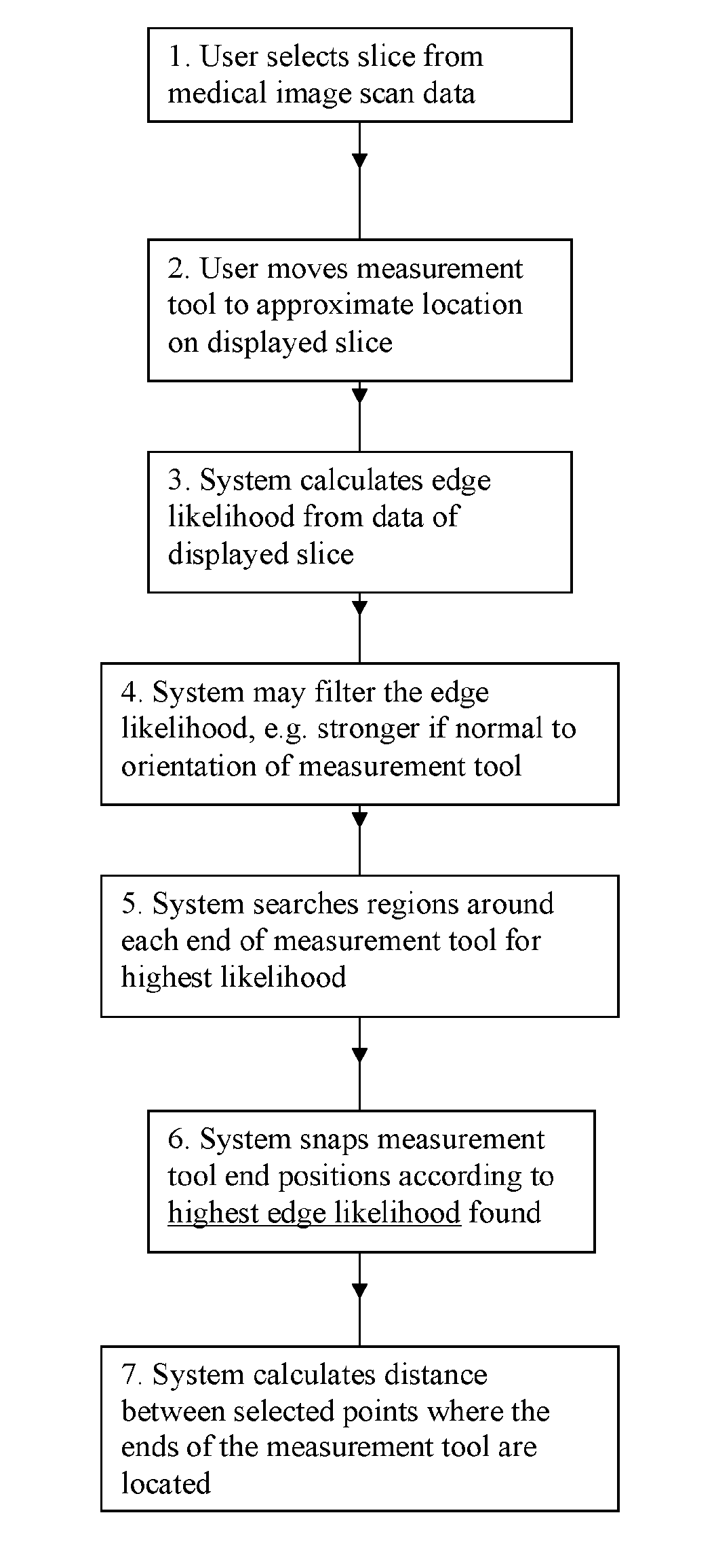
[0066]The invention provides a method of measuring a parameter of a structure on a medical image. Such images are obtained through a variety of digital scanning processes, such as ultrasound scans, CT scans and MRI scans.

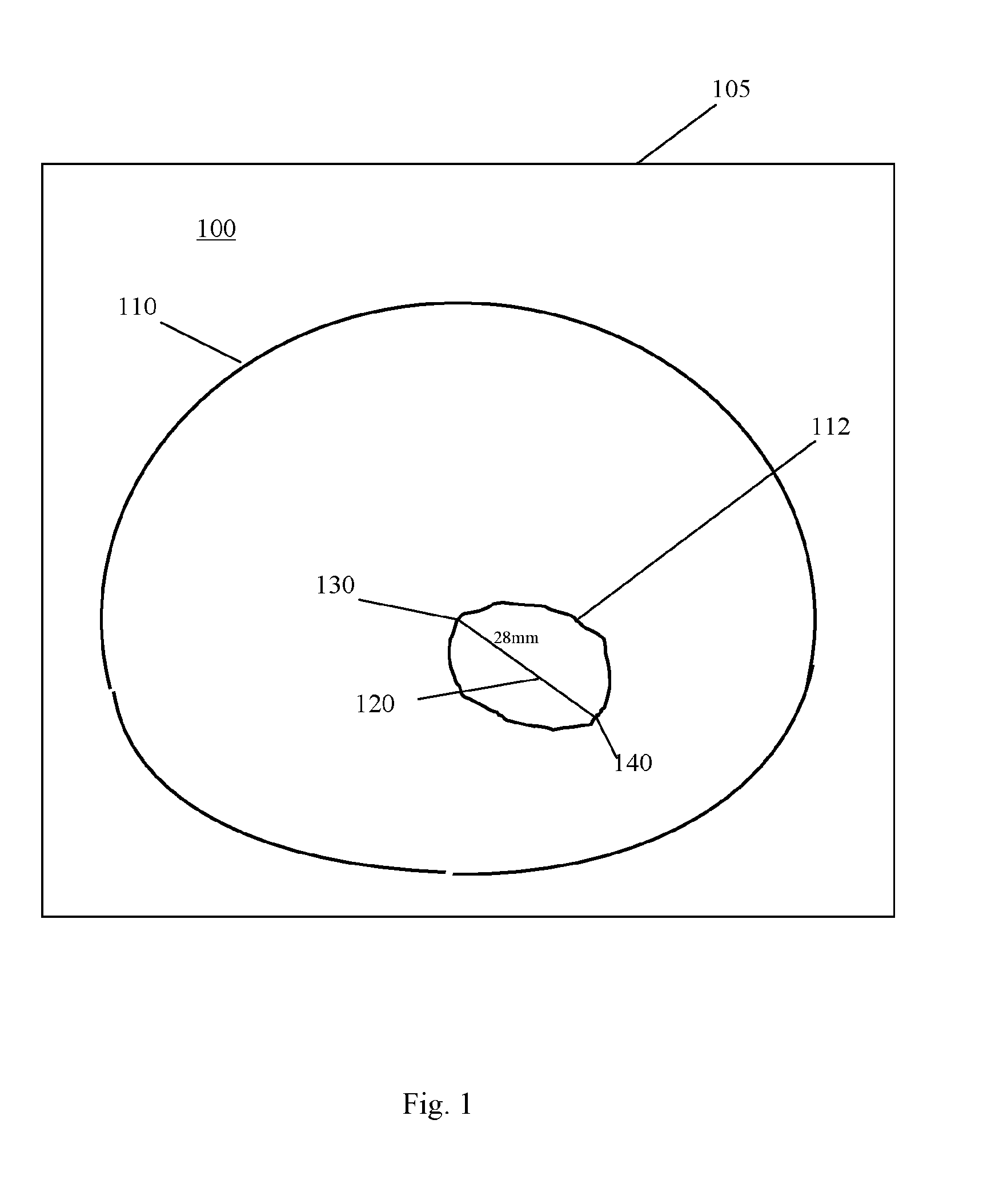
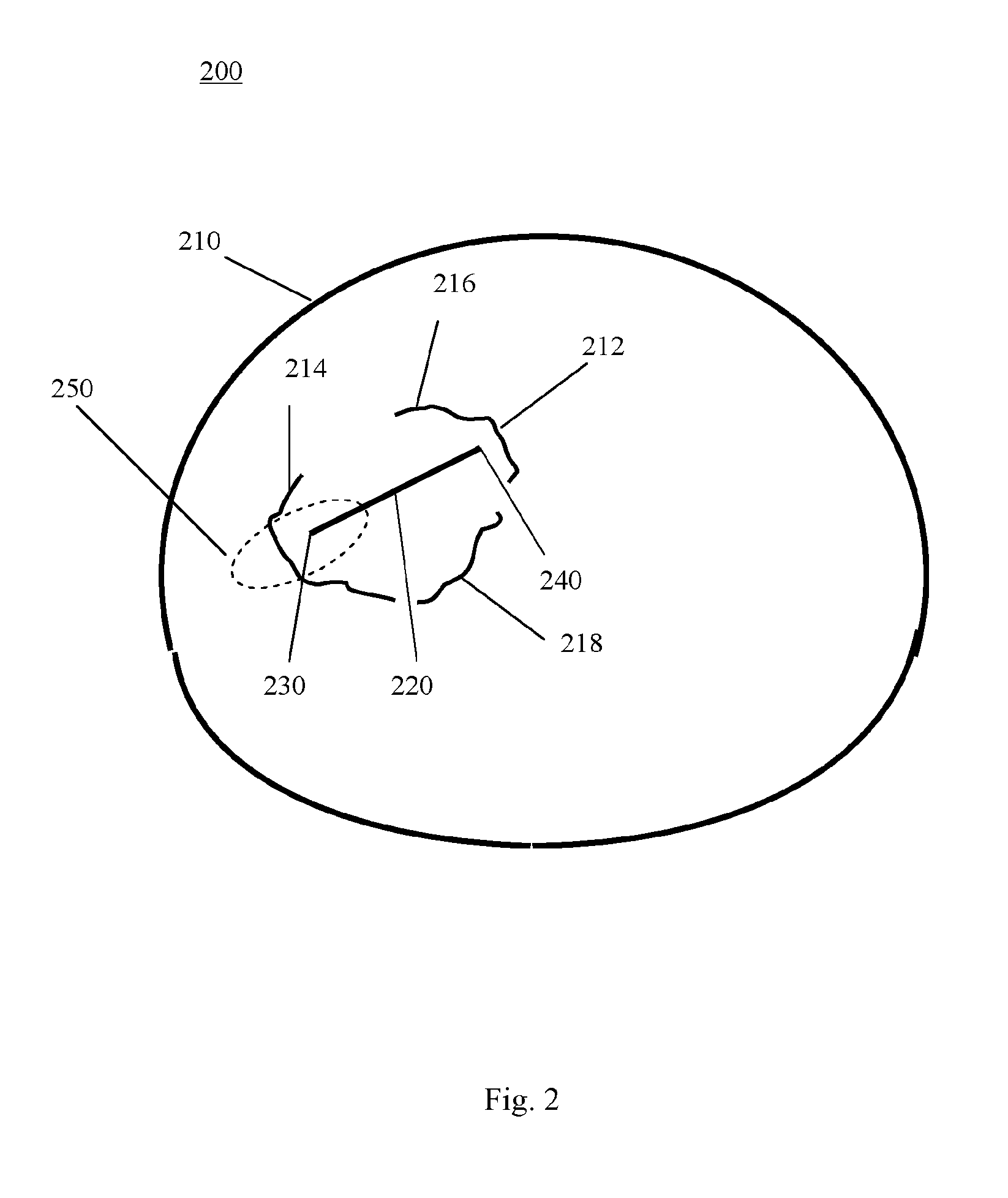
[0067]Tumours or anatomical features are examples of structures for which measurements may be sought. A ‘structure’ might include a single entity, a group of essentially separate objects, or a gap bordered by two or more objects or edges.

[0068]The medical image is displayed as a ‘soft copy’. Normally, the image will have been obtained by a 3-d scan. For an image that has been captured in 3-d, the display will be of a particular ‘slice’ through the 3-d image. If only a 2-d image was captured, that 2-d image is displayed. Henceforth, reference will be made to the image ‘slice’, to cover both these possibilities.

[0069]A measurement tool comprising at least two ends is displayed on the image slice. When the measurement tool is visible on the displayed image slice, a use...


PUM


Abstract

A method of measuring a parameter of a structure (212) on a medical image (200) comprises a measurement tool (220) displayed on a slice of the image. An automated point detection function identifies a point within at least one region (250) of the image that optimises the placement of the respective end of the measurement tool (220). The region may be 2-d or 3-d. The identified point(s) are then used to calculate the parameter, which may be distance, angle, area or volume. The measurement tool (320) may move to the identified points (330, 340). A system (2000), computer program and computer-readable medium are also provided. The invention may make the measurement of tumours, and measurements of the spacing within or between various structures on a medical image, more accurate and/or more consistent.

Description

CROSS-REFERENCE TO RELATED APPLICATIONS[0001]This application is a U.S. national counterpart application of international application serial No. PCT / EP2012 / 056098 filed Apr. 5, 2012, which claims priority to GB Patent Application No. 1105725.4 filed Apr. 5, 2011.FIELD OF THE INVENTION[0002]The present invention concerns the measurement of structures within images, the images being generated in the field of medicine.BACKGROUND OF THE INVENTION[0003]A variety of technologies can be used to provide medical images. Typical modern sources of medical images are ultrasound scans, CT scans and MRI scans.[0004]Most medical imaging involves images of humans. However, images may also be obtained of non-human animals, particularly as part of medical research projects. Medical images need to be read, analysed and reviewed by specialists.[0005]Medical images may include information about a wide variety of anatomical features, structures, spaces and distances. For example, an image may show variou...

Claims


Application Information

Patent Timeline
IPC IPC(8): G06T7/00
CPCG06T7/0051G06T7/0012A61B6/463A61B6/467A61B8/467G06T19/00G06T2210/41G06T7/60G06T2207/20101G06T2207/30096A61B8/461A61B8/469G06T7/50G06T7/62
Inventor KADIR, TIMORGOODING, MARK
Owner MIRADA MEDICAL
Why Eureka
Try Eureka