Catheter with ultrasound sensor and method for creating a volume graphic by means of the catheter

a catheter and ultrasound sensor technology, applied in the field of catheters with ultrasound sensors, can solve the problems of difficult to obtain an overview for the observer, difficult to reproduce individual stenose details for instance with the same high local resolution

Active Publication Date: 2014-02-06
SIEMENS HEALTHCARE GMBH
View PDF6 Cites 61 Cited by
  • Summary
  • Abstract
  • Description
  • Claims
  • Application Information

AI Technical Summary

Benefits of technology

[0006]The object of the present invention is to make it possible to more...

Problems solved by technology

The disadvantage of IVUS technology however is that the doctor is only shown a cross-section of the blood vessel very restricted in its location.
This makes it difficult for the observer to obtain an overview.
It is disadvantageous how...

Method used


Image

  • Catheter with ultrasound sensor and method for creating a volume graphic by means of the catheter
  • Catheter with ultrasound sensor and method for creating a volume graphic by means of the catheter
  • Catheter with ultrasound sensor and method for creating a volume graphic by means of the catheter

Examples


Embodiment Construction

[0032]The examples represent preferred embodiment variants of the invention.

[0033]In the examples explained below, the components of the embodiment variants and the steps of the method described each represent features of the invention to be considered individually, independently of one another, which each also develop the invention independently of one another and are thus also to be regarded individually or in a combination other than that shown as a component of the invention. Furthermore the described embodiment variants are also able to be supplemented by further features of the invention which have already described.

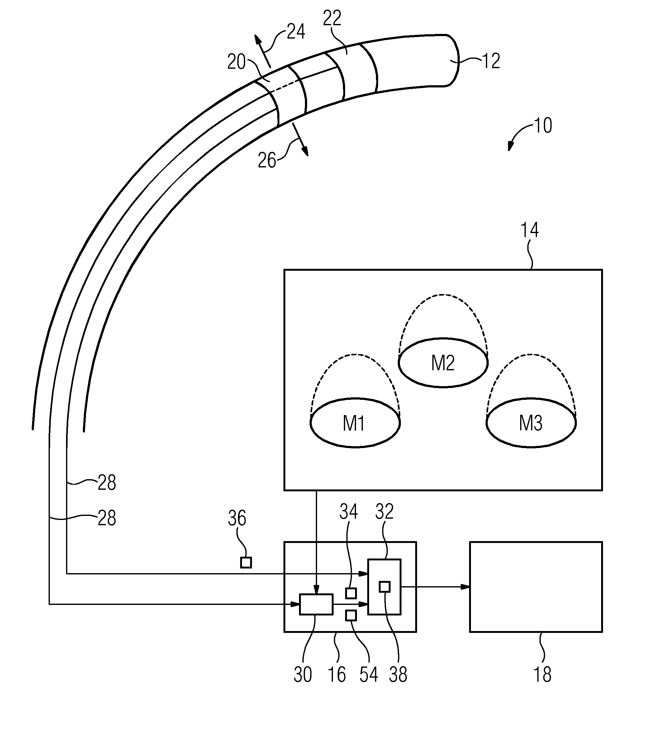
[0034]FIG. 1 shows an imaging system 10 having a catheter 12, a localization unit 14, an evaluation unit 16 and a display unit 20. The imaging system 10 can for example be embodied to create a three-dimensional model, i.e. a volume model, from a blood vessel in a body of a human or an animal.

[0035]Only a tip of the catheter 12 is shown in FIG. 1, which in accordanc...


PUM


Abstract

A method is provided for creating a volume graphic via a catheter for introduction into a vessel which has an ultrasound unit, which is embodied to transmit ultrasound, to measure an echo of the transmitted ultrasound, and to create image data for a two-dimensional cross-section of the vessel on the basis of the measured echo. The method involves creating image data of a cross-section with the ultrasound unit at a plurality of different positions on a movement track of the catheter, and, in addition to the image data, creating position data for the current position of the ultrasound unit with a localization device. The method further involves creating the volume graphic from the image data of all cross-sections in which the cross-sections are arranged relative to one another in accordance with the position data.

Description

CROSS REFERENCE TO RELATED APPLICATIONS[0001]The present application claims priority to German Patent Office application No. 102012213456.5 DE filed Jul. 31, 2012, the entire content of which is hereby incorporated herein by reference.FIELD OF INVENTION[0002]The invention relates to a catheter with an ultrasound sensor for an intravascular ultrasound (IVUS) examination. The invention also includes a method for recording an image of a vessel such as a blood vessel for example with a catheter, which is to be introduced into the vessel for said purpose. Finally the invention also includes an imaging system.BACKGROUND OF INVENTION[0003]IVUS examination is a medical modality which makes it possible, from the inside of a blood vessel, to examine the inner side of the blood vessel and its wall. For this purpose an ultrasound unit, which is embodied to transmit ultrasound and to measure an echo of the transmitted ultrasound, can be disposed on the tip of the catheter to be introduced into t...

Claims


Application Information

Patent Timeline
IPC IPC(8): A61B8/12
CPCA61B8/12A61B8/4254A61B8/466A61B8/483A61B8/5261A61B8/5292
Inventor JIANG, YUAN
Owner SIEMENS HEALTHCARE GMBH
Why Eureka
Try Eureka