Image processing device for finding corresponding regions in two image data sets of an object

a technology of image processing and object, applied in image analysis, image enhancement, instruments, etc., can solve the problems of high cost, high computational complexity, and inability to find corresponding regions in two image data sets of objects, so as to improve the speed and confidence of the workflow

Active Publication Date: 2014-02-27
MEVIS MEDICAL SOLUTIONS +1
View PDF6 Cites 32 Cited by
  • Summary
  • Abstract
  • Description
  • Claims
  • Application Information

AI Technical Summary

Benefits of technology

[0004]Due to the different nature of the images acquired with different modalities and the physiological variability of tissue, the spatial correlation of different modalities becomes a difficult and time-consuming task even for well trained experts. If a finding has been identified in one modality, but is not immediately apparent in a second modality, the reader has to orient himself by means of anatomical landmarks which are visible in both modalities. The nipple, pectoral muscle, skin surface, major blood vessels or components of the glandular disc may serve as landmarks and help radiologist in finding an approximate corresponding position in a second modality where he / she can perform a local search for additional subtle signs of a tissue disorders. In either situation, the reader has to develop a mental model that takes into account the underlying technical principals of each modality in order to translate positions of findings from one modality to the other modality. Furthermore, a certain level of interaction with the image data is required, e.g. for selecting a correct image slice or changing the orientations of a reformation plane if the modality provides a three-dimensional image volume which is displayed as a two-dimensional image.
[0005]Computer tools which support the reader in finding corresponding regions in mammography and MRI images of the same breast promise to improve this workflow in terms of speed and confidence. The article “An intensity-based approach to X-ray mammography—MRI registration” by Mertzanidou et al., Proc. SPIE Medical Imaging: Image Processing, 7623-106, 2010, describes an approach for translating positions in MRI to approximate positions in a CC mammogram of the sane breast. Central component is a finite element model (FEM) of the biomechanics of the breast. Using a FEM that is adapted to the individual patient using a segmentation of the breast in the MRI image, the same compression force applied in mammography is applied in a simulation to the MRI image. Subsequently, MRI intensities are mapped to X-ray attenuation values and a two-dimensional projection image is simulated from the deformed MRI dataset. The simulated X-ray mammogram resembles the real mammogram and can therefore be aligned with the latter using conventional intensity-based image registration algorithms. This last step completes the processing pipeline which allows for translating three-dimensional positions in MRI to the corresponding two-dimensional position in CC mammograms. In the opposite direction, a two-dimensional position in the CC mammogram can only be translated into a line in the MRI due to the missing depth information in the X-ray projection image. This rather sophisticated approach has the disadvantage that FEM are computational very demanding and require reasonable approximations of the biomechanical properties of breast tissue. Additionally, the method requires a detailed segmentation not only of the pectoral muscle and skin surface but also of the fatty and glandular tissue within the breast for the purpose of estimating reasonable X-ray attenuation values from MRI intensities.

Problems solved by technology

The visual appearance of a finding in different modalities is usually highly variable and to a certain extent unpredictable due to the individual tissue physiology, but also because of the different technical principals of each imaging technique.
Due to the different nature of the images acquired with different modalities and the physiological variability of tissue, the spatial correlation of different modalities becomes a difficult and time-consuming task even for well trained experts.
This rather sophisticated approach has the disadvantage that FEM are computational very demanding and require reasonable approximations of the biomechanical properties of breast tissue.

Method used


Image

  • Image processing device for finding corresponding regions in two image data sets of an object
  • Image processing device for finding corresponding regions in two image data sets of an object
  • Image processing device for finding corresponding regions in two image data sets of an object

Examples


Embodiment Construction

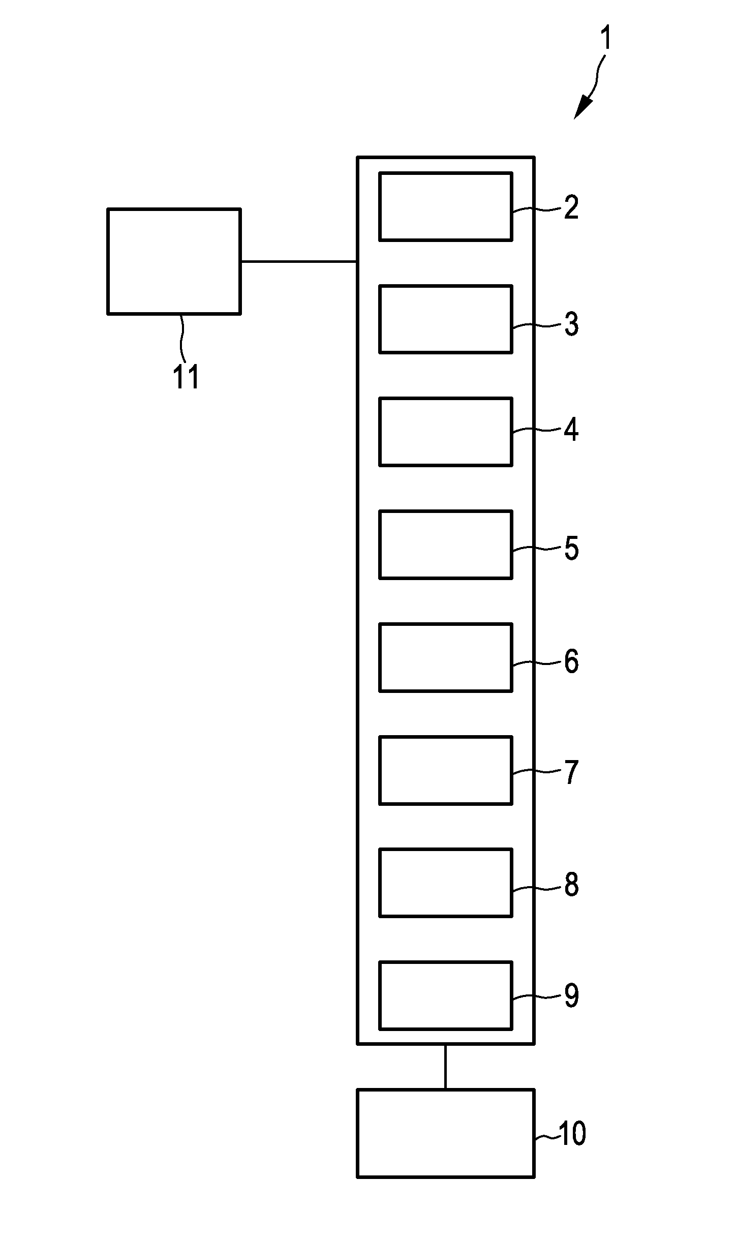
[0099]FIG. 1 shows schematically and exemplarily an embodiment of an image processing device for finding corresponding regions in two image data sets of an object. The image processing device 1 comprises an image providing unit 2 for providing a first image data set of the object and a second image data set of the same object. In this embodiment, the object is a breast, and one of the first image data set and the second image data set comprises a three-dimensional MR image of the breast and the other of the first image data set and the second image data set comprises a CC mammography image and an MLO mammography image of the breast. The CC mammography image and the MLO mammography image are projection images with different projection directions, wherein the breast is compressed in the respective projection direction between two parallel plates. In other embodiments, instead of the MLO mammography image an ML mammography image can be provided. In general, the first image data set and...


PUM


Abstract

The invention relates to an image processing device (1) for finding corresponding first and second regions in two image data sets of an object. In a first image data set a source line and in a second image data set a corresponding target line are determined depending on reference regions detectable in both image data sets. A first region in the first image data set is projected onto the source line, thereby dividing the source line into two source sub-lines and defining a source ratio as the ratio of the length of one of the source sub-lines to the length of the entire source line. A second region in the second image data set is then determined such that a projection of the second region onto the target line leads to a corresponding target ratio which is similar to the source ratio.

Description

FIELD OF THE INVENTION[0001]The invention relates to an image processing device, image processing method and image processing computer program for finding corresponding regions in two image data sets of an object.BACKGROUND OF THE INVENTION[0002]Breast cancer detection and diagnosis benefit significantly from multi-modal imaging. Mammography (MG) is the first line modality for population screening. For surveillance of women which are known to have an increased risk of developing breast cancer, e.g. due to a family history of cancer or genetic predisposition, additional modalities such as contrast-enhanced magnetic resonance imaging (MRI) are utilized and have been widely integrated into regular healthcare provision programs. Both modalities provide complementary diagnostic information. Several signs of breast cancer like micro calcifications can be identified in MG images but barely in MRI. Other tumors can be detected in mammograms, but the full spatial extent of the tissue disorde...

Claims


Application Information

Patent Timeline
Patent Type & Authority Applications(United States)
IPC IPC(8): G06T7/00
CPCG06T7/0014G06T7/0012G06T2207/10072G06T2207/10104G06T2207/10108G06T2207/10132G06T2207/20068G06T2207/30068G06T7/33
Inventor TWELLMANN, THORSTENHAHN, HORSTZOHRER, FABIANFILIPPATOS, KONSTANTINOS
Owner MEVIS MEDICAL SOLUTIONS
Why Eureka
Try Eureka