
Catheter Placement Detection System and Method for Surgical Procedures

a catheter placement and detection system technology, applied in the field of catheter placement detection system and surgical procedures, can solve the problems of prostrate brachytherapy not providing a clear image of catheter placement, the insertion path and final position of the catheter cannot be assumed to be along straight lines extending from the template, and the fundamental limitations of the use of rectally inserted ultrasonic probes during catheter placement procedures

Inactive Publication Date: 2014-12-04
WILLIAM BEAUMONT HOSPITAL
View PDF3 Cites 42 Cited by
  • Summary
  • Abstract
  • Description
  • Claims
  • Application Information

AI Technical Summary

Benefits of technology

The patent describes a system for accurately tracking the placement of catheters in brachytherapy procedures, such as prostate brachytherapy. The system uses electromagnetic technology to track the position of the catheters in real-time, allowing for improved accuracy and efficiency in the placement of the catheters. The system can also use ultrasonic imaging to confirm the position of the catheters, but this can be time-consuming. The invention aims to provide a detection system that provides higher accuracy and reduces evaluation time for verifying catheter placement.

Problems solved by technology

However, due to structural characteristics of the catheters, their tips, and density variations in the human tissue, the insertion paths and final positions of the catheters cannot be assumed to be along straight lines extending from the template.
Unfortunately, the ultrasonic procedure used for human prostrate brachytherapy does not provide a clear image of catheter placement.
There are numerous artifacts in the image reconstruction and, moreover, there are fundamental limits in the use of a rectally inserted ultrasonic probe during catheter placement procedures.
For a real-time ultrasound guided HDR prostate implant procedure, catheter reconstruction has always been challenging and time consuming.
This is due in part to many factors including high speckle noise, inter-needle interference, artifacts from calcifications, hyper-echoic tissues, and coil markers for external beam treatment.
Furthermore, the catheters are always not straight.
They are often curved either inadvertently, or intentionally to reduce normal tissue dose and increase conformity, making the reconstruction of catheter geometry even more difficult.

Method used


Image

  • Catheter Placement Detection System and Method for Surgical Procedures
  • Catheter Placement Detection System and Method for Surgical Procedures
  • Catheter Placement Detection System and Method for Surgical Procedures

Examples


Embodiment Construction

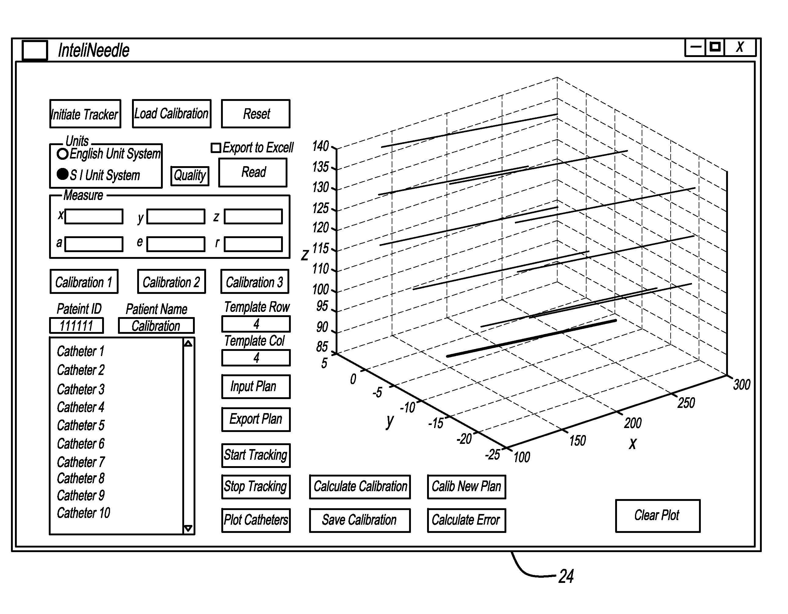
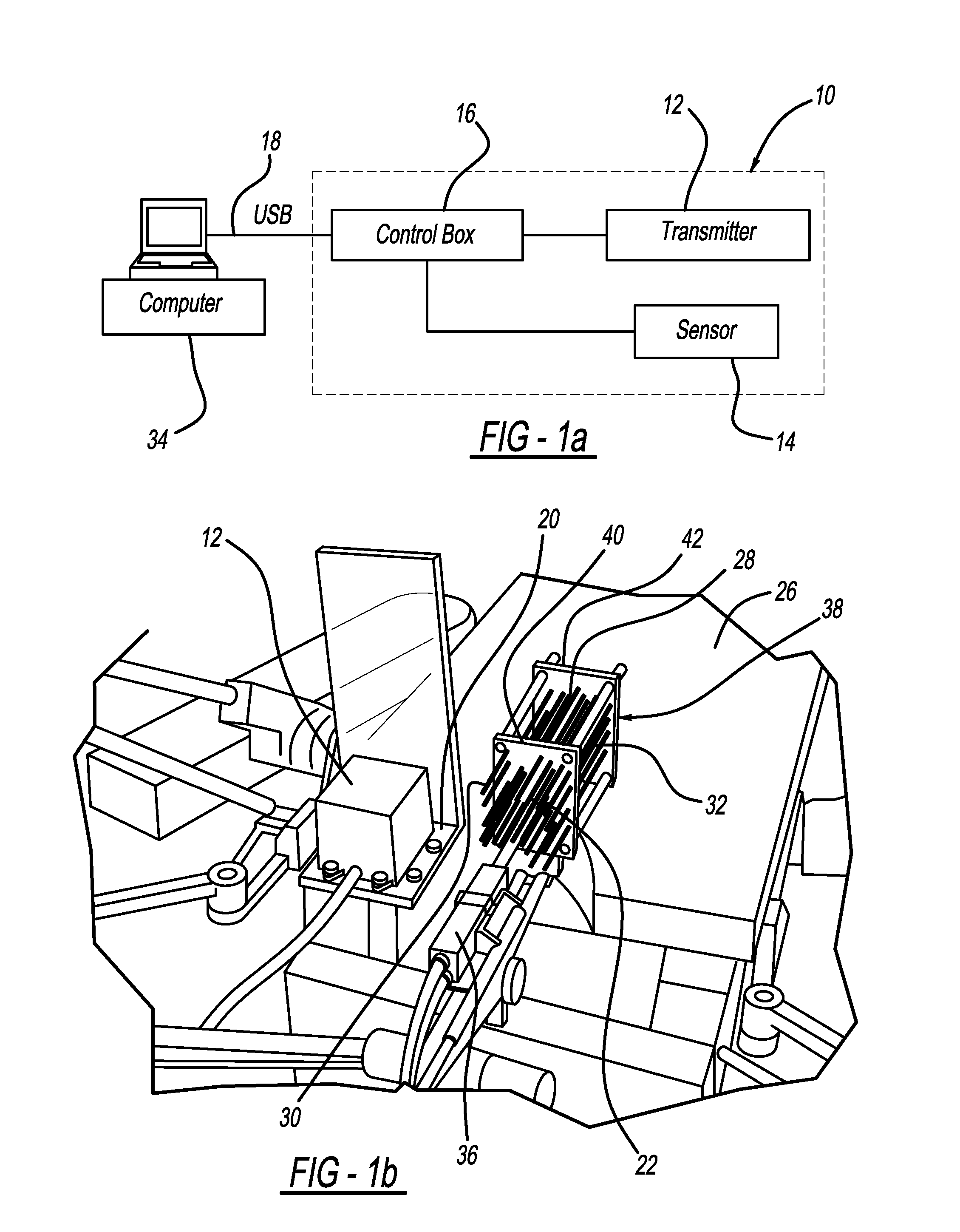
[0012]In accordance with this invention, an electromagnetic tracking system 10 is employed. The tracking system10 as shown in FIG. 1(a) utilizes a transmitter unit 12, preferably one using so-called passive magnetic DC technology (e.g. products available from Ascension Technology Corporation including their “3D Guidance driveBAY”, or “3D Guidance trakSTAR” systems). It is also possible to other tracking systems 10 in accordance with this invention, including those using passive magnetic AC technology. Tracking system 10 include the transmitter 12 mentioned previously, along with one or more miniature sensors 14 which are small enough in size to be inserted into brachytherapy catheters 22 (catheters 22 may also be referred to as “needles”), shown in FIG. 1(b). The system 10 allows the relative position between the transmitter 12 and sensor 14 to be detected and displayed. Catheters 22 have a distal end 28, proximal end 30, and a hollow lumen 32 therebetween.

[0013]Systems utilizing pa...


PUM


Abstract

In order to increase the accuracy and speed of catheter reconstruction in surgical procedures such as an HDR prostate implant procedure, an automatic tracking system is provided preferably using an electromagnetic tracking device. The system uses a transmitter with a sensor used for catheter position. Due to substantial interference in the electromagnetic field from the surgical table, implant stepper / stabilizer etc, a calibration algorithm using a scattered data interpolation scheme is implemented to correct tracking location errors. The invention includes methods and systems used to carry out the methods.

Description

FIELD OF THE INVENTION[0001]This invention relates to methods and systems usable in human and animal surgical procedures. For example, the invention is applicable in the field of human brachytherapy treatment procedures.BACKGROUND OF THE INVENTION[0002]In typical brachytherapy surgical procedures, a physician inserts a number of hollow catheters into a target structure within the human body. The number and location of the catheters is determined by a treatment plan, prescribed by a physician based on imaging studies usually done prior to treatment and many other factors. Often, a grid-like guide template structure is used as a guide for catheter insertion having insertion passages arranged in an orthogonal grid pattern. After inserting a number of such catheters at the prescribed loading position and depth, radioisotope sources are either placed permanently in the tissue as “seeds” (low dose rate or LDR brachytherapy), or are loaded into the catheters and are moved robotically insid...

Claims


Application Information

Patent Timeline
IPC IPC(8): A61N5/10A61B5/06
CPCA61N5/103A61N2005/1024A61N5/1007A61B5/065A61B5/061A61B2017/00274A61B2017/00725A61B17/3468A61B34/20A61B2034/2051
Inventor ZHOU, JUN
Owner WILLIAM BEAUMONT HOSPITAL
Learn More