Atherosclerosis characterization using a multi-contrast MRI sequence

a multi-contrast, mri-sequence technology, applied in the field of imaging methods, can solve problems such as compromising evaluation accuracy and misregistration between image sets

Active Publication Date: 2015-02-19
CEDARS SINAI MEDICAL CENT
View PDF3 Cites 5 Cited by
  • Summary
  • Abstract
  • Description
  • Claims
  • Application Information

AI Technical Summary

Problems solved by technology

Unfortunately, misregistration between image sets due to inter-scan motion often compromises evaluation accuracy.

Method used


Image

  • Atherosclerosis characterization using a multi-contrast MRI sequence
  • Atherosclerosis characterization using a multi-contrast MRI sequence
  • Atherosclerosis characterization using a multi-contrast MRI sequence

Examples


example 1

Methods

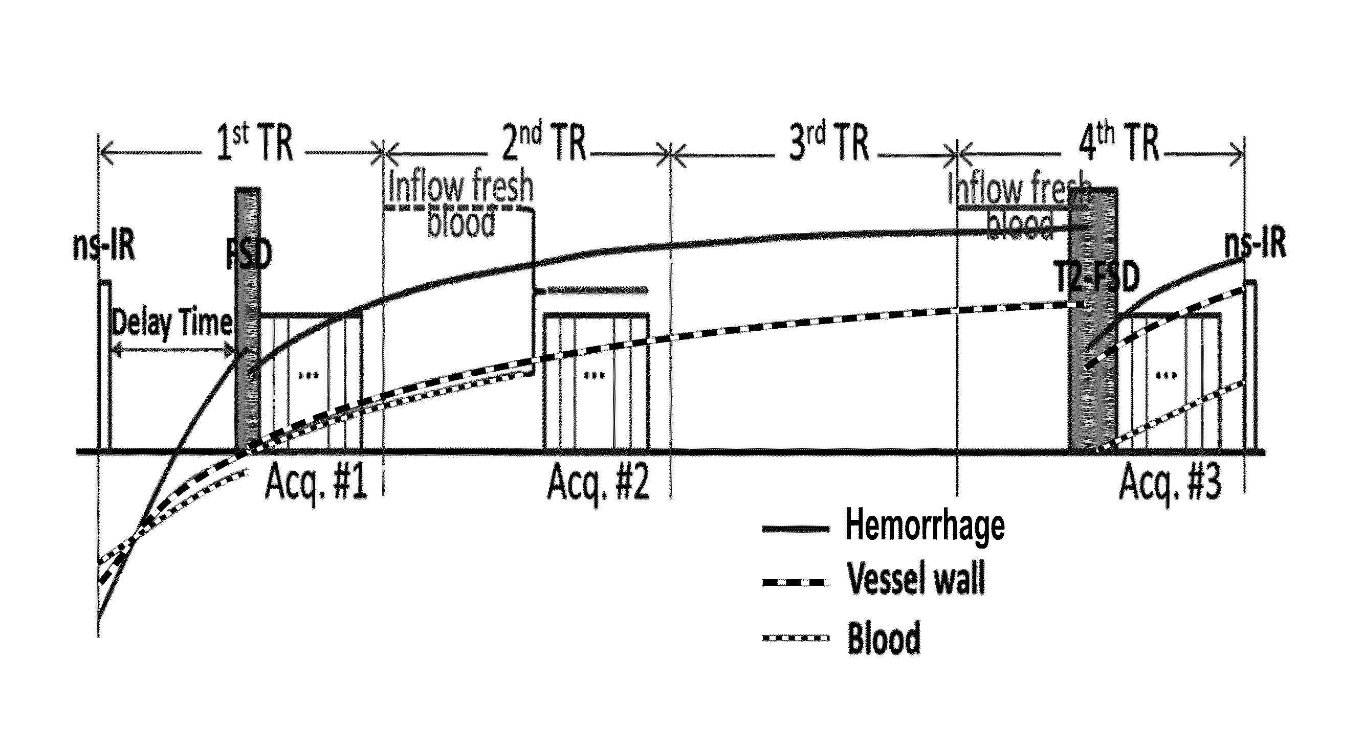
[0034]The inventive “MATCH” (Multicontrast Atherosclerosis Characterization) sequence uses a low-flip-angle gradient echo-based MRI acquisition technique combined with specialized magnetization preparative schemes, and multiple co-registered 3D image sets with different contrast weightings are collected in an interleaved fashion.

[0035]As shown in FIG. 1, the interleaved acquisition of the inventive method consists of 4 repetition times (TRs). The first TR provides hyper T1-weighted (T1-w) contrast at the vessel wall by using a nonselective inversion pulse and a blood-suppressing FSD preparation. The second TR provides grey-to-bright blood lumen that is secondary to both blood T1-recovery and in-flow fresh blood. The third TR is for the vessel wall spins to continue to recover. Finally, the fourth TR provides T2-weighted (T2-w) contrast at the vessel wall by using a long-duration FSD preparation. The three contrasts are aimed to identify the intra-plaque hemorrhage, juxtalumin...

example 2

Results & Discussion

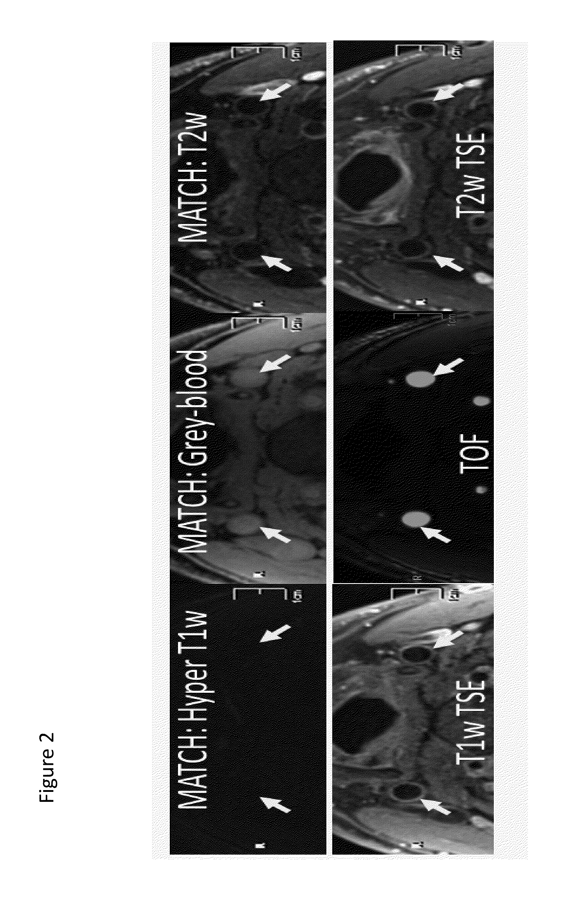
[0037]With the optimized imaging parameters based on computer simulations, the “MATCH” technique described above was capable of providing nulled normal vessel wall on the first contrast weighting, iso-intense or brighter blood on the second contrast weighting, and dark-blood wall on the third contrast weighting, as observed in the healthy volunteer scans (FIG. 2). When applied to clinical cases, MATCH yielded focal signal voids for juxtaluminal calcification (FIG. 3) and hyper-intense depiction for hemorrhage (FIG. 4), both of which were confirmed by conventional protocol. The three 3D image sets that were obtained were spatially co-registered, markedly facilitating plaque assessment.

[0038]As demonstrated above, MATCH is a very useful technique for accurate plaque characterization. One of skill in the art would readily appreciate that mechanical improvements such as isotropic resolution and fast imaging would further improve the clinical value of the inventive te...


PUM


Abstract

The present invention relates to imaging and characterizing atherosclerotic lesions. The invention utilizes a low-flip-angle gradient echo-based MRI acquisition technique combined with specialized magnetization preparative schemes (i.e. non-selective inversion and FSD), and multiple co-registered 3D image sets with different contrast weightings are collected in an interleaved fashion. Using the inventive method, a single scan allows for comprehensive assessment of atherosclerotic plaque within just a few minutes.

Description

FIELD OF THE INVENTION[0001]The present invention generally relates to imaging methods, and more specifically those used for detecting and analyzing atherosclerosis.BACKGROUND[0002]The following description includes information that may be useful in understanding the present invention. It is not an admission that any of the information provided herein is prior art or relevant to the presently claimed invention.[0003]The conventional MRI protocol for investigating atherosclerotic plaques involves a series of scans that provide multiple contrast weightings (e.g. T1-weighted, T2-weighted, and bright-blood, etc.) for resolving high-risk plaque characteristics. Unfortunately, misregistration between image sets due to inter-scan motion often compromises evaluation accuracy.[0004]Considering the state of the art, there is clearly a need for improved MRI methods that allow for more efficient and accurate assessment of atherosclerotic plaque.SUMMARY OF THE INVENTION[0005]In various embodimen...

Claims


Application Information

Patent Timeline
Patent Type & Authority Applications(United States)
IPC IPC(8): A61B5/055G01R33/54G01R33/50G01R33/483G01R33/28
CPCA61B5/055G01R33/4835G01R33/543G01R33/50G01R33/281G01R33/5602G01R33/5607G01R33/5635A61B5/02007
Inventor LI, DEBIAOFAN, ZHAOYANG
Owner CEDARS SINAI MEDICAL CENT
Why Eureka
Try Eureka