Orientation-Aware Average Intensity Histogram to Indicate Object Boundary Depth in Ultrasound Images

an average intensity, ultrasound image technology, applied in image analysis, image enhancement, instruments, etc., can solve the problems of not being able to image the full object, the ultrasound image formed from one pass of the ultrasound wand is typically very narrow, and the noise is typical

Active Publication Date: 2015-02-26
SEIKO EPSON CORP
View PDF0 Cites 11 Cited by
  • Summary
  • Abstract
  • Description
  • Claims
  • Application Information

AI Technical Summary

Benefits of technology

[0031]The above objects may also be met in a method/mechanism/system of defining and characterizing candidate

Problems solved by technology

A problem with ultrasound images is that they are typically very noisy, due in part to the great many tissues and fluids of differing densities and types encountered by a sound wave as it propagates and dissipates through its downward and upward paths through an observed body.
Another problem with ultrasound images is that they are constructed by moving an ultrasound wand over the surface of a target tissue area, but the resultant ultrasound image formed from on

Method used


Image

  • Orientation-Aware Average Intensity Histogram to Indicate Object Boundary Depth in Ultrasound Images
  • Orientation-Aware Average Intensity Histogram to Indicate Object Boundary Depth in Ultrasound Images
  • Orientation-Aware Average Intensity Histogram to Indicate Object Boundary Depth in Ultrasound Images

Examples


Embodiment Construction

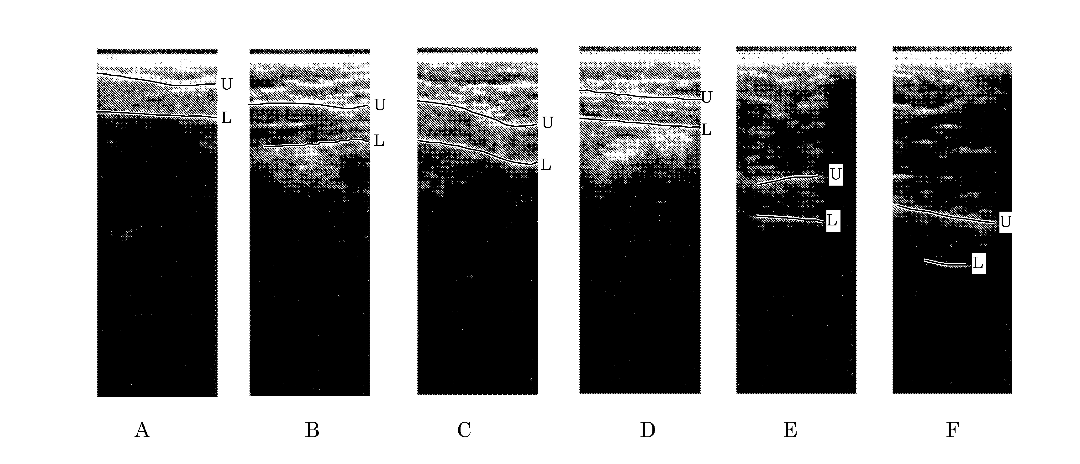
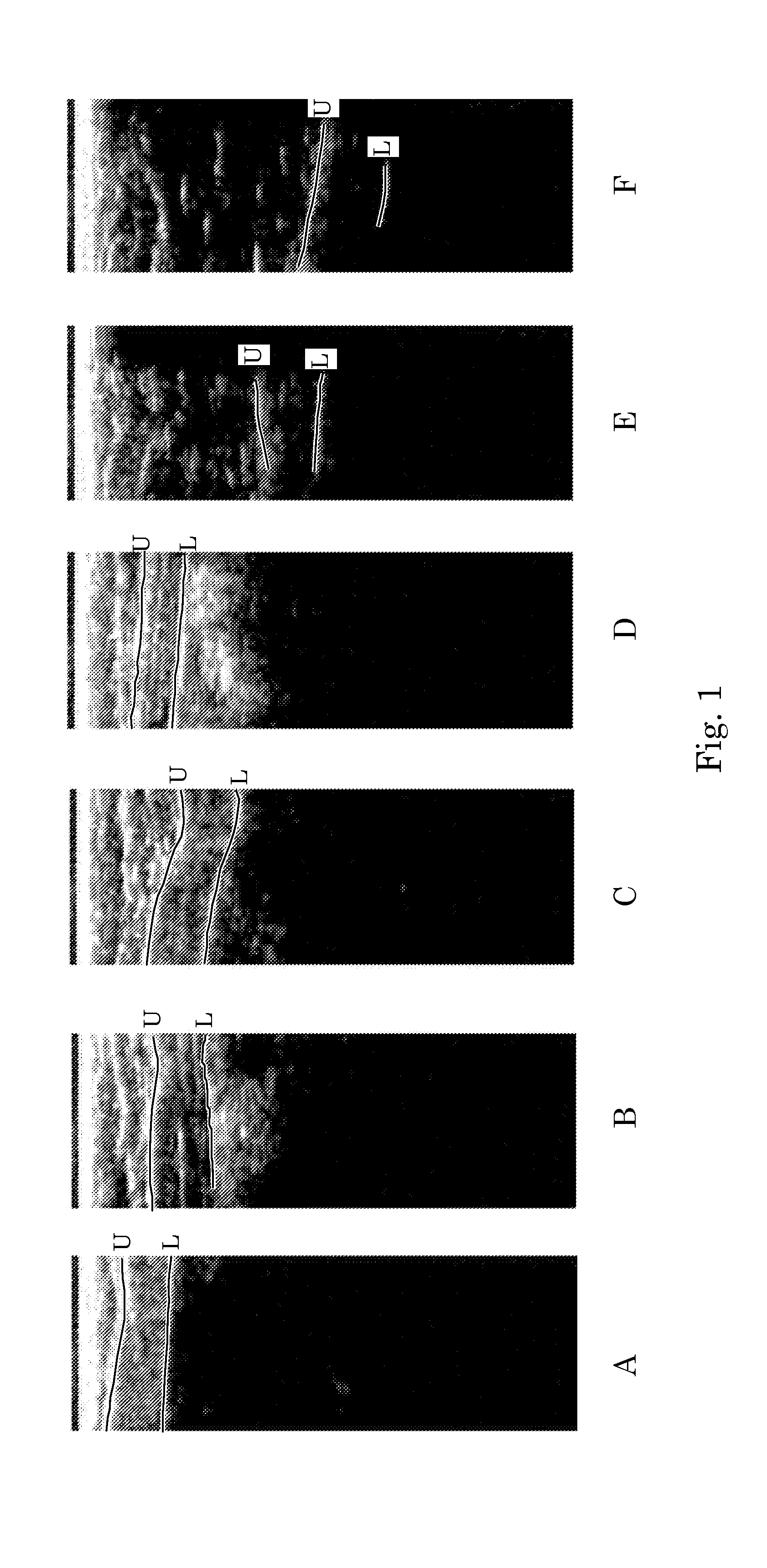
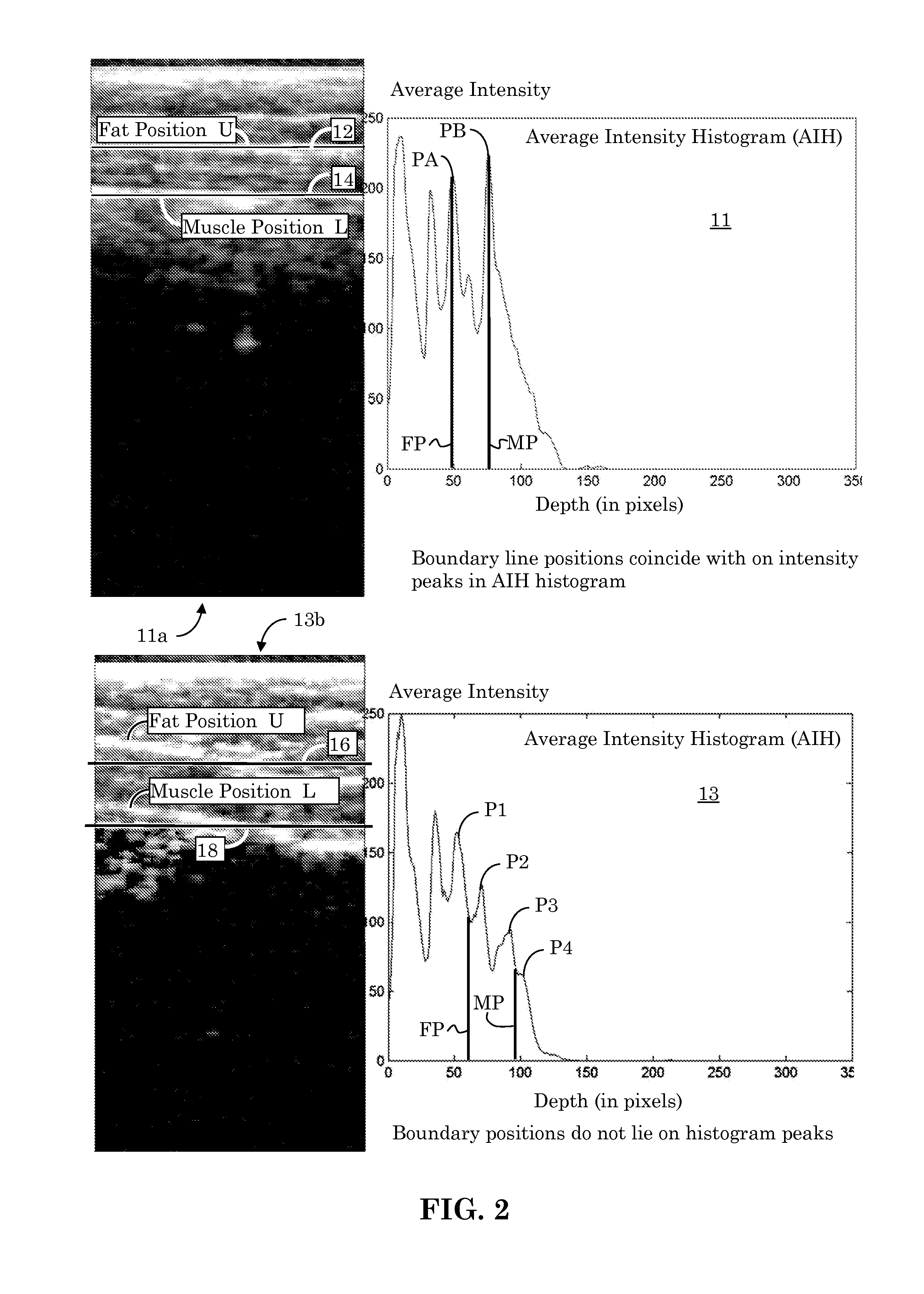
[0050]Measuring object boundary depths, such as the boundary depth of a fat layer (or fatty tissue layer) and / or the boundary depth of a muscle layer (or muscle tissue layer) using ultrasound images is important for health care, medical diagnosis, obesity control, general fitness, etc. Boundary localization of objects, or of different tissue types, within ultrasound images is important for determination of the boundary depth of tissue layers.

[0051]Identifying these boundary depths, however, is challenging due to ultrasound images being highly noisy. Further complicating matters is that fat and muscle boundaries can be at various depths and shapes across different people. The identification of boundary lines is further complicated due to the shapes and image texture of fat and muscle boundaries both being highly variable.

[0052]FIG. 1 provides multiple examples A, B, C, D, E, and F of ultrasound images of an abdominal area. Bright pixels typically delineate the boundary regions betwee...


PUM


Abstract

Linear candidate lines at different angles are used to determine an average intensity for each pixel level of an ultrasound image. The resultant average intensities are collected into a histogram, and the histogram is used to determine the depth positions of tissue boundary lines within an ultrasound image.

Description

BACKGROUND[0001]1. Field of Invention[0002]The present invention relates to the field of ultrasound imaging. More specifically, it relates to detecting the depths of inter-tissue boundaries within an ultrasound image.[0003]2. Description of Related Art[0004]Ultrasound imaging refers to the imaging of structures below a subject's surface (such as the imaging of internal human organs below a skin surface) by the sending of sound waves of known frequency into the interior of the subject and observing any sound waves that bounce back. By monitoring how long it takes for sound waves to bounce back from an internal structure, it is possible to estimate the depth and shape of the structure within the subject. It is also possible to discern some characteristics about the internal structure based on how the sound waves are absorbed, dispersed or deflected.[0005]Ultrasonography, or diagnostic sonography, refers to the use of ultrasound imaging techniques for imaging subcutaneous body structur...

Claims


Application Information

Patent Timeline
IPC IPC(8): G06T7/00G06K9/46
CPCG06T7/0012G06K9/4647G06T7/0051G06T7/0085G06T2207/10132G06T2207/20068G06T7/12G06V10/267G06V10/507
Inventor XIAO, JINGWU, CHENYU
Owner SEIKO EPSON CORP
Why Eureka
Try Eureka