Cell image segmentation method and a nuclear-to-cytoplasmic ratio evaluation method using the same

Active Publication Date: 2015-03-19
COGNINU TECH CO LTD
View PDF22 Cites 22 Cited by
  • Summary
  • Abstract
  • Description
  • Claims
  • Application Information

AI Technical Summary

Benefits of technology

[0025]Another objective of this disclosure is to obtain high effici

Problems solved by technology

Regarding conventional biopsy methodology and pathology examination, conventional methodology was achieved primarily through physical biopsy, which needs to remove a tissue from a living subject that is invasive, time-consuming, high-cost for preparation of abundant samples and evaluation at the microscopic level and also must be followed by complicated procedures including fixing, embedding, sectioning, and staining to determine the status of diseases.
In addition to being painful to the patients, these invasive procedures may also result in risk of infection or even spreading of the cancer cells.
Without tissue removal, optical in vivo virtual biopsy can not only conquer the tedious stages in conventional invasive biopsy procedure, but also save time and cost for conventional pathology examination.
It is time consuming, highly subjective, and also has inconsistent accuracy, especially for a huge mass amount of data to be analyzed.
Illumination and noise are also factors to make image thresholding have undesired segmentation results even with adap

Method used


Image

  • Cell image segmentation method and a nuclear-to-cytoplasmic ratio evaluation method using the same
  • Cell image segmentation method and a nuclear-to-cytoplasmic ratio evaluation method using the same
  • Cell image segmentation method and a nuclear-to-cytoplasmic ratio evaluation method using the same

Examples


Embodiment Construction

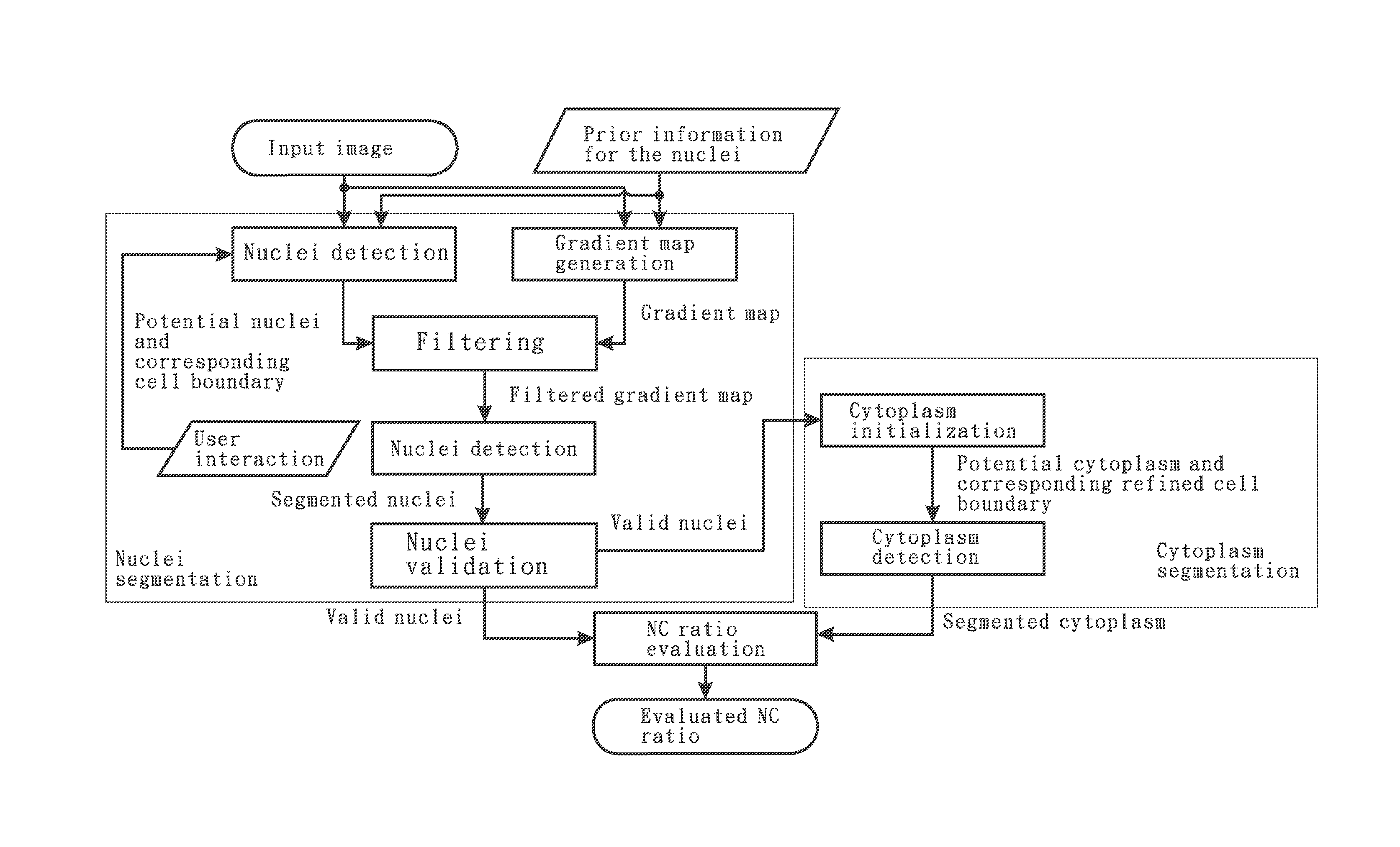
[0119]Cell Segmentation and NC Ratio Analysis

[0120]In this disclosure, we focus on the processing and analysis procedure applied to images and image sequences with application to biomedical imaging. When observing images or image sequences, there are four basic descriptors, including shape, size, color and texture, perceived by human eyes. For making it possible to understand and analyze these basic descriptors by computers or machines, some mathematical models and their corresponding physical meanings will be understood and utilized to transform observable descriptors into informative features. For the application of Nuclear-to-Cytoplasmic ratio (NC ratio) analysis in biomedical imaging, biopsy technique is required to acquire the cells or tissues to be analyzed and cell segmentation which can isolate and select individual nucleus and its cytoplasm is then performed for NC ratio analysis. Subsequently, the evaluated NC ratios or cell information are discussed and interpreted with m...


PUM


Abstract

A cell image segmentation method includes receiving a cell image, performing a nuclei initialization step to find an internal marker and an external marker to obtain a potential nuclei and a potential cell boundary, calculating a gradient map of the received cell image, performing a filtering step on the gradient map to generate a filtered gradient map, performing a nuclei detection step to obtain a segmented nuclei, and performing a nuclei validation step to obtain a valid nuclei. The nuclei initialization step includes performing a blob detection step to obtain a nuclei candidate, an outlier removal step to obtain the internal marker, a distance transform step to obtain a distance map, and a cell boundary initialization step to obtain the external marker. In another embodiment, a nuclear-to-cytoplasmic ratio evaluation method using the above cell image segmentation method is proposed.

Description

BACKGROUND OF THE INVENTION[0001]1. Field of the Invention[0002]The present disclosure generally relates to a cell image segmentation method and a nuclear-to-cytoplasmic ratio evaluation method using the cell image segmentation method and, more particularly, to a cell image segmentation method for cell segmentation and nuclear-to-cytoplasmic ratio analysis, as well as a nuclear-to-cytoplasmic ratio evaluation method using the same.[0003]2. Description of the Related Art[0004]Regarding conventional biopsy methodology and pathology examination, conventional methodology was achieved primarily through physical biopsy, which needs to remove a tissue from a living subject that is invasive, time-consuming, high-cost for preparation of abundant samples and evaluation at the microscopic level and also must be followed by complicated procedures including fixing, embedding, sectioning, and staining to determine the status of diseases. In addition to being painful to the patients, these invasiv...

Claims


Application Information

Patent Timeline
IPC IPC(8): G06K9/00G06T7/00
CPCG06K9/0014G06T7/0091G06T7/0012G06T2207/20152G06T2207/20192G06T2207/30024G06T7/11G06T7/12G06T7/155G06V20/695
Inventor LEE, GWO GIUNLIN, HUAN-HSIANGTSAI, CHENG-SHIUNCHEN, CHUN-FU
Owner COGNINU TECH CO LTD
Why Eureka
Try Eureka