Method for preparing patient-specific glioblastoma animal model, and uses thereof

a glioblastoma and mouse model technology, applied in the field of preparing patient-specific glioblastoma animal models, can solve the problems of difficult treatment, high incidence, and unreported effective treatment methods other than radiotherapy for gliomas, and achieve safe and effective patient-specific glioblastoma

Active Publication Date: 2015-04-16
SAMSUNG LIFE PUBLIC WELFARE FOUND
View PDF1 Cites 1 Cited by
  • Summary
  • Abstract
  • Description
  • Claims
  • Application Information

AI Technical Summary

Benefits of technology

[0007]The present invention provides a method for producing a glioblastoma mouse model, comprising the steps of: (a) dividing a glioblastoma tissue, isolated from a patient, into or more sections, and collecting one or more pieces from each of the sections; (b) dissociating a mixture of the collected pieces into glioblastoma cells as single cells; and (c) orthotopically transplanting a graft sample, which contains the glioblastoma cells obtained in step (b), into the brain of an immunodeficient mouse. The present invention also provides a mouse model prod

Problems solved by technology

Gliomas are malignant tumors accounting for about 60% of primary brain tumors, have high incidence and are difficult to treat.
Thus, effective treatment methods other than radiotherapy for gliomas have not yet reported.
However, in the case of such brain tumors, the delivery of therapeutic drugs is not easy due to the presence of blood-brain barriers.
Particularly, because of the insufficient understanding of brain neurobiology, the development of therapeutic agents is slowly progressing.
Further, glioblastoma represents an aggressive variant compared to other brain tumors, and thus can cause lethal results within several weeks, if it is not treated within a short time.
However, the screening of molecular markers that can represent clinical results is still insufficient.
Particularly, gene expression analysis is performed at the transcriptome level, and thus cannot indicate the expression level of protein in actual cancer tissue.
In the above paper, a mouse model was produced using only a graft sample prepared from the piece of a specific port

Method used


Image

  • Method for preparing patient-specific glioblastoma animal model, and uses thereof
  • Method for preparing patient-specific glioblastoma animal model, and uses thereof
  • Method for preparing patient-specific glioblastoma animal model, and uses thereof

Examples


example 1

Glioblastoma (GBM) Patients and the Primary Culture of Cell Cultures Derived from the Patients

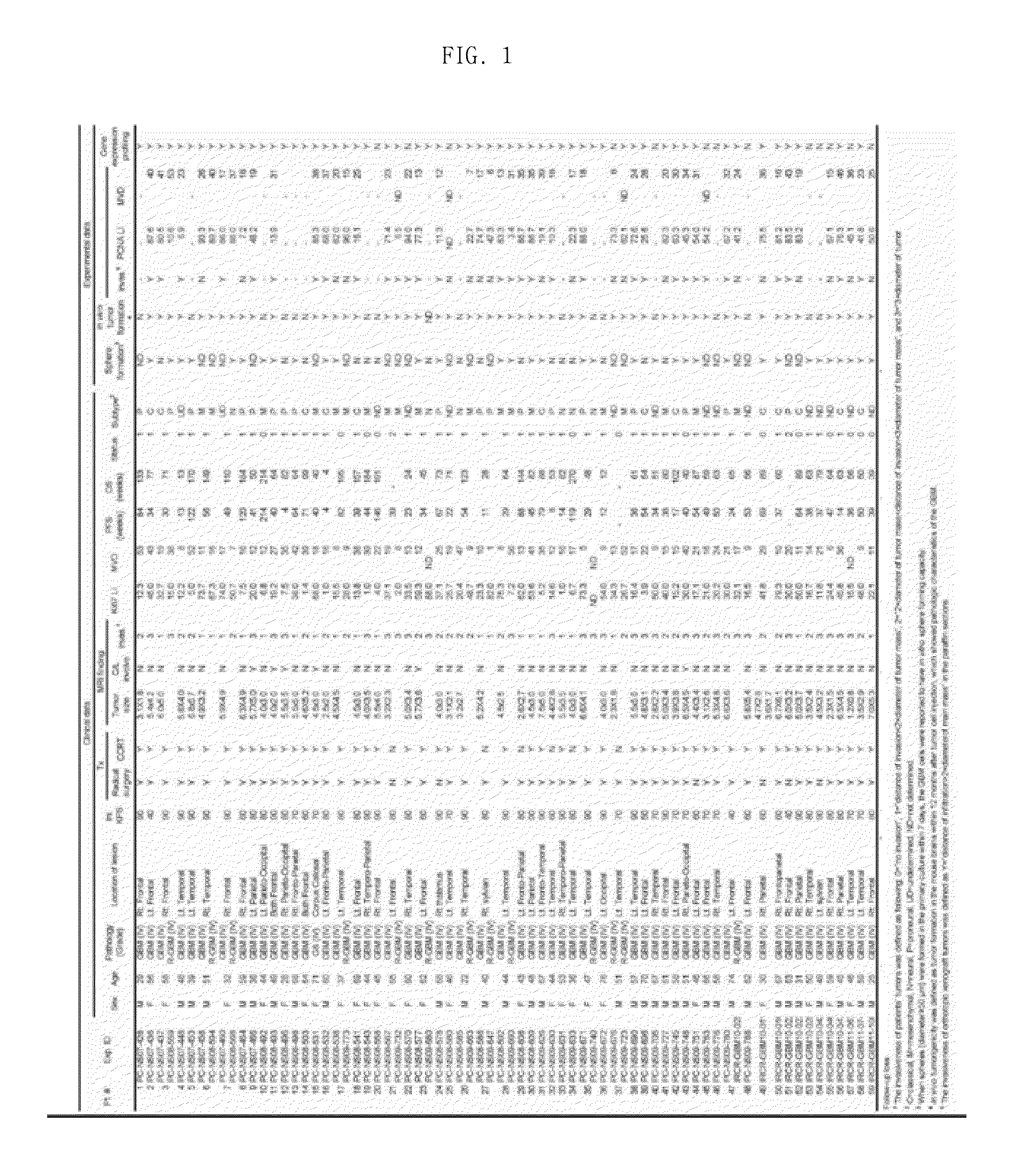
[0059]Following informed consent, surgical samples and clinical records were obtained from 58 GBM patients who had brain tumor removal surgery at the Samsung Medical Center (Seoul, Korea), and data thereon are shown in FIG. 1. Specifically, FIG. 1 shows the clinical data of glioblastoma patients and experimental results for primarily cultured glioblastoma (GBM) cells derived from the patients.

[0060]The obtained surgical samples were classified as GBM based on WHO criteria by examination of pathologists. Parts of the surgical samples were enzymatically dissociated into single cells, and the dissociated GBM cells were cultured in the “NBE” condition. Specifically, the dissociated GBM cells were cultured in Neurobasal media with N2 and B27 supplements (0.53× each; Invitrogen) and bFGF and EGF (25 ng / ml each; R&D Systems). Alternatively, acutely dissociated GBM cells in the NBE condition were p...

example 2

Construction of Orthotopic Xenograft Mouse Model

[0061]Animal experiments were approved by the Institutional Review Boards of the Samsung Medical Center and conducted in accordance with the “National Institutes of Health Guide for the Care and Use of Laboratory Animals” (NIH publication 80-23), and an orthotopic xenograft mouse model was produced in the following manner.

[0062]The production of the orthotopic xenograft mouse model was largely divided into the steps of: 1) preparing a sample (to be transplanted into a mouse) from a glioblastoma tissue isolated from a glioblastoma patient; and 2) transplanting the sample into a mouse. The procedure for the production of the orthotopic xenograft mouse model was as follows.

[0063](1) Step of Preparing Homogenized Graft Sample from Tissue Isolated from Glioblastoma Patient

[0064]A sample to be transplanted into a mouse was prepared using a fresh tissue within 2-3 hours after isolation from a glioblastoma patient, and the preparation was perf...

example 3

Tissue Microarray and Immunohistochemistry

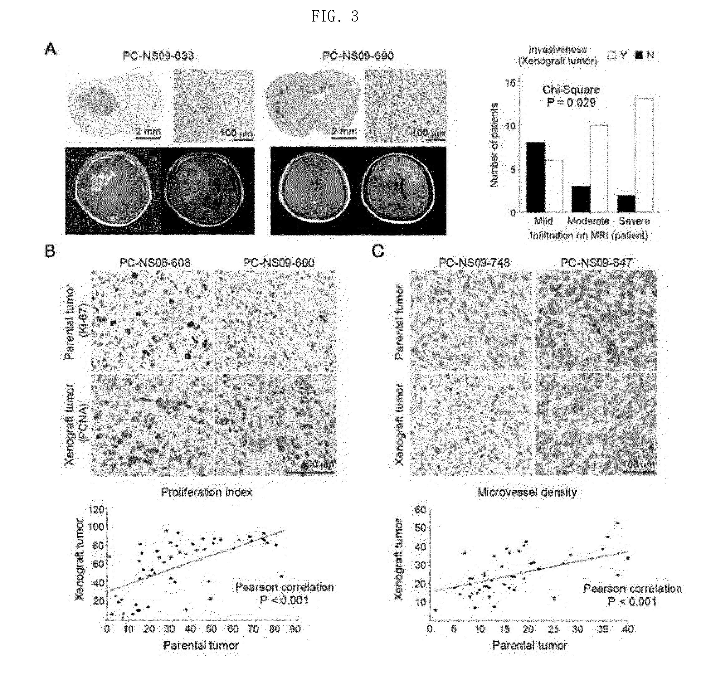
[0079]A tissue microarray was performed on parental glioblastoma and orthotopic xenograft tumors (PC-NS07-464, PC-NS08-493, PC-NS08-532, PC-NS08-559, PC-NS08-608, PC-NS09-626, PC-NS09-630, PC-NS09-633, PC-NS09-660, PC-NS09-690, and PC-NS09-696) derived therefrom.

[0080]Immunohistochemistry was performed using protein-specific antibodies (Ki-67, PCNA (DAKO), DLL3 (Santa Cruz), SOX2, Olig2, MAP2 (Abcam), MBP, PDGFA, EGFR (Santa Cruz), CHI3L1, TOP2A (LifeSpan Bioscience) and CD31, and BD Pharmingen for parental tumor).


PUM


Abstract

Provided is a method for producing a glioblastoma mouse model and the mouse model produced thereby, the method including the steps of: (a) dividing a glioblastoma tissue, isolated from a patient, into 4 or more sections, and collecting one or more pieces from each of the sections; (b) dissociating a mixture of the collected pieces into glioblastoma cells as single cells; and (c) orthotopically transplanting a graft sample containing the glioblastoma cells obtained in step (b), into the brain of an immunodeficient mouse. Further provided are a method of screening a glioblastoma therapeutic agent using the mouse model and a method of providing information for selection of a patient-specific glioblastoma therapy using the mouse model. The glioblastoma mouse model shows the same genetic, morphological and pathological characteristics as those of the parental tumor, and it allows screening patient-specific glioblastoma therapeutic agent or selecting safer and more effective patient-specific glioblastoma therapy.

Description

TECHNICAL FIELD[0001]The present invention relates to a method for producing a glioblastoma mouse model, comprising the steps of: (a) dividing a glioblastoma tissue, isolated from a patient, into or more sections, and collecting one or more pieces from each of the sections; (b) dissociating a mixture of the collected pieces into glioblastoma cells as single cells; and (c) orthotopically transplanting a graft sample, which contains the glioblastoma cells obtained in step (b), into the brain of an immunodeficient mouse; a glioblastoma mouse model produced by the method; a method for screening a glioblastoma therapeutic agent using the mouse model; and a method for providing information for selection of a patient-specific glioblastoma therapy using the mouse model.BACKGROUND ART[0002]Gliomas are malignant tumors accounting for about 60% of primary brain tumors, have high incidence and are difficult to treat. Thus, effective treatment methods other than radiotherapy for gliomas have not...

Claims


Application Information

Patent Timeline
IPC IPC(8): A61K49/00A01K67/027
CPCA61K49/0008A01K67/0271A01K2227/105A01K2267/0331A01K2267/0356G01N33/15A01K67/027
Inventor NAM, DO HYUNJIN, JU YUONJOO, KYEUNG MIN
Owner SAMSUNG LIFE PUBLIC WELFARE FOUND
Why Eureka
Try Eureka