Wearable external ventricular drain system

a technology of external ventricular drain and wearable, which is applied in the direction of wound drain, functional valve types, transportation and packaging, etc., can solve the problems of increased intracranial pressure, reduced patient safety, severe brain damage or death, etc., and achieve the effect of reducing the potential for patient harm

Active Publication Date: 2015-04-23
INFINIVATION BIOMEDICAL
View PDF21 Cites 10 Cited by
  • Summary
  • Abstract
  • Description
  • Claims
  • Application Information

AI Technical Summary

Benefits of technology

[0013]In accordance with the foregoing objects and advantages, the present invention comprises a wearable EVD system having a ventricular catheter that is placed in the ventricle of the brain to drain CSF and a valve located in line with the patient's ear. A transducer may be also provided level with the patient's ventricle and connected to a monitor that continuously or periodically measures the intracranial pressure. The valve and transducer are firmly secured at the level of the ventricle with a support headband and / or ear clip. The valve may be either an adjustable orifice valve or a spring-loaded needle valve or any other adjustable valve. CSF that drains through the valve is collected in the drip chamber for periodic measurement and then emptied into the drainage bag. In a simpler embodiment, the CSF can collect directly into the drainage bag and the drip chamber may be left out. The drip chamber and drainage may be attached to the patient inferior to the cranium via securement straps. As a result, no IV pole is needed and a patient is free to sit up or move about while the EVD system is in place without disrupting drainage of CSF, eliminating the potential for patient harm.
[0014]The present invention will be more fully understood and appreciated by reading the following Detailed Description in conjunction with the accompanying drawings, in which:
[0015]FIG. 1 is a schematic of a wearable external ventricular drain system according to the present invention;
[0016]FIG. 2 is a schematic of an adjustable needle valve for a wearable external ventricular drain system according to the present invention; and
[0017]FIG. 3 is a schematic of an adjustable orifice valve for a wearable external ventricular drain system according to the present invention.

Problems solved by technology

It results in increased intracranial pressure (“ICP”) and can cause severe brain damage or death if the pressure is not relieved.
The greatest issue with the current EVD system is that the zero level of the drain must be level with the patient's ventricle at all times. If the drain is not level with the patient's ventricle, more or less back pressure will be applied to the brain, and thus more or less CSF than desired will flow into the drip chamber.
This requirement causes decreased safety to the patient and increased nursing time necessary to level the drain, as patients are prone to frequently adjusting the positions and elevations of their heads.
For example, many ICU patients with hydrocephalus have neurological deficits and cannot always remember that they have an EVD.
These patients are likely to sit up quickly, which results in over-drainage of CSF into the EVD.
Over-drainage can be very hazardous to the patient, possibly causing increased headache, collapse of ventricles, decreased cerebral perfusion, hemorrhage, and death.
In addition, if the patient adjusts his / her bed from an elevated position to a horizontal position, under-drainage of CSF would occur.
Under-drainage of CSF can lead to a dangerous over-accumulation of fluid in the brain, possibly causing headaches, nausea, brain damage, brain herniation, and death.
Conventional EVD systems thus result in decreased mobility and independence for ICU patients, which lead to poorer outcomes and loss of function while the patients are hospitalized.
Only properly trained nurses can adjust the EVD drain, so this requirement prevents other health care workers or family members from being able to assist the patients to reposition their heads, or move about the room or hospital.
This procedure adds the risk of the patient tripping, and limits the amount of time that the patient can be out of the bed, since the EVD must be adjusted or turned off and closely monitored in order for the patient to stand.

Method used


Image

  • Wearable external ventricular drain system
  • Wearable external ventricular drain system
  • Wearable external ventricular drain system

Examples


first embodiment

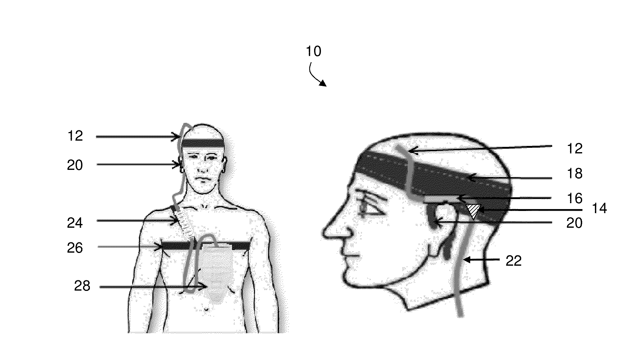
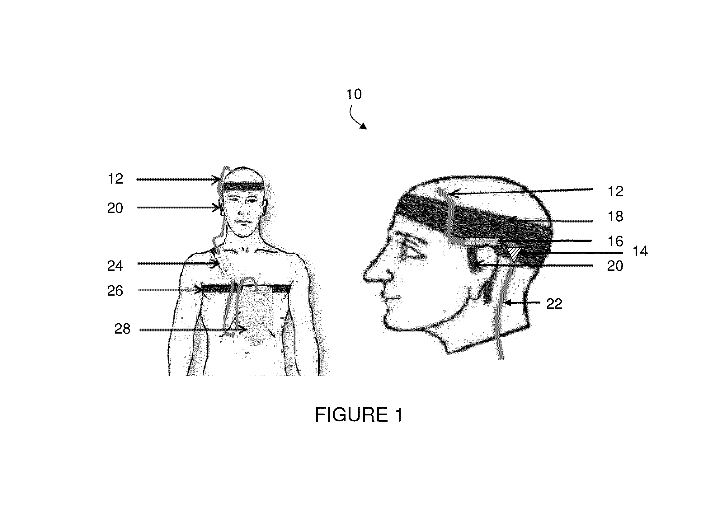
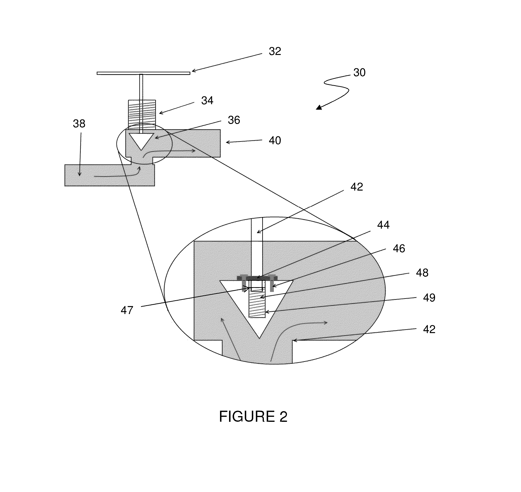
[0022]The present invention encompasses at least three embodiments for valve 14, although one or more alternative valves may be employed instead. As seen in FIG. 2, valve 14 is an adjustable, spring-loaded needle valve. Needle valves are capable of sensitive adjustments and high degrees of attainable resolution. This high resolution is critical, since using the device necessitates operating within a small pressure range. A needle valve according to the present invention is preferably adjustable from 0-30 mmHg in increments of 1 mmHg. Needle valve 30 comprises an adjustment knob 32 having advancement threads 34 for advancing or retracting a needle in the flow path having a fluid inlet 38 and a fluid outlet 40. As further seen in FIG. 2, needle 36 is adapted to engage a needle seat 42 positioned between fluid inlet 38 and fluid outlet 40, thereby adjusting the amount of fluid that can flow from fluid inlet 38 to fluid outlet 40. When needle 36 is positioned far away from needle seat 4...

second embodiment

[0024]valve 14 comprises an orifice valve 50 that is adjustable both in diameter and length. The back pressure provided by an orifice is provided by both its diameter and its length according to the Hagen-Poiseuille equation:

ΔP=8μLQπr4

[0025]As seen in FIG. 3, a series of rotating cylinders 52, each having multiple orifices of varying diameters, are aligned along a common axis X-X. Orifices are preferably offset from the axis of rotation and positioned to align with a fluid pathway by a fluid input tube 56 to a fluid outlet 58. As seen in FIG. 4, each cylinder 52 has a small orifice 60 and large orifice 62 that may be selectively aligned along fluid pathway and configures to provide a certain back pressure according to the Hagen-Poiseuille equation. For example, aligning the small orifices 62 of four cylinders 52, each providing 1 mmHg back pressure, with the large orifices 62 of four remaining cylinders 52, will provide a total of 4 mmHg back pressure. Each cylinder 52 may be sealed...

third embodiment

[0026]In a third embodiment, valve 14 may comprise an array of orifices of various specific diameters and lengths that provide a range of back pressures according to the Hagen-Poiseuille equation. A moveable shutter that selectively exposes a predetermined single orifice or array of orifices may be used to allow a user to adjust the specific amount of back pressure for system 10.


PUM


Abstract

A wearable EVD system having a ventricular catheter and transducer supported proximately to a patient's ear by a mount, such as supporting headband or ear clip. An adjustable orifice valve or a spring-loaded needle valve is used to control the amount of CSF that drains into a drip chamber suspended on the patient for periodic measurement and emptying into a similarly located drainage bag, thereby avoiding the need for an IV pole and allowing the patient more mobility without disrupting drainage of CSF.

Description

BACKGROUND OF THE INVENTION[0001]1. Field of the Invention[0002]The present invention relates to external ventricular drain systems and, more particularly, to a wearable external ventricular drain system.[0003]2. Description of the Related Art[0004]Hydrocephalus is a medical condition characterized by an excess accumulation of fluid in the brain. It results in increased intracranial pressure (“ICP”) and can cause severe brain damage or death if the pressure is not relieved. An external ventricular drain (“EVD”) is used to drain excess cerebrospinal fluid (“CSF”) in cases of temporary hydrocephalus such as brain hemorrhage and shunt malfunction in order to provide a therapeutic reduction in ICP.[0005]The currently existing EVD system consists of a thin soft tube (ventricular catheter) that is inserted under sterile procedure into the ventricle of the brain. The distal end of the ventricular catheter is connected to the EVD tubing which leads to a drip chamber with an adjustable heigh...

Claims


Application Information

Patent Timeline
Patent Type & Authority Applications(United States)
IPC IPC(8): A61M1/00
CPCA61M1/0019A61M27/006A61M2205/3523A61M2205/3561A61M2209/088A61M2210/0693A61M2230/00A61M1/70A61M1/743Y10T137/7932Y10T137/7935F16K1/2071F16K1/38F16K5/10F16K15/063F16K17/04
Inventor STEBBINS, KRISTENPENNELL, II, THOMAS J.
Owner INFINIVATION BIOMEDICAL
Why Eureka
Try Eureka