Computer aided diagnosis for detecting abdominal bleeding with 3D ultrasound imaging

Inactive Publication Date: 2015-07-23
HER MAJESTY THE QUEEN AS REPRESENTED BY THE MINIST OF NAT DEFENCE OF HER MAJESTYS CANADIAN GOVERNMENT
View PDF6 Cites 9 Cited by
  • Summary
  • Abstract
  • Description
  • Claims
  • Application Information

AI Technical Summary

Problems solved by technology

Second, inner body fluids are detected by the system w

Method used


Image

  • Computer aided diagnosis for detecting abdominal bleeding with 3D ultrasound imaging
  • Computer aided diagnosis for detecting abdominal bleeding with 3D ultrasound imaging
  • Computer aided diagnosis for detecting abdominal bleeding with 3D ultrasound imaging

Examples


Embodiment Construction

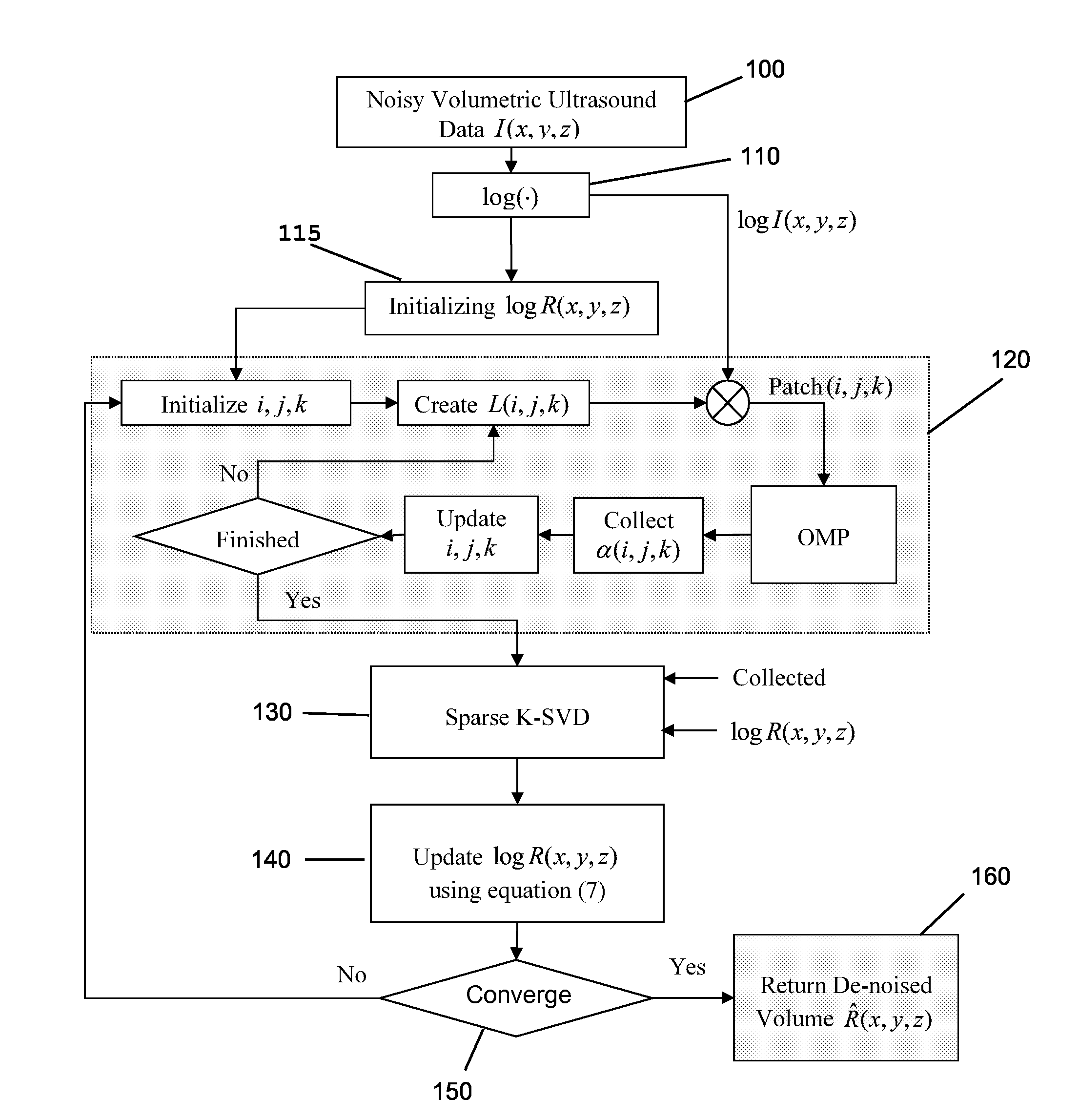
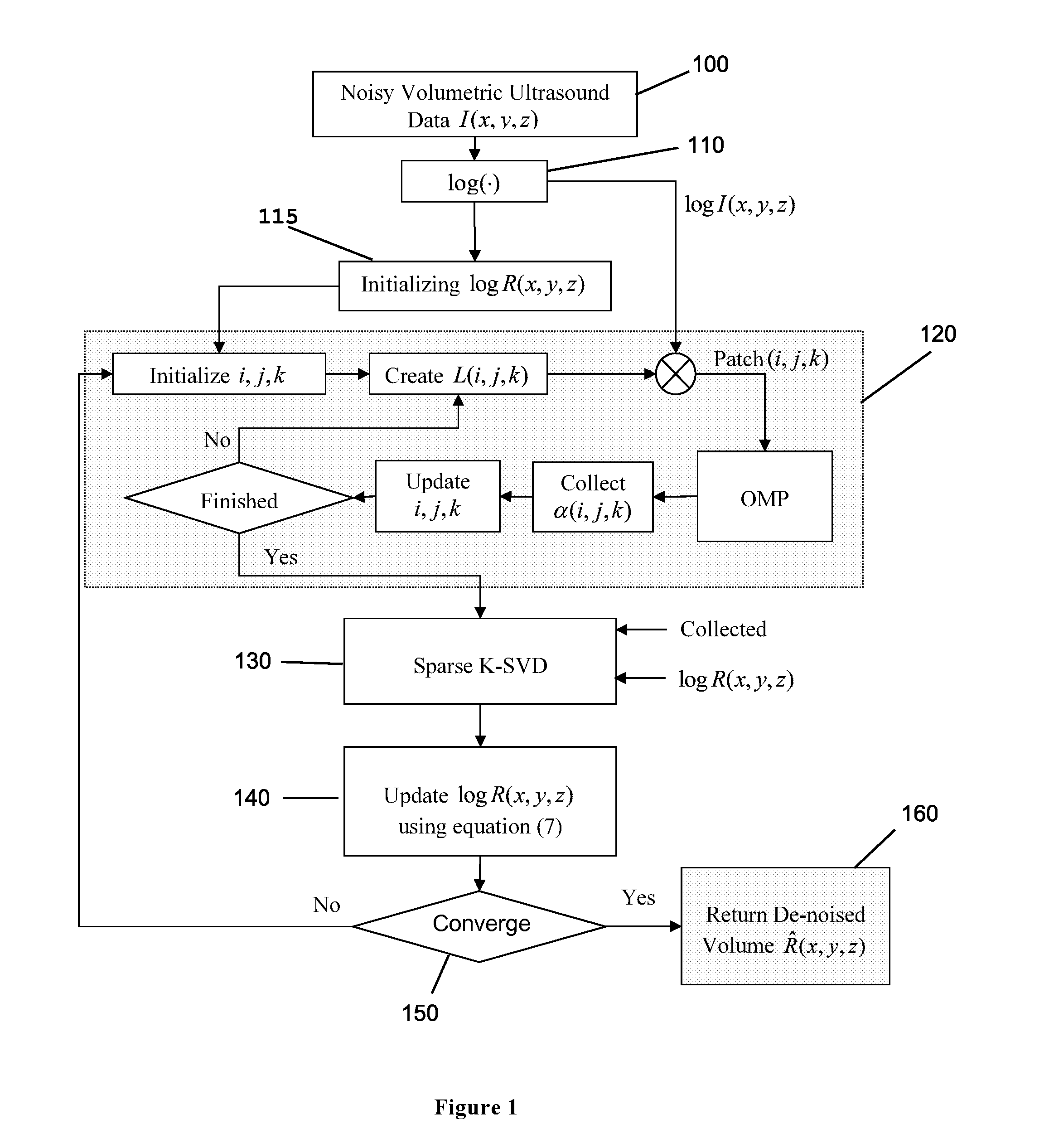
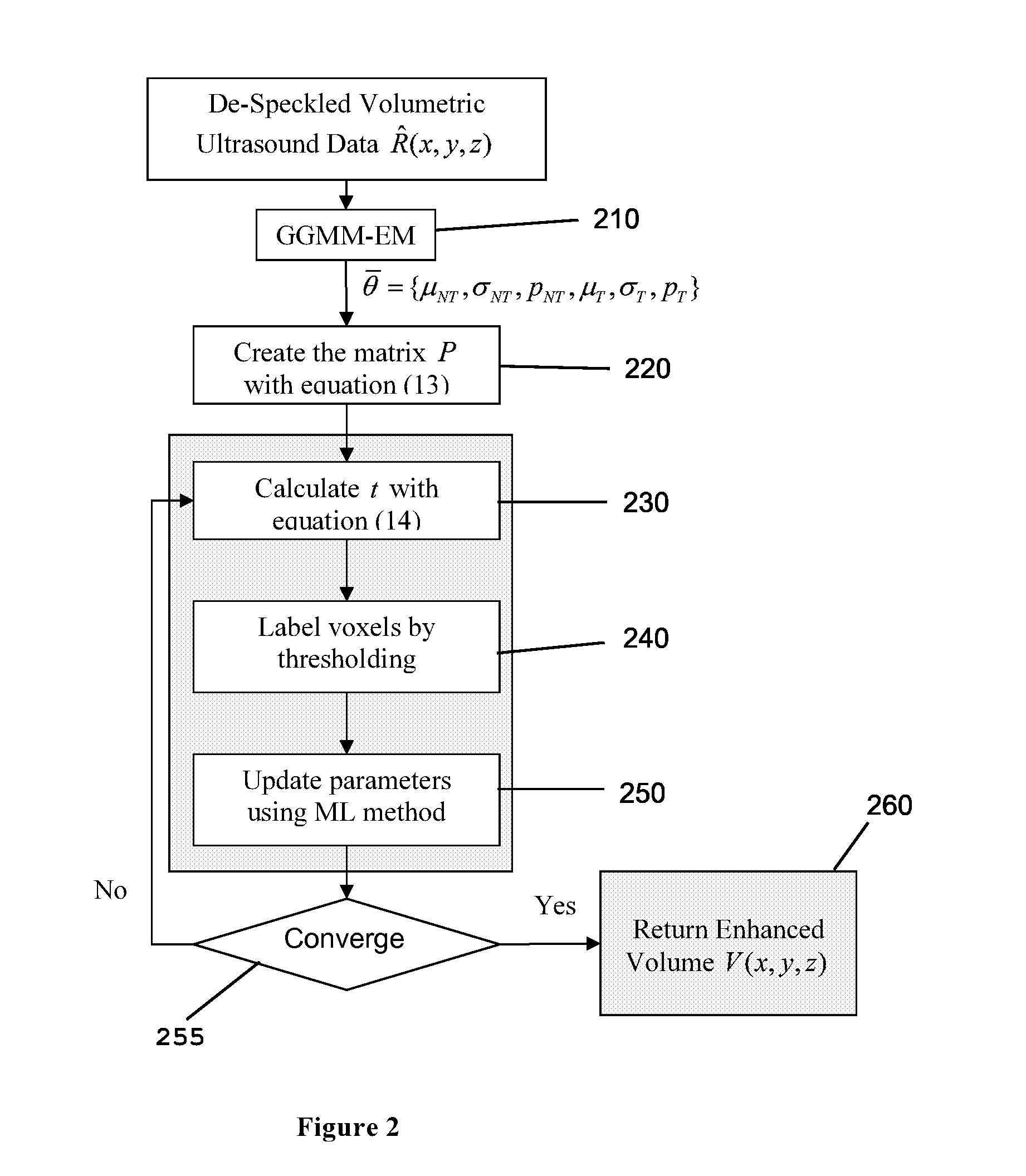
[0036]The task of object detection in 3D medical images has been investigated by researchers, due to its implications for medical diagnosis. The procedural steps of object detection in 3D volumes consist of preprocessing tasks, manual adjustment, 3D segmentation and classification. The aim of the preprocessing tasks is to put more emphasis on valuable information and to reduce the effect of unwanted interfering signals. Some examples of such tasks are denoising, edge refinement, contrast enhancement and volume clipping. As a second step, user intervention can be applied to boost the segmentation results. The 3D segmentation task can be considered to be the main part of an anatomical organ detection procedure. The selected 3D segmentation method for ultrasound volumes has to be robust against intensity variability, speckle noise and discontinuities among object walls. Finally, the classification step labels the segmented region as internal bleeding area, kidney, liver or other organs...


PUM


Abstract

The presented invention provides a detection method for free fluid in the human body. The preprocessing procedure is applied to reduce the speckle noise and enhance tissue volumetric pixels (voxels) intensity levels to make tissue and non-tissue voxels more distinguishable from each other. An initial surface selection step provides flexibility to either manually or automatically selecting a seed point to segment fluid regions in the volumetric data. A kidney and liver organ detection procedure provides a method for determining whether the detected fluid region is a normal fluid-carrying body organ or if it is due to a medical condition as a free fluid region. A combinational approach using a 3-dimensional Snake and Level-Set is utilized to accurately detect fluid regions in the volumetric ultrasound data.

Description

FIELD OF THE INVENTION[0001]The present invention relates to a diagnosis system for analyzing 3D ultrasound images to detect the size and location of internal organs and free fluid.BACKGROUND[0002]The rapid diagnosis of invisible internal injury in austere and hostile front-line environments remains a challenge for medical personnel. The availability of a portable real-time 3-Dimensional (3D) ultrasound imaging system with automated diagnostic capabilities for detecting non-visible internal abdominal bleeding, pneumothorax, hematothorax and facilitating image guided operations is helpful, if not essential, in supporting triage and medical decisions for trauma patients.[0003]In trauma patients, inner-body fluids can accumulate at the lowest position of the concavities within the body. The site of fluid accumulation depends on the source of the bleeding and the position of the patient. In general, patients are examined on the bed lying flat on the back. In such conditions, inner-body ...

Claims


Application Information

Patent Timeline
IPC IPC(8): A61B8/08A61B5/02
CPCA61B8/5223A61B8/08A61B8/5269A61B8/483A61B5/02042A61B8/463G06T2207/10132G06T2207/30096G06T7/11G06T7/62G06T7/136G16H50/30
Inventor STERGIOPOULOS, STERGIOSSHEK, PANGPLATANIOTIS, KONSTANTINOSMARSOUSI, MAHDI
Owner HER MAJESTY THE QUEEN AS REPRESENTED BY THE MINIST OF NAT DEFENCE OF HER MAJESTYS CANADIAN GOVERNMENT
Why Eureka
Try Eureka