
Devices applicable to tissue(s) which facilitates confocal microscopy, optical microscopy, spectroscopy and/or imaging

a tissue and optical microscopy technology, applied in the field of tissue and tissue application of microscopy to anatomical structures, can solve the problems of inaccurate and/or incomplete removal of cancer, labor-intensive pathology preparation, inaccurate and/or incomplete removal, etc., and achieve the effect of facilitating rapid pathology technologies

Active Publication Date: 2015-08-20
MEMORIAL SLOAN KETTERING CANCER CENT
View PDF4 Cites 4 Cited by
  • Summary
  • Abstract
  • Description
  • Claims
  • Application Information

AI Technical Summary

Benefits of technology

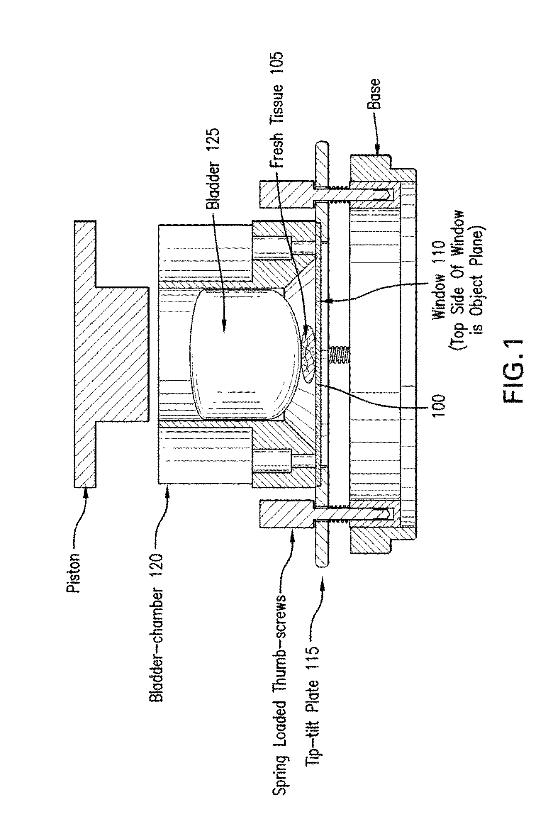
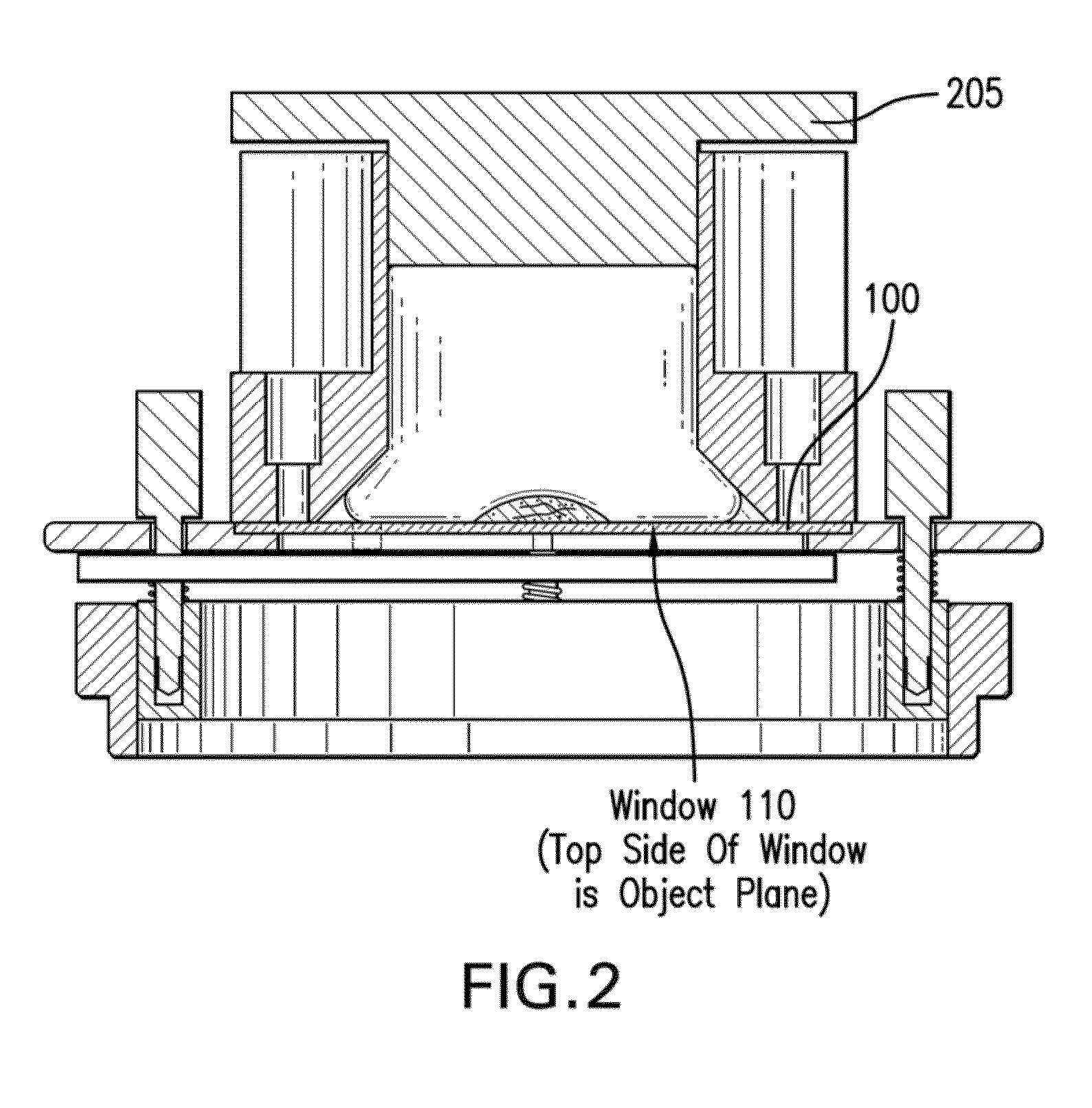
The patent describes a method for analyzing samples using a special arrangement that includes an inflatable device. This device can be used to apply force to the sample to make its surface lie flat against the arrangement. This helps to ensure that the entire imaging surface is parallel to the imaging plane, which is important for accurate imaging. The technical effect of this method is that it enables faster and more accurate pathology analysis at the bedside, using optical imaging techniques.

Problems solved by technology

However, a preparation of the pathology can be labor-intensive, and can be time-consuming.
The processing of excised tissue and preparation of thin sections for pathology can take hours (e.g., for frozen sections) or days (e.g., for fixed sections), which can result in insufficient sampling of tissue, and inaccurate and / or incomplete removal of cancer.

Method used


Image

  • Devices applicable to tissue(s) which facilitates confocal microscopy, optical microscopy, spectroscopy and/or imaging
  • Devices applicable to tissue(s) which facilitates confocal microscopy, optical microscopy, spectroscopy and/or imaging
  • Devices applicable to tissue(s) which facilitates confocal microscopy, optical microscopy, spectroscopy and/or imaging

Examples


Embodiment Construction

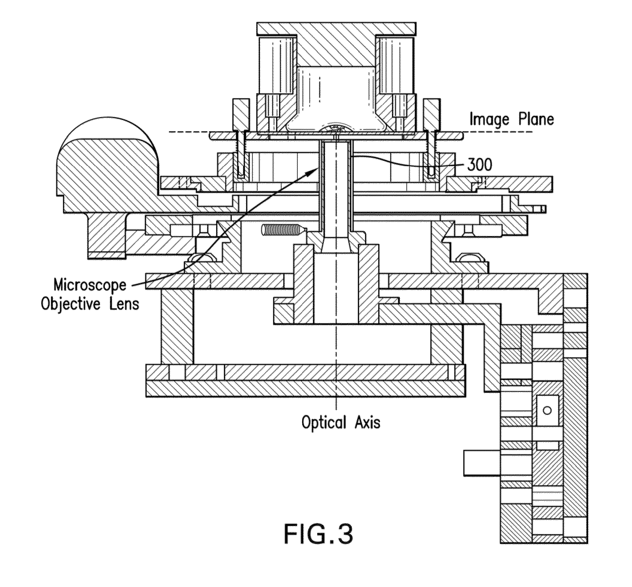
[0007]Indeed, one of the objects of certain exemplary embodiments of the present disclosure can be to address the exemplary problems described herein above, and / or to overcome the exemplary deficiencies commonly associated with the prior art as, for example, described herein. Accordingly, for example, provided and described herein are certain exemplary embodiments of exemplary devices according to the present disclosure which can be applicable to tissue(s) which facilitates confocal microscopy, optical microscopy and / or imaging.

[0008]Due to the three-dimensional (“3D”) topography and irregular shapes and sizes of fresh surgically excised, or biopsied, tissue, mounting the tissue for imaging large areas with a scanning confocal microscope, or other modalities, as mentioned above, can be challenging due to the following problems:[0009]a. Sag, for example, bending of the desired tissue surface (e.g., imaging plane) to be imaged.[0010]b. Tissue stability during imaging and mosaicing pro...


PUM


Abstract

Exemplary embodiments of apparatus and method for facilitating an analysis of a sample(s) can be provided. For example, using a first arrangement(s), it can be possible to receive the sample(s) thereon. Further, for example, using a second arrangement(s), it can be possible to cause a force to be applied on a portion(s) of the sample(s) such that a surface(s) of the sample(s) can be flattened against a section(s) of the first arrangement(s).

Description

CROSS-REFERENCE TO RELATED APPLICATIONS[0001]This application relates to and claims priority from U.S. Patent Application No. 61 / 682,407, filed on Aug. 13, 2012, the entire disclosure of which is incorporated herein by reference.STATEMENT OF FEDERAL SUPPORT[0002]The present disclosure was made with U.S. Government support under grant numbers R01EB002715 and R01 EB012466 from the National Institute of Health. Thus, the Government has certain rights to the disclosure described and claimed herein.FIELD OF THE DISCLOSURE[0003]Exemplary embodiments of the present disclosure relate to imaging and application of microscopy to anatomical structures, and more particular to devices applicable to any type of tissue(s) which can facilitate confocal microscopy, optical microscopy and / or imaging.BACKGROUND INFORMATION[0004]A complete and accurate surgical excision of cancers, while preserving as much of the surrounding normal tissue as intact as possible, can be guided by the examination of patho...

Claims


Application Information

Patent Timeline
IPC IPC(8): G01N1/28
CPCG01N1/286G02B21/34G02B21/0088
Inventor ABEYTUNGE, SANJEETOLEDEO-CROW, RICARDORAJADHYAKSHA, MILIND
Owner MEMORIAL SLOAN KETTERING CANCER CENT
Learn More