Method and apparatus for unsupervised segmentation of microscopic color image of unstained specimen and digital staining of segmented histological structures

a color image and specimen technology, applied in the field of unsupervised segmentation of microscopic color image of unstained specimen and digital staining of segmented histological structures, can solve the problems of fluorescent dyes, fluorescent probes cannot be employed to stain cell nuclei, shrinkage and/or other types of morphological changes,

Inactive Publication Date: 2015-09-24
RUDJER BOSKOVIC INST
View PDF0 Cites 13 Cited by
  • Summary
  • Abstract
  • Description
  • Claims
  • Application Information

AI Technical Summary

Benefits of technology

[0053]The novelty of proposed method for unsupervised segmentation of color microscopic image of unstained specimen and digital staining of segmented histological structures in relation to state-of-the-art is in empirical kernel map based nonlinear mapping of the color image of unstained specimen onto high-dimensional space. This mapping embeds spectral profiles of the histological structures from 3-dimensional space to space with much higher dimensionality (for example 150-dimensi

Problems solved by technology

Staining, however, involves few hours of preprocessing of the specimen, during that some chemical effects can be added to the nature of the cells or tissues, causing their shrinkage and/or other type of morphological changes.
For example, in studying effects of DNA damage to cell viability, fluorescent probes cannot be employed to stain the cell nuclei because the viability of the cultures must not be comprised.
When studying the inhibitory effects of compounds designed to block the replication of cancerous cells, fluorescent dyes, due to their toxicity, cannot be used to mark their nuclei.
Subcellular localization of the genetically encoded proteins imposes constraints on cell recognition

Method used


Image

  • Method and apparatus for unsupervised segmentation of microscopic color image of unstained specimen and digital staining of segmented histological structures
  • Method and apparatus for unsupervised segmentation of microscopic color image of unstained specimen and digital staining of segmented histological structures
  • Method and apparatus for unsupervised segmentation of microscopic color image of unstained specimen and digital staining of segmented histological structures

Examples


Embodiment Construction

[0061]Embodiments will now be described with reference to the accompanying figures. The terminology used in the description presented herein is not intended to be interpreted in any limited or restrictive manner, simply because it is being utilized in conjunction with a detailed description of certain specific embodiments. Furthermore, embodiments may comprise several novel features, no single one of which is solely responsible for its desirable attribute or which is essential to practicing the embodiments herein described.

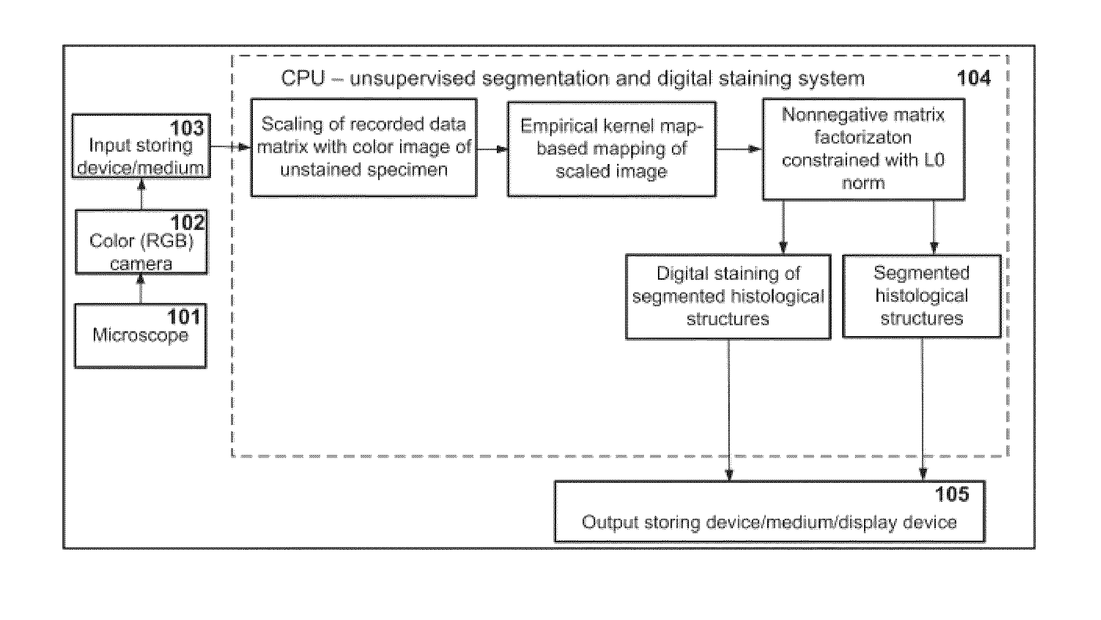
[0062]A schematic block-diagram of a device for unsupervised segmentation of color microscopic image of unstained specimen and digital staining of segmented histological structures, that is defined by equation [II] and employing methodology of empirical kernel map-based nonlinear mapping and nonnegativity and l0-norm constrained matrix factorization, according to an embodiment of the present invention is shown in FIG. 1. The device consists of: light microscope 10...


PUM


Abstract

The invention relates to a computing device-implemented method and apparatus for unsupervised segmentation of microscopic color image of unstained specimen and digital staining of segmented histological structures. Image of unstained specimen is created by light microscope 101, recorded by color camera 102 and stored on computer-readable medium 103. The invention is carried out by a computing device 104 comprised of: computer-readable medium for storing and computer for executing instructions of the algorithm for unsupervised segmentation of microscopic color image of unstained specimen and digital staining of segmented histological structures. Segmented histological structures and digitally stained image are stored and displayed on the output storing and display device 105 in order to establish diagnosis of a disease. The invention is an improvement over the prior art as it is characterized by the: (i) shortening of slide preparation process; (ii) reduction of intra-histologist variation in diagnosis; (iii) elimination of adding chemical effects on specimen; (iv) elimination of altering morphology of the specimen; (v) simplification of histological and intra-surgical tissue analysis; (vi) being significantly cheaper than existing staining techniques; (vii) being harmless to the user because toxic chemical stains are not used; (viii) discrimination of several types of histological structures present in the specimen; (ix) usage of the same specimen for more than one analysis.

Description

FIELD OF THE INVENTION[0001]The invention relates to a computing device-implemented method and apparatus for unsupervised segmentation of microscopic color image of unstained specimen and digital staining of segmented histological structures. Segmented histological structures and digitally stained image are displayed in order to diagnose a disease. Some benefits of application of the invention, when compared against existing staining techniques, are: (i) shortening of slide preparation process; (ii) reduction of variation in diagnosis between histologist; (iii) total elimination of adding chemical effects on a specimen; (iv) elimination of morphological changes of a specimen e.g. shrinkage; (v) simplification of histological and intra-surgical tissue analysis; (vi) being significantly cheaper; (vii) harmless to the user because toxic chemical stains are not used; (viii) discrimination of several types of histological structures present in the specimen; (ix) using the same specimen f...

Claims


Application Information

Patent Timeline
IPC IPC(8): G06F19/26G01N33/483G16B45/00
CPCG01N33/4833G06F19/26G16B45/00G06V20/695G06V10/7715G06F18/2133
Inventor KOPRIVA, IVICAPOPOVIC-HADZIJA, MARIJANAHADZIJA, MIRKOARALICA, GORANA
Owner RUDJER BOSKOVIC INST
Why Eureka
Try Eureka