Device, system and method for tumor detection and/or monitoring
a tumor detection and/or monitoring technology, applied in the field of tumor detection and/or monitoring devices, can solve problems such as cell starvation of oxygen
- Summary
- Abstract
- Description
- Claims
- Application Information
AI Technical Summary
Benefits of technology
Problems solved by technology
Method used
Image
Examples
first embodiment
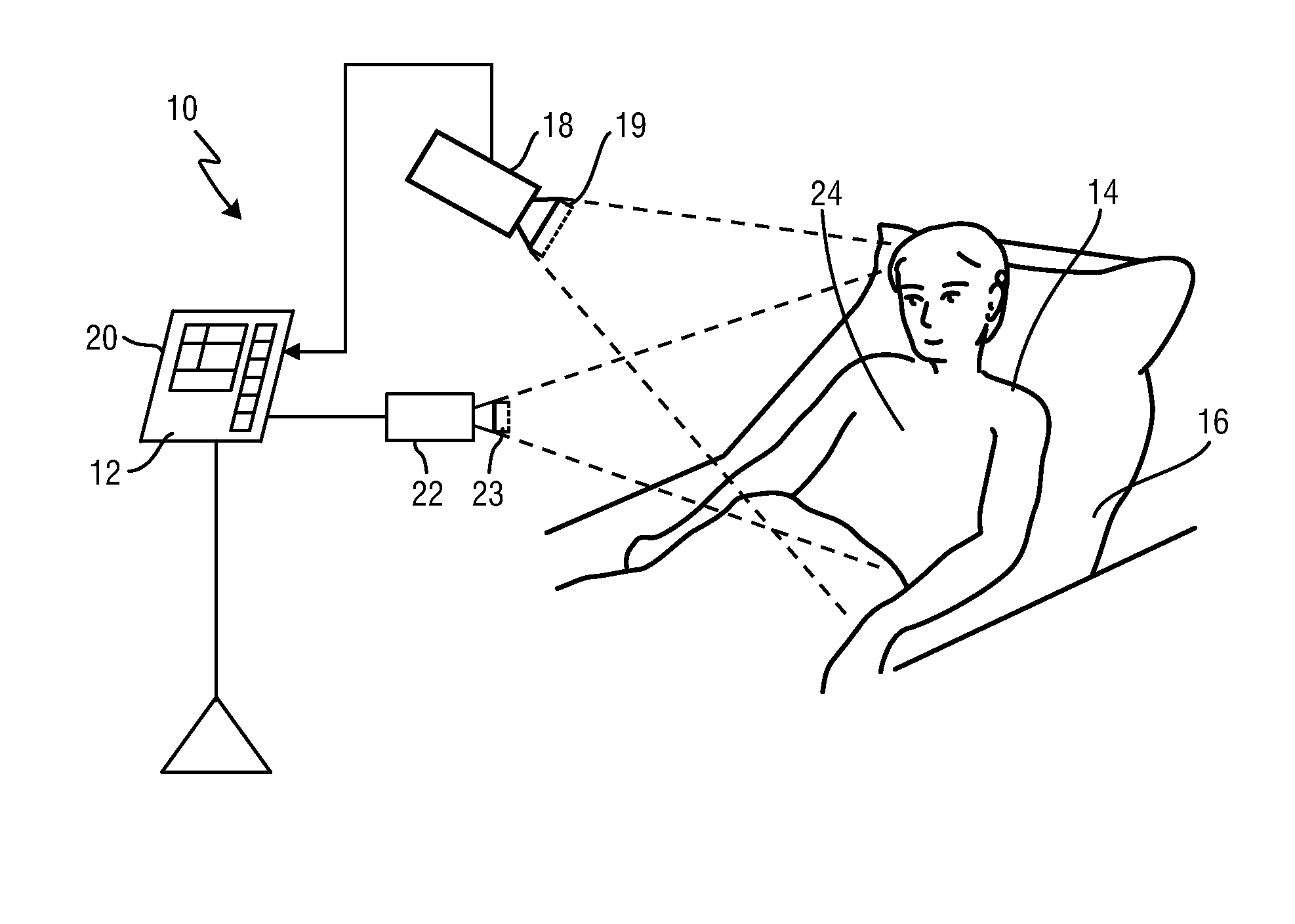
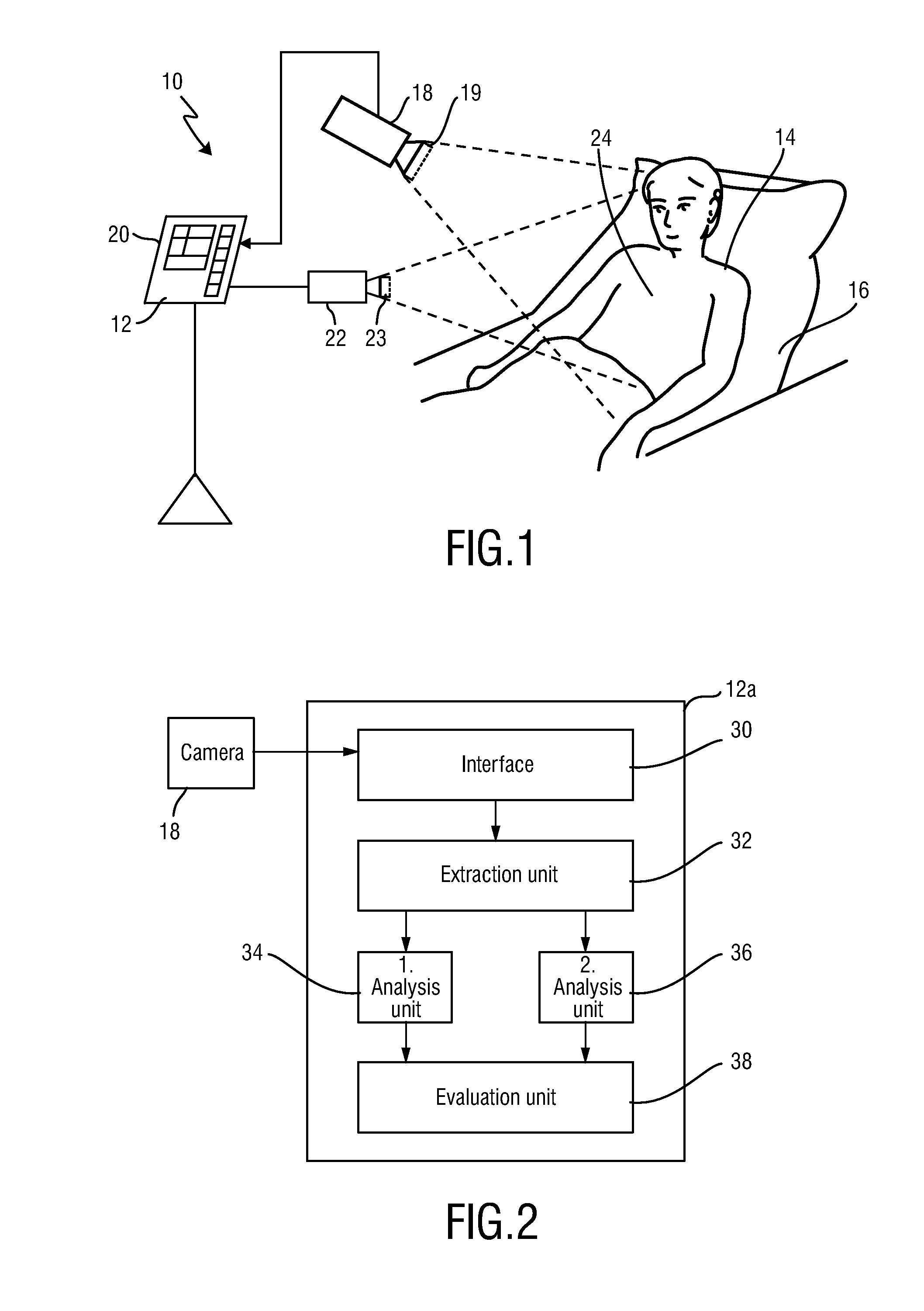
[0050]FIG. 1 shows a schematic diagram of a system 10 including a device 12 for detecting and / or monitoring of a tumor of a subject 14 according to the present invention. The subject 14, in this example a patient, lies in a bed 16, e.g. in a hospital or other healthcare facility, but may also be a neonate or premature infant, e.g. lying in an incubator, or person at home or in a different environment. Image frames of the subject 14 are captured by means of a camera 18 (also generally referred to as detection unit, or as imaging unit or camera-based or remote PPG sensor) including a suitable photosensor. The camera 18 forwards the recorded image frames to the device 12, where the image frames will be processed as explained in more detail below. The device 12 preferably comprises an interface 20 for displaying the determined information and / or for providing medical personnel with an interface to change settings of the device 12 and / or other elements of the system 10. Such an interface...
second embodiment
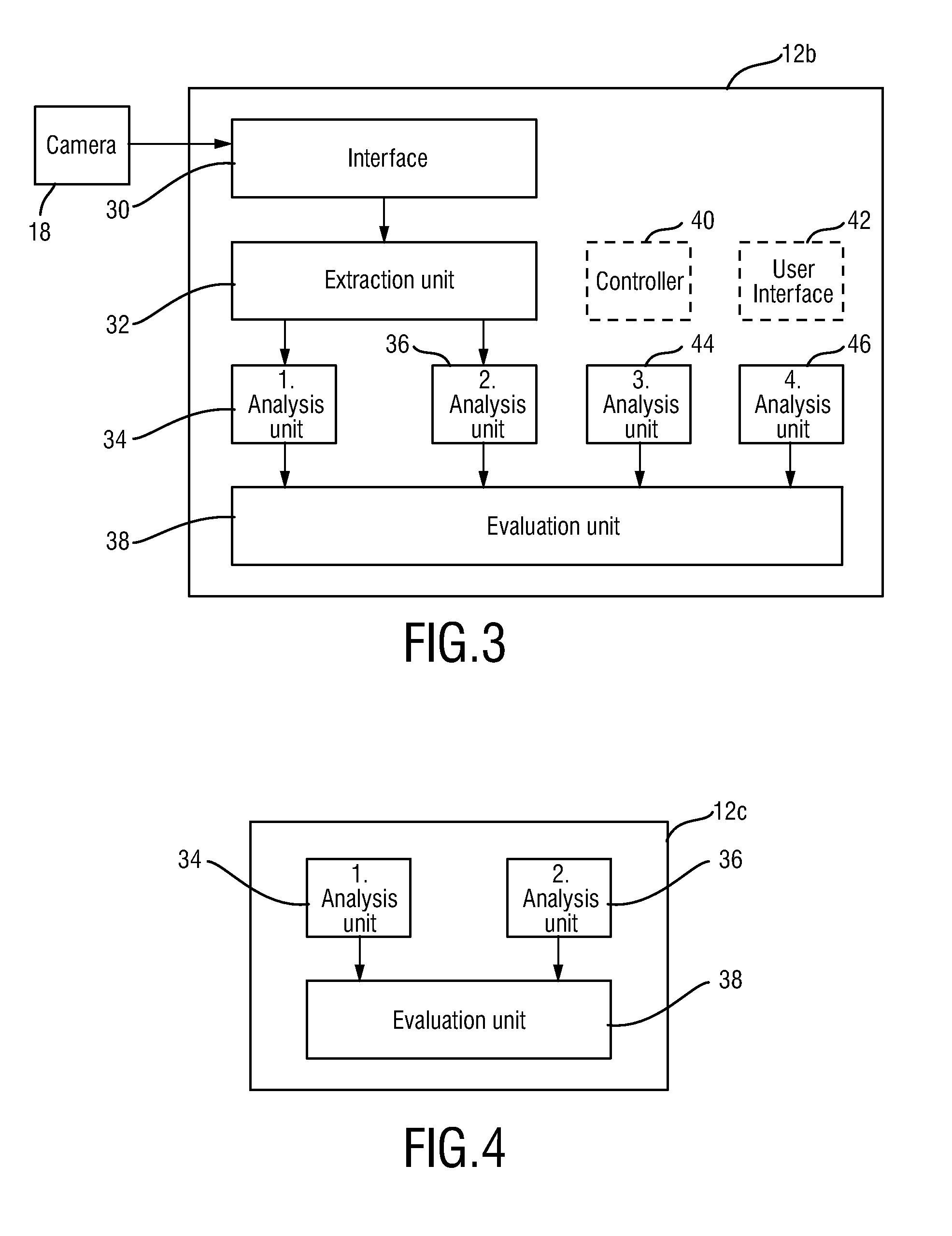
[0061]In a preferred embodiment, an SpO2 map is obtained by the second analysis unit. This SpO2 map, in particular changes of this SpO2 map, are analyzed by inducing changes of oxygen supply (e.g. by holding a breath, or by reducing oxygen content in breathing air) and monitoring the spatial distribution of changes of SpO2. For this purpose, as exemplarily shown in the device 12b depicted in FIG. 3, a controller 40 and / or user interface 42 for inducing changes of oxygen supply to the subject 14 may be provided. For instance, the controller 40 may control the oxygen content in the breathing air provided to the subject 14 (e.g. via a facial mask). Additionally or alternatively, the user interface may provide instructions to the subject 14 for controlled breathing, e.g. to hold the breath for some time and to deeply inhale after said time.
[0062]The present invention uses an analysis of spatial non-uniformity of SpO2 changes. Therefore, in order to provide such analysis, SpO2 needs to b...
third embodiment
[0067]Another embodiment of a system 10a is schematically depicted in FIG. 5. Instead of an imaging unit and an illumination unit it comprises one or more contact pulse oximeter sensors 50, 52, 54 placed at the subject's body for obtaining the PPG signals representing electromagnetic radiation reflected from skin of the subject 12, in particular in the red and infrared wavelength ranges. Said pulse oximeter sensors 50, 52, 54 are preferably similar or identical to conventional sensors used for obtaining SpO2 information in reflective mode. The device 12 may be configured as the third embodiment shown in FIG. 4 since the sensors 50, 52, 54 may directly provide the PPG signals. Thus, generally the same principle as discussed above for use with PPG signals derived from image data acquired by a camera (i.e. in a contactless way) can be used with PPG signals obtained by pulse oximeter sensors (i.e. in a contact way).
[0068]Generally, the present invention can be applied for detection and / ...
PUM
 Abstract
Description
Claims
Application Information
 - R&D
- Intellectual Property
- Life Sciences
- Materials
- Tech Scout
- Unparalleled Data Quality
- Higher Quality Content
- 60% Fewer Hallucinations
Browse by: Latest US Patents, China's latest patents, Technical Efficacy Thesaurus, Application Domain, Technology Topic, Popular Technical Reports.
© 2025 PatSnap. All rights reserved.Legal|Privacy policy|Modern Slavery Act Transparency Statement|Sitemap|About US| Contact US: help@patsnap.com


