Real-time scene-modeling combining 3D ultrasound and 2d x-ray imagery

a technology of image material and 3d ultrasound, applied in the field of apparatus for visualizing image material, can solve the problems of insufficient intuitiveness for the operator to readily understand the spatial relationship between the two, and the soft tissue is barely visibl

Active Publication Date: 2015-10-22
KONINKLJIJKE PHILIPS NV
View PDF6 Cites 26 Cited by
  • Summary
  • Abstract
  • Description
  • Claims
  • Application Information

AI Technical Summary

Benefits of technology

[0022]In one embodiment, the apparatus affords the further functionality of allowing the user to manually adjust the position the marker along the line and in the ultrasound image data set. In one embodiment, an automatic adjustment algorithm is used instead based on the similarity of image contents (in the X-ray image and the ultrasound image data) in respective neighborhoods of the respective markers.

Problems solved by technology

One of the challenges of image-guided medical, in particular surgical, procedures is to efficiently use the information provided by the many imaging techniques the patient may have been through before and during the intervention.
However, soft-tissues are barely visible in these images, and furthermore, these images are projections which do not give a direct access to the volumetric geometry of the intervention scene.
It has been found however that the the usual way of displaying both ultrasound and X-ray real-time streams, such as in Applicant's US WO 2011 / 070492, are at times not intuitive enough for the operator to readily understand the spatial relationship between the two modalities when carrying out the intervention, oftentimes under great stress.

Method used


Image

  • Real-time scene-modeling combining 3D ultrasound and 2d x-ray imagery
  • Real-time scene-modeling combining 3D ultrasound and 2d x-ray imagery
  • Real-time scene-modeling combining 3D ultrasound and 2d x-ray imagery

Examples


Embodiment Construction

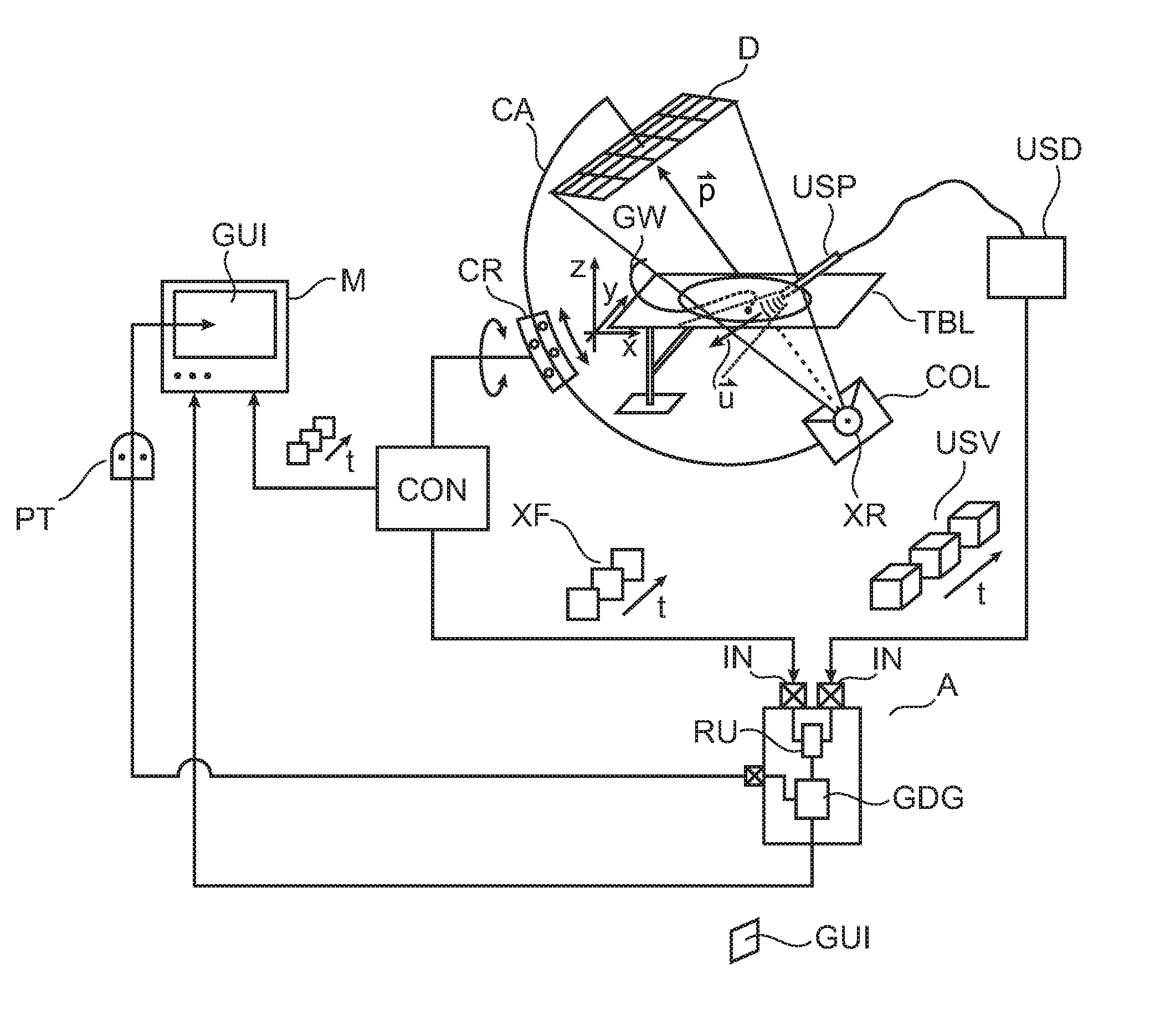
[0033]With reference to FIG. 1 there is shown an arrangement for multimodal image-guided support of minimal invasive interventions such as mitral clipping.

[0034]Broadly, said arrangement comprises an X-ray imager 100, an examination table TBL and ultrasound imaging equipment USD. Specifically, the arrangement allows acquiring images from both, the ultrasound imager and the X-ray imager. As will be explained below in more detail, the two image streams are combined in a manner so as to harness as user desired the most suitably one of the two streams for relevant anatomic information.

[0035]During the intervention a patient PAT is disposed on examination table TBL. A medical device such as a guidewire GW is introduced through an entry point into the patient's body and the interventional radiologist faces the challenge of navigating said guide-wire through the complex maze of patient's vasculature to arrive at a lesioned site. An example for such an intervention that relies on multimodal...


PUM


Abstract

An apparatus for visualizing image material in a multimodal imaging environment. Images (XF, USV) from an X-ray imager (100) and an ultrasound probe (USP) are fused into an interactive graphics display (GUI) to form a 3D scene, where, alongside to a 3D rendering (USS) of an ultrasound volume (USV) an X-Ray projection plan (XF) is displayed in perspective within the same scene. The rendering is according to a user selected view (UV) which is changeable upon user interaction with the interactive graphics display (GUI).

Description

FIELD OF THE INVENTION[0001]The invention relates to an apparatus for visualizing image material in a multimodal imaging environment, to a method of visualizing image material in a multimodal imaging environment, to an imaging system, to a computer readable medium, and to a computer program product.BACKGROUND OF THE INVENTION[0002]One of the challenges of image-guided medical, in particular surgical, procedures is to efficiently use the information provided by the many imaging techniques the patient may have been through before and during the intervention.[0003]For example, in cardiology, physicians often have access to real-time X-ray images acquired by a C-arm. These images have a very good spatial and temporal accuracy thus enabling to follow precisely the progression of even thin catheters and other interventional tools. However, soft-tissues are barely visible in these images, and furthermore, these images are projections which do not give a direct access to the volumetric geom...

Claims


Application Information

Patent Timeline
Patent Type & Authority Applications(United States)
IPC IPC(8): G06T15/20G06T3/40G06T7/00
CPCG06T15/205G06T7/0024G06T3/40G06T2207/10136G06T2207/20104G06T2207/20221G06T2207/30204G06T2200/04G06T2200/08G06T2207/10081A61B6/4441A61B6/461A61B6/487A61B6/503A61B6/5247A61B8/0883A61B8/12A61B8/466A61B8/483A61B8/5261G06T2207/10121G06T15/20G06T2210/41G06T2207/30048A61B34/25A61B2090/364A61B2090/376A61B2090/3784F04C2270/041G06T7/30
Inventor FLORENT, RAOULNEMPONT, OLIVIER PIERRECATHIER, PASCAL YVES FRANCOISNIJHOF, NIELS
Owner KONINKLJIJKE PHILIPS NV
Why Eureka
Try Eureka