
Suite of coordinating diagnostic medical simulators for live training and evaluation

a diagnostic medical simulator and simulator technology, applied in the field of live training and evaluation of diagnostic medical simulators, can solve the problems of not being able to use in environments too noisy to permit hearing the characteristic sounds, and possibly less accurate, and unable to achieve calibration

Active Publication Date: 2015-10-22
KBPORT LLC
View PDF2 Cites 9 Cited by
  • Summary
  • Abstract
  • Description
  • Claims
  • Application Information

AI Technical Summary

Benefits of technology

The present invention provides a simulator for training and evaluating sphygmomanometers. It can be used with any manikin brand and can even be used with live simulated patients. The purpose of the simulator is for live training, meaning it should simulate blood pressure readings instead of simply displaying the actual pressure. The goal of using the simulator for evaluation is to provide a way to objectively evaluate the trainee's performance.

Problems solved by technology

They are used by trained practitioners, and cannot be used in environments too noisy to permit hearing the characteristic sounds.
Aneroid sphygmomanometers are considered safer than mercury based, although possibly less accurate.
A major cause of departure from calibration is mechanical jarring.
In addition to the digital oscillometric monitors drawbacks where they cannot be used, the overall accuracy of such devices has been questioned.
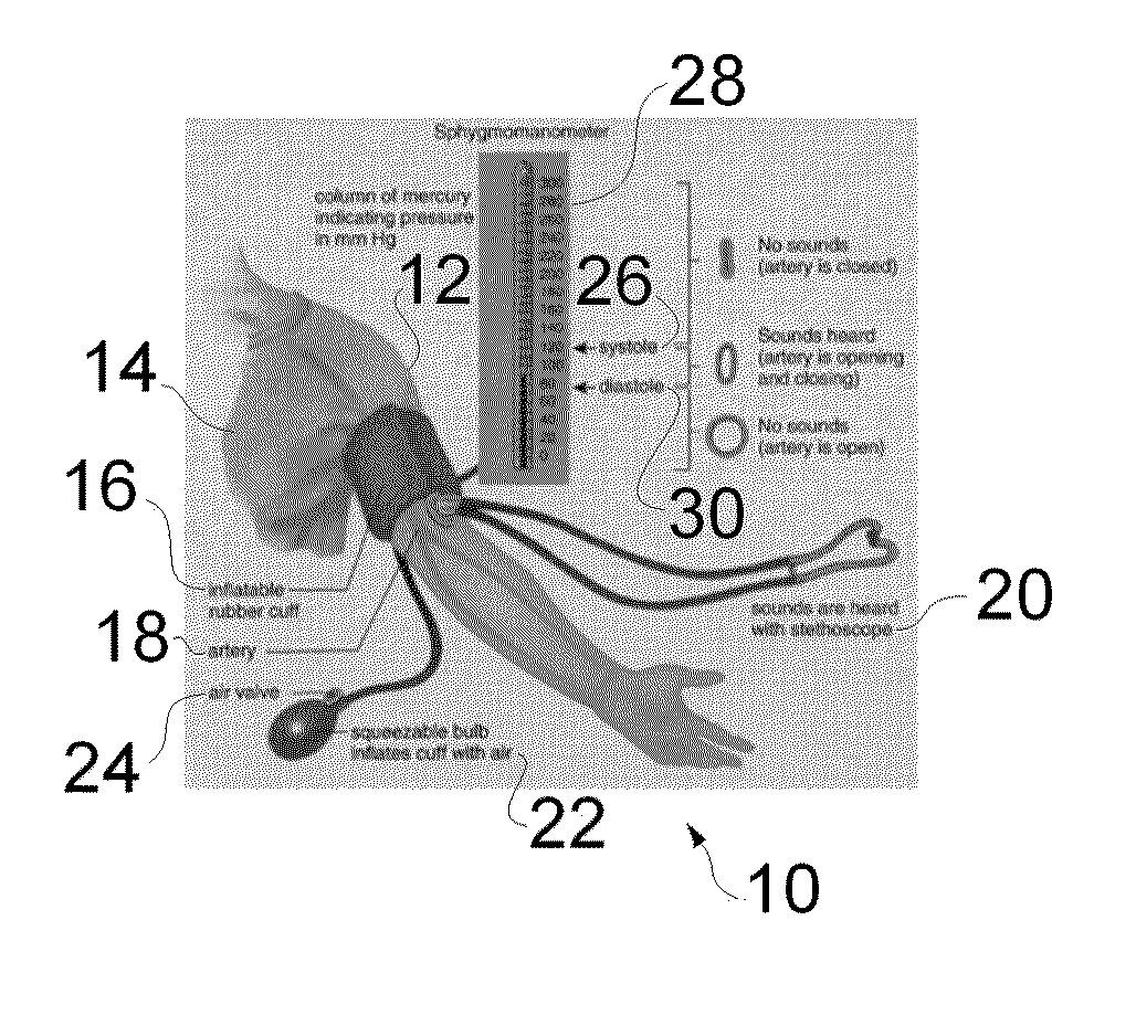
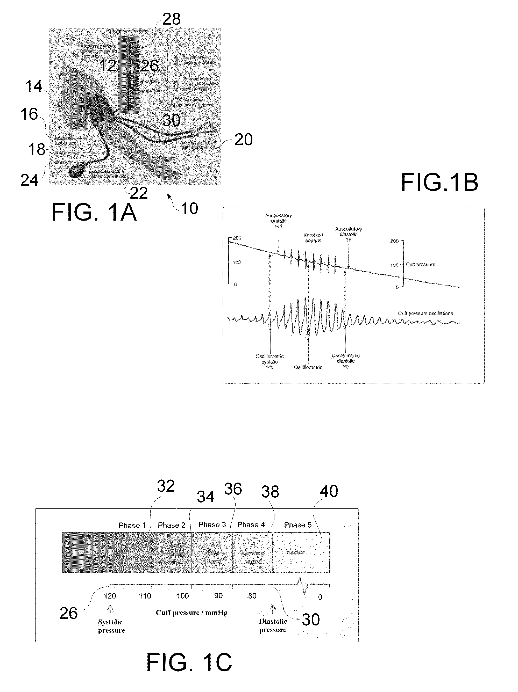
Too small a cuff 16 results in too high a pressure, while too large a cuff 16 results in too low a pressure.
The electronically generated sounds are digitally recorded.” While these simulators provide effective tools for supplying the trainees with a wide range of blood pressures to obtain and provide a method of verifying the accuracy of the trainee's results, they do not provide the real live aspects of interacting with a human regardless of how “lifelike” the systems become.
However this training technique offers very little variation in the blood pressures that the trainees will experience (in general the class room subjects have an average blood pressure) and does not allow the teacher to easily verify the results of a particular trainee or to present a trainee with a desired blood pressure to measure.
This live training technique also has the advantage of having trainees work with live subjects with all the aspects and nuances of interacting with live subjects, but it does not allow the trainee to actually obtain abnormal pressures (barring an actual abnormal condition of the actor) and lessens the realism of the training event as the trainee must disregard the obtained values and imagine some other imaginary set of values.
This method also fails to allow the trainer to validate the accuracy of an abnormal blood pressure measurement obtained by the trainee.
The concept is similar to today's conventional pulse oximetry but suffered due to unstable photocells and light sources and the method was not used clinically.
This use was limited to pulmonary functions due to cost and size.
Patients that are grossly overweight or severely underweight raise serious medical concerns.
There are detrimental health effects to the patient in being moderately over or underweight for long durations.
Rectal temperature is generally considered the most accurate, however this method may be considered unpleasant, or embarrassing; also, if not taken the correct way, rectal temperature-taking can be uncomfortable and in some cases painful for the patient.
A forehead thermometer uses a band thermometer which is applied to the patient's brow, wherein it is typically a band coated with different temperature-sensitive markings, and is considered reliable only to give an indication of fever, but is not considered accurate otherwise.
Further, in given scenarios the desired temperature output of a patient may exhibit radical change.
Programming each simulator can greatly add to effectiveness and realism of the training event, but separately programming these distinct devices can become burdensome and time consuming, needlessly increasing the costs of developing and running specific scenarios.

Method used


Image

  • Suite of coordinating diagnostic medical simulators for live training and evaluation
  • Suite of coordinating diagnostic medical simulators for live training and evaluation
  • Suite of coordinating diagnostic medical simulators for live training and evaluation

Examples


example 1

[0127]A standardized patient is brought to the simulated weight scale, the system detects the proximity of the standardized patient to the simulated weight scale and automatically uploads the patient scenario to the scale.

example 2

[0128]A standardized patient is brought into an examination room containing a wide variety of simulated diagnostic equipment such as a by cuff, pulse ox, etc. The system detects that the patient is in the room containing the equipment and uploads the patient scenario to all of the nearby simulated diagnostic equipment.

[0129]There are multiple ways of getting the standardized patients location within the environment such as RFID, NFC, Video recognition and so on. During facility setup, a map is generated of the environment and the location of various simulated diagnostic equipment. One preferred approach—Medical simulation facilities are usually outfitted with a wide variety of audio / video recording equipment. In addition to the standard microphones normally used, we will strategically place microphones capable of operating in the ultrasonic range above human hearing. A piece of standardized patient worn equipment (probably the vest) will have a wireless transceiver and an ultrasonic...


PUM


Abstract

A medical training system comprises a controller configured to program a plurality of medical device simulators, and a plurality of medical device simulators coupled to the controller, including at least two of a sphygmomanometer simulator, pulse simulator, thoracic cavity simulator, scale and stadiometer simulator, pulse oximetry simulator, and thermometer simulator. The plurality of medical device simulators form a suite of coordinating

Description

RELATED APPLICATIONS[0001]This application claims the benefit of U.S. Provisional Patent Application Ser. No. 61 / 931,790, filed Jan. 27, 2014 entitled “Diagnostic Medical Simulators for Live Training and Evaluation.”[0002]This application is a continuation in part of U.S. patent application Ser. No. 14 / 281,506 entitled Universal Sphygmomanometer Simulator for Live Training and Evaluation” which published as US 2014-0342332 A1 on Nov. 20, 2014 and which publication is incorporated herein by reference in its entirety. U.S. patent application Ser. No. 14 / 281,506 claims the benefit of U.S. Provisional Patent Application Ser. No. 61 / 824,939, filed May 17, 2013 entitled “Universal Sphygmomanometer Simulator for Live Training and Evaluation.”BACKGROUND INFORMATION[0003]1. Field of the Invention[0004]The present invention relates to a suite of coordinating diagnostic medical simulators for live training and evaluation, such as a sphygmomanometer simulator, pulse simulator, thoracic cavity s...

Claims


Application Information

Patent Timeline
IPC IPC(8): G09B23/28A61B5/1455A61B5/022
CPCG09B23/28A61B5/14551A61B5/022G09B23/288G16H50/50
Inventor OLMSTEAD, CLIFFORD D.MILLER, CHARLES G.WOODS, JERRYDHINGRA, SUKHTEJ
Owner KBPORT LLC
Learn More