Methods and kits for detecting melanoma

a technology for detecting kits and melanoma, applied in the field of new methods and kits for detecting melanoma, and differentially methylated regulatory elements associated with melanoma, can solve the problems of large disagreement between pathologists regarding the diagnosis of melanoma and benign diseases, death of diseases, and difficult early diagnosis

Inactive Publication Date: 2015-12-31
THE UNIV OF NORTH CAROLINA AT CHAPEL HILL
View PDF0 Cites 5 Cited by
  • Summary
  • Abstract
  • Description
  • Claims
  • Application Information

AI Technical Summary

Problems solved by technology

However, if diagnosed in late stages, it is a deadly disease.
Early diagnosis is difficult due to the overlap in clinical and histopathological features of early melanomas and benign nevi, especially benign atypical nevi (Strauss et al., 2007, Br. J. Dermatol. 157, 758-764).
Moreover, there is a sizeable disagreement amongst pathologists regarding the diagnosis of melanoma and benign diseases such as compound melanocytic nevi or Spitz nevi.
In fact, many nevi, especially atypical or dysplastic nevi, are difficult to distinguish from melanoma, even by expert pathologists (Farmer et al., 1996, Hum. Pathol. 27, 528-531).
This results in a quandary for clinicians who not only biopsy but re-excise with margins large numbers of benign atypical nevi in the population (Fung, 2003, Arch. Dermatol. 139, 1374-1375), at least, in part, due to lack of confidence in the histopathologic diagnosis.
This high rate of misdiagnosis is problematic on many levels.
The false positives lead to unnecessary costly medical interventions, e.g., overly large excisions, high-dose interleukin-2 or interferon alpha, and needless stress for the patients.
The false negatives mean increased likelihood of a presentation with more severe disease, which as discussed above, dramatically increases the risk of a poor clinical outcome and risk of death.
However, excisional biopsy with such broad margins may not be appropriate for sites such as the face, ears, fingers, palms, or soles of the feet.
The utility of CGH in a clinical setting is limited because it currently requires approximately a microgram of DNA and about a month for results.
However, FISH is better for detecting amplifications than deletions so some information from CGH is lost.
16, 267-273; Wu et al., 2007, J. Dermatopathol. 29, 534-537), which limits their usefulness for melanoma diagnosis.
Moreover, there is an unmet medical need for improved melanoma diagnosis.

Method used


Image

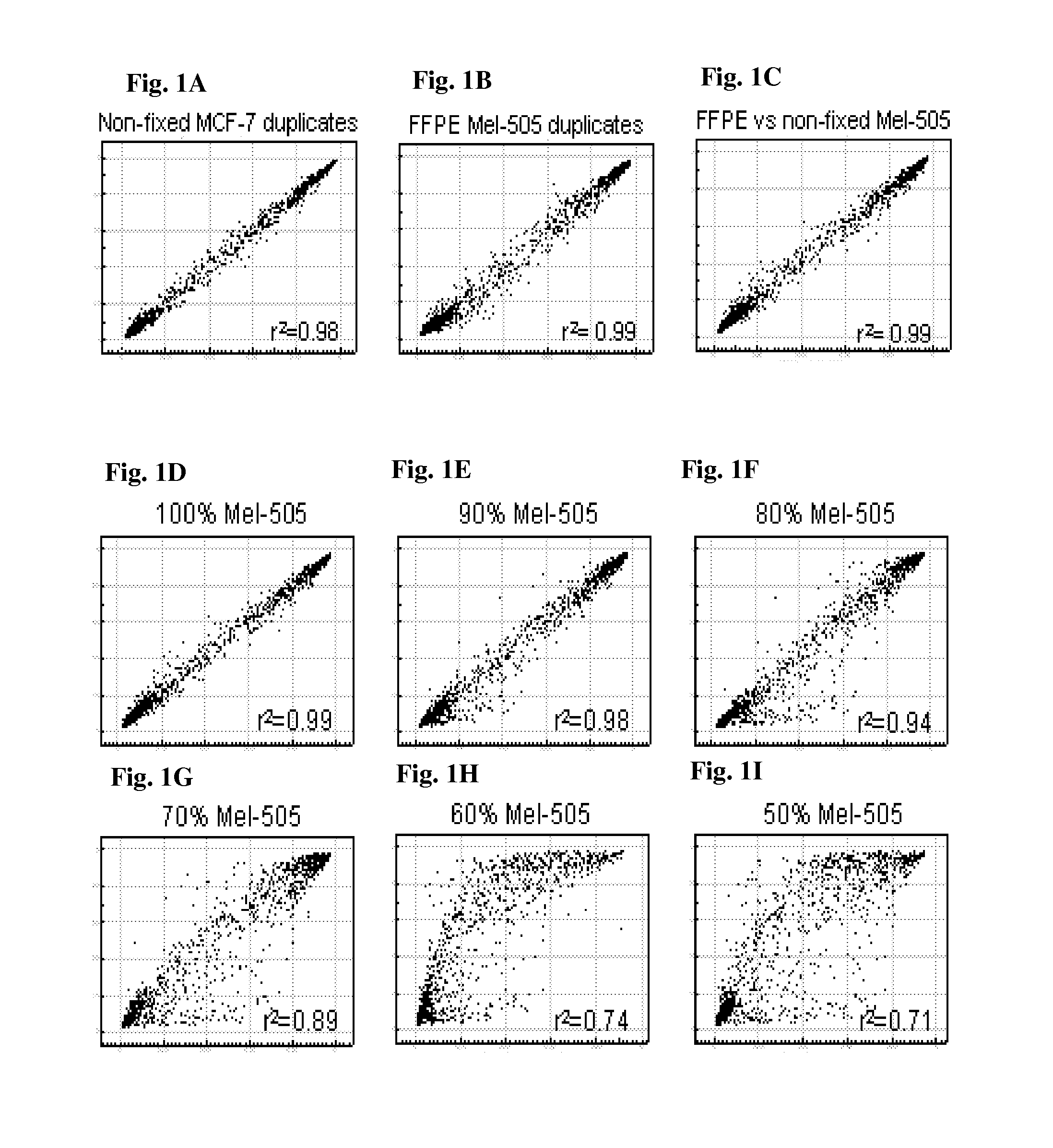
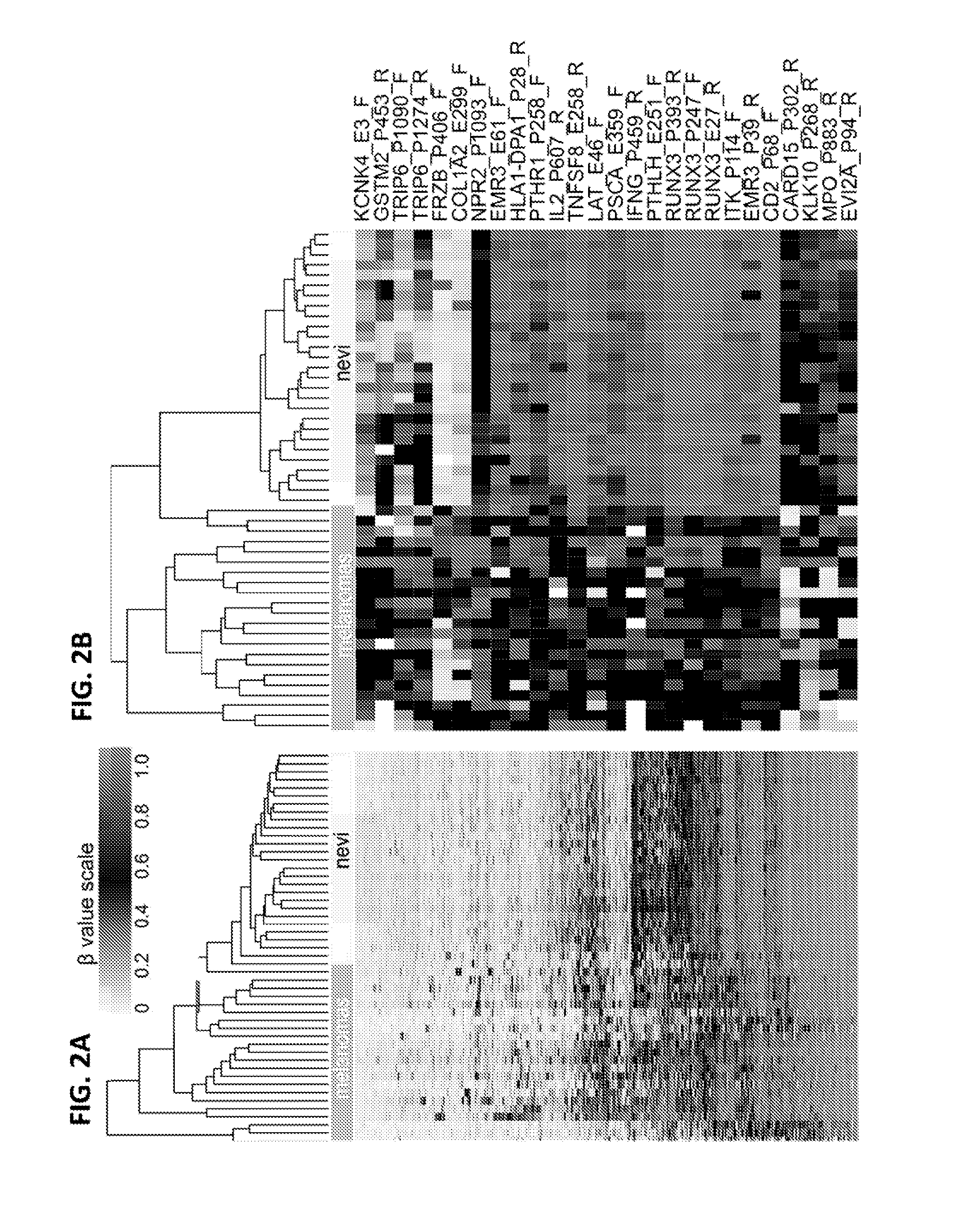
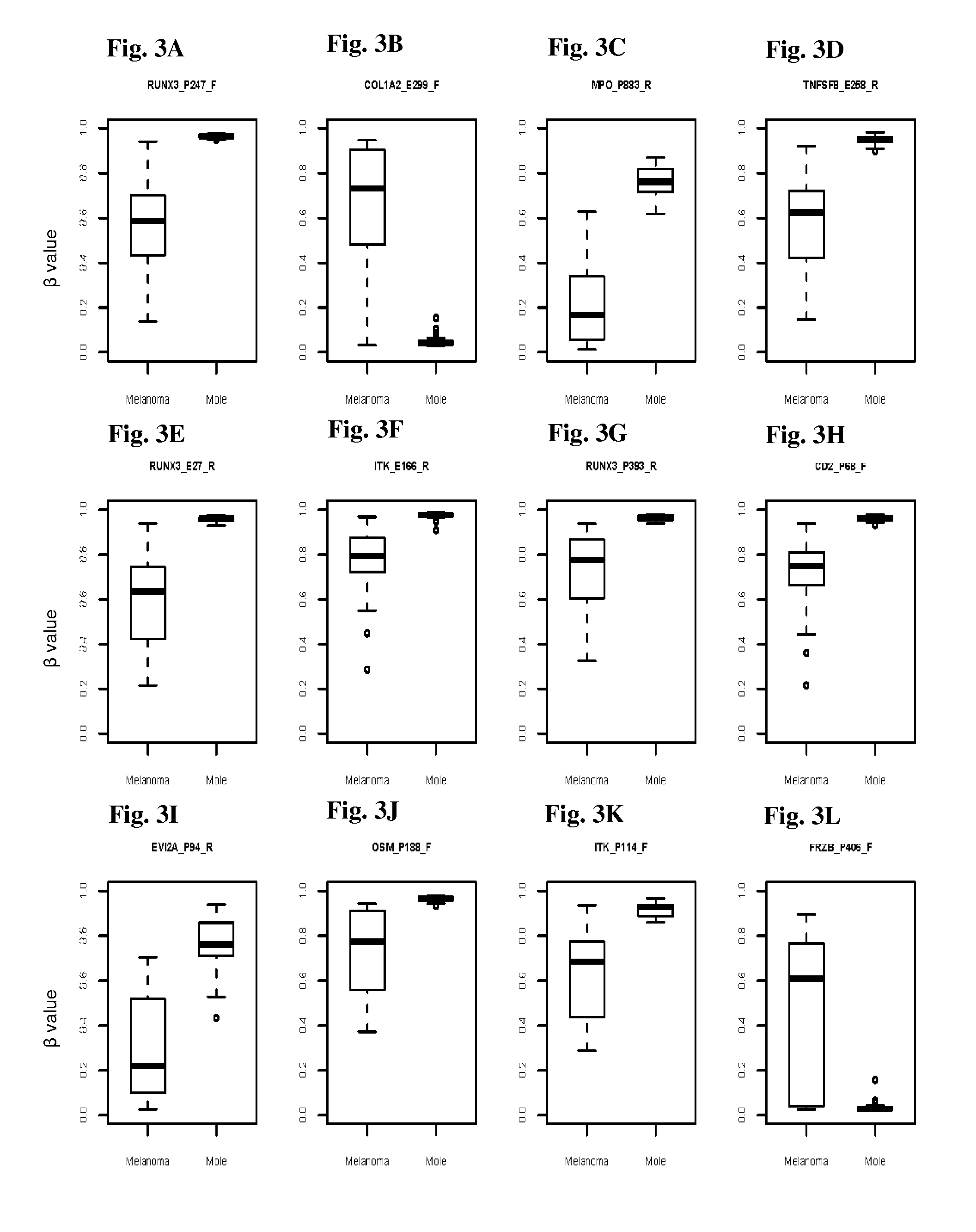
  • Methods and kits for detecting melanoma
  • Methods and kits for detecting melanoma
  • Methods and kits for detecting melanoma

Examples


Embodiment Construction

5.1. Definitions

[0029]The term “melanoma” refers to malignant neoplasms of melanocytes, which are pigment cells present normally in the epidermis, in adnexal structures including hair follicles, and sometimes in the dermis, as well as extracutaneous sites such as the mucosa, meninx, conjuctiva, and uvea. Sometimes it is referred to as “cutaneous melanoma” or “malignant melanoma.” There are at least four types of cutaneous melanoma: lentigo maligna melanoma (LMM), superficial spreading melanoma (SSM), nodular melanoma (NM), and acral lentiginous melanoma (ALM). Cutaneous melanoma typically starts as a proliferation of single melanocytes, e.g., at the junction of the epidermis and the dermis. The cells first grow in a horizontal manner and settle in an area of the skin that can vary from a few millimeters to several centimeters. As noted above, in most instances the transformed melanocytes produce increased amounts of pigment so that the area involved can be seen by the clinician.

[003...


PUM

PropertyMeasurementUnit
methylation sensitive high resolution melting assayaaaaaaaaaa
mass spectrometryaaaaaaaaaa
fluorescence in situ hybridizationaaaaaaaaaa

Abstract

This invention is directed to a method for detecting melanoma in a tissue sample by measuring a level of methylation of one or more regulatory elements differentially methylated in melanoma and benign nevi. The invention provides methods for detecting melanoma, related kits, and methods of screening for compounds to prevent or treat melanoma.

Description

RELATED APPLICATION[0001]This application claims the benefit of U.S. Provisional Patent Application No. 61 / 382,623, filed Sep. 14, 2010 entitled “Methods and Kits for Detecting Melanoma” naming Nancy Thomas et al. as inventors with Attorney Docket No. UNC10001USV. The entire contents of which are hereby incorporated by reference including all text, tables, and drawings.STATEMENT REGARDING FEDERALLY SPONSORED RESEARCH OR DEVELOPMENT[0002]This invention was made at least in part with government support under grant number 1R21 CA134368-01 awarded by the National Cancer Institute. The United States Government has certain rights in the invention.1. FIELD OF THE INVENTION[0003]This invention relates generally to the discovery of novel differentially methylated regulatory elements associated with melanoma. The invention provides methods for detecting melanoma, related kits, and methods of screening for compounds to prevent or treat melanoma.2. BACKGROUND OF THE INVENTION2.1. Skin Cancer an...

Claims


Application Information

Patent Timeline
Patent Type & Authority Applications(United States)
IPC IPC(8): C12Q1/68
CPCC12Q1/6886C12Q2600/136C12Q2600/154C12Q2600/156C12Q2600/16G01N33/6881
Inventor THOMAS, NANCYDORSEY, KATHLEENEDMISTON, SHARONGROBEN, PAMELA
Owner THE UNIV OF NORTH CAROLINA AT CHAPEL HILL
Why Eureka
Try Eureka