Method for rendering tissue transparent, reagent for rendering tissue transparent, and tissue observation method
a tissue and reagent technology, applied in the field of tissue rendering, can solve the problems of limited observation depth, accompanied by great labor, and difficulty in fluorescence image acquisition, and achieve the effect of simple operation
- Summary
- Abstract
- Description
- Claims
- Application Information
AI Technical Summary
Benefits of technology
Problems solved by technology
Method used
Image

Examples
example 1
Rendering Rat Spinal Cord Transparent
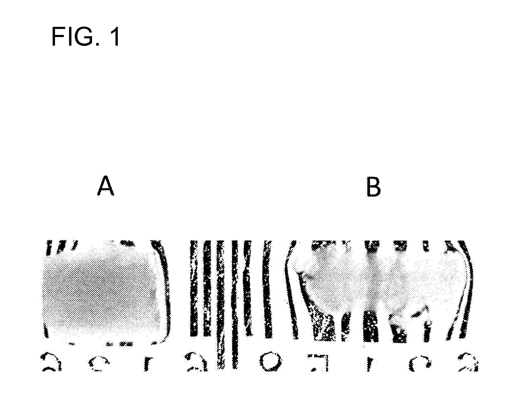
[0090]After perfusion fixation with 4% paraformaldehyde buffer solution, rat spinal cord was removed and further immersed in the same solution for 24 hours for fixation. The fixed spinal cord (3 mm in diameter) was immersed in the solution of thiodiethanol:glycerol:sucrose=20:40:40, the solution of 50:40:10, and the solution of 70:25:5 as pretreatment solutions each for 24 hours in that order, and then immersed in the final solution of 90:5:5 for 24 hours for transparentization.
[0091]The results are shown in B of FIG. 1. A shows the results obtained using the Scale method described in Non Patent Literature 2. The method for rendering tissue transparent according to the present invention (B) could render the spinal cord transparent with a high degree of transparency compared to the Scale method (A). Whereas the Scale method (A) had a problem that the spinal cord was swollen 2-fold, the method for rendering tissue transparent according to the prese...
example 2
Rendering Rat Brain Transparent
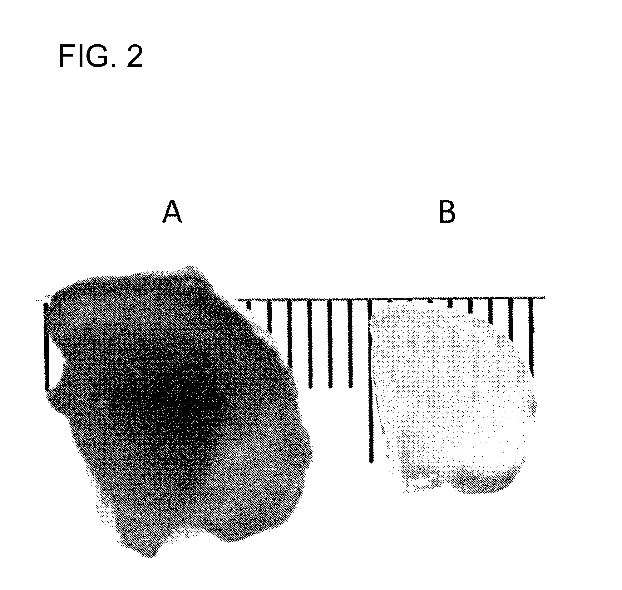
[0092]Rat brain (tissue thickness: 6 mm) was fixed and rendered transparent by the procedure described in Example 1.
[0093]The results are shown in B of FIG. 2. A shows the results obtained using the Scale method described in Non Patent Literature 2. The method for rendering tissue transparent according to the present invention (B) could render the brain transparent with a high degree of transparency compared to the Scale method (A). Whereas the Scale method (A) had a problem that the brain markedly expanded, and easily collapsed when pressed with a finger, the method for rendering tissue transparent according to the present invention (B) did not cause swelling or weakening.
example 3
Fluorescent Observation of Rat Spinal Cord
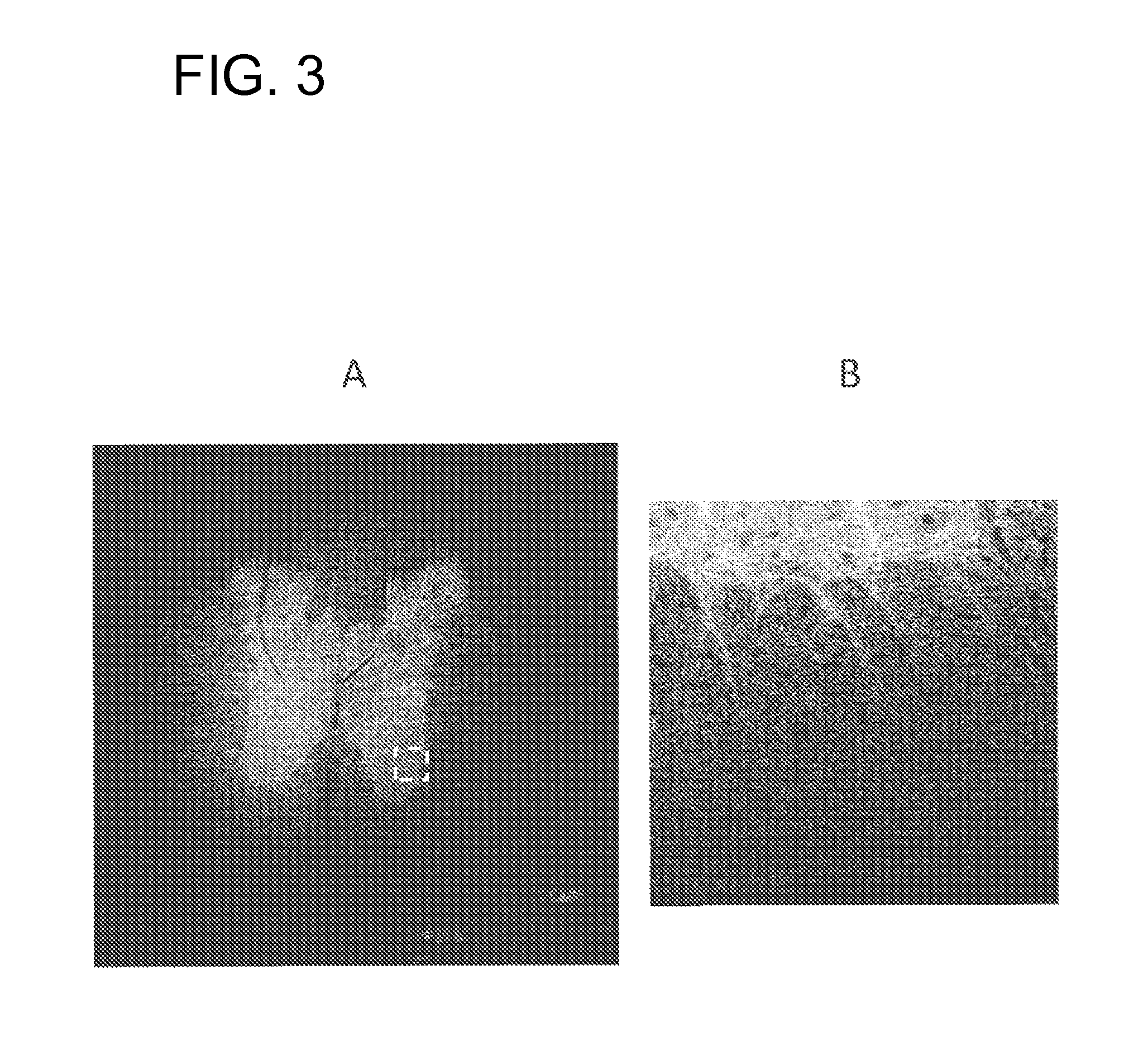
[0094]A transgenic rat was prepared, in which a fluorescent protein, VENUS, is expressed in nerve axons. The transgenic rat was prepared by the method described in Non Patent Literature 3 (“Visual properties of transgenic rats harboring the channelrhodopsin-2 gene regulated by the thy-1.2 promoter.” PLoS ONE, 2009, Vol. 4, No. 11, e7679). The spinal cord fixed and rendered transparent by the procedure described in Example 1 was observed using a confocal microscope (Zeiss, LSA-700).
[0095]The fluorescence image obtained is shown in A of FIG. 3. B is an enlarged image of the region enclosed by a dotted line in A. The tissue observation method according to the present invention enabled the observation of nerve axons with high accuracy. The disappearance or attenuation of a fluorescence signal of the fluorescent protein could be suppressed even under conditions of a relatively high concentration (90% by volume) of thiodiethanol.
PUM
| Property | Measurement | Unit |
|---|---|---|
| tissue transparent | aaaaa | aaaaa |
| refractive index | aaaaa | aaaaa |
| thickness | aaaaa | aaaaa |
Abstract
Description
Claims
Application Information
 - R&D Engineer
- R&D Manager
- IP Professional
- Industry Leading Data Capabilities
- Powerful AI technology
- Patent DNA Extraction
Browse by: Latest US Patents, China's latest patents, Technical Efficacy Thesaurus, Application Domain, Technology Topic.
© 2024 PatSnap. All rights reserved.Legal|Privacy policy|Modern Slavery Act Transparency Statement|Sitemap