
Magnetic resonance imaging (MRI) apparatus and method of processing mr image

a magnetic resonance imaging and apparatus technology, applied in the field of magnetic resonance imaging (mri) apparatus and a processing method of an mr image, to achieve the effect of more accurate detection

Active Publication Date: 2016-04-21
SAMSUNG ELECTRONICS CO LTD
View PDF1 Cites 26 Cited by
  • Summary
  • Abstract
  • Description
  • Claims
  • Application Information

AI Technical Summary

Benefits of technology

This patent text describes a method for improving the detection and tracking of white matter hyperintensity (WMH) regions in magnetic resonance imaging (MRI). This is achieved by using a combination of a Fluid Attenuated Inversion Recovery (FLAIR) image and a T1 weighted image to separate gray matter and white matter regions. Additionally, this method can accurately detect changes in the degree ofWMH over time by normalizing signal intensity in the MRI image over time. Overall, this improves the accuracy and reliability of identifying and monitoring brain abnormalities associated with various neurological disorders.

Problems solved by technology

Also, the exemplary embodiments are not required to overcome the disadvantages described above, and an exemplary embodiment may not overcome any of the problems described above.

Method used


Image

  • Magnetic resonance imaging (MRI) apparatus and method of processing mr image
  • Magnetic resonance imaging (MRI) apparatus and method of processing mr image
  • Magnetic resonance imaging (MRI) apparatus and method of processing mr image

Examples


Embodiment Construction

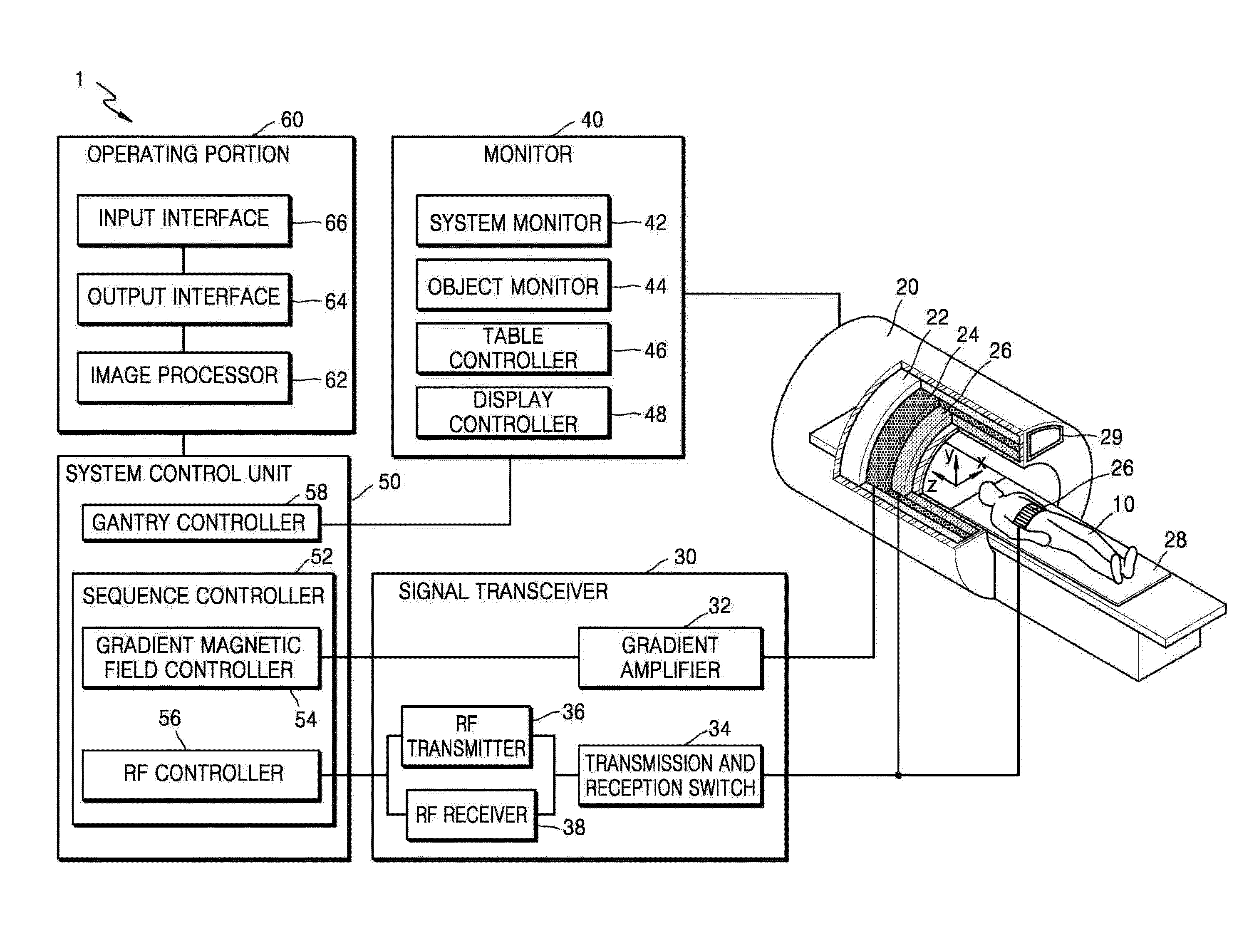
[0040]Exemplary embodiments are described in greater detail below with reference to the accompanying drawings.

[0041]In the following description, like drawing reference numerals are used for like elements, even in different drawings. The matters defined in the description, such as detailed construction and elements, are provided to assist in a comprehensive understanding of the exemplary embodiments. However, it is apparent that the exemplary embodiments may be practiced without those specifically defined matters. Also, well-known functions or constructions may not be described in detail because they would obscure the description with unnecessary detail.

[0042]In the present specification, an “image” may refer to multi-dimensional data composed of discrete image elements (e.g., pixels in a two-dimensional (2D) image and voxels in a three-dimensional (3D) image). For example, the image may be a medical image of an object captured by an X-ray apparatus, a computed tomography (CT) appar...


PUM


Abstract

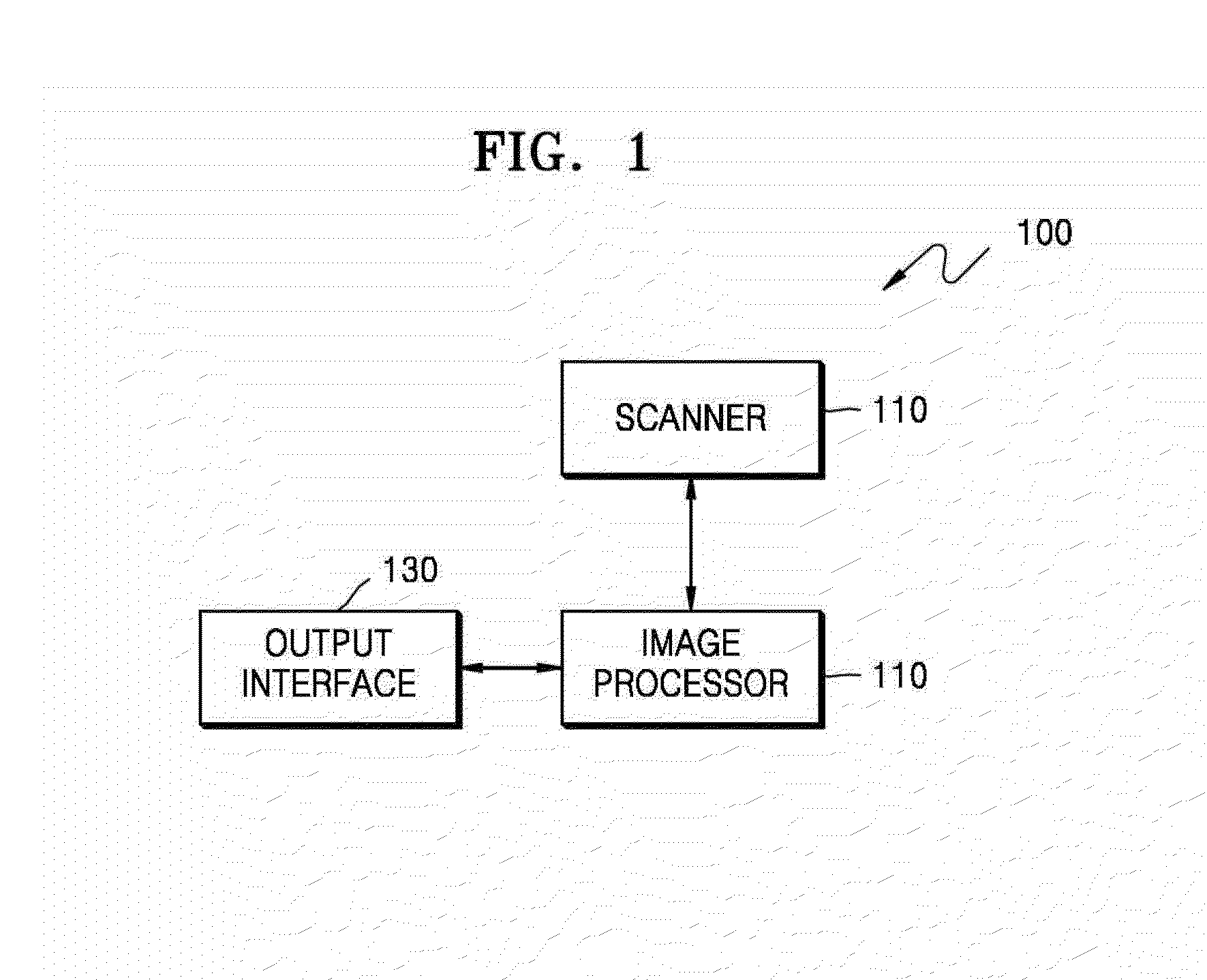
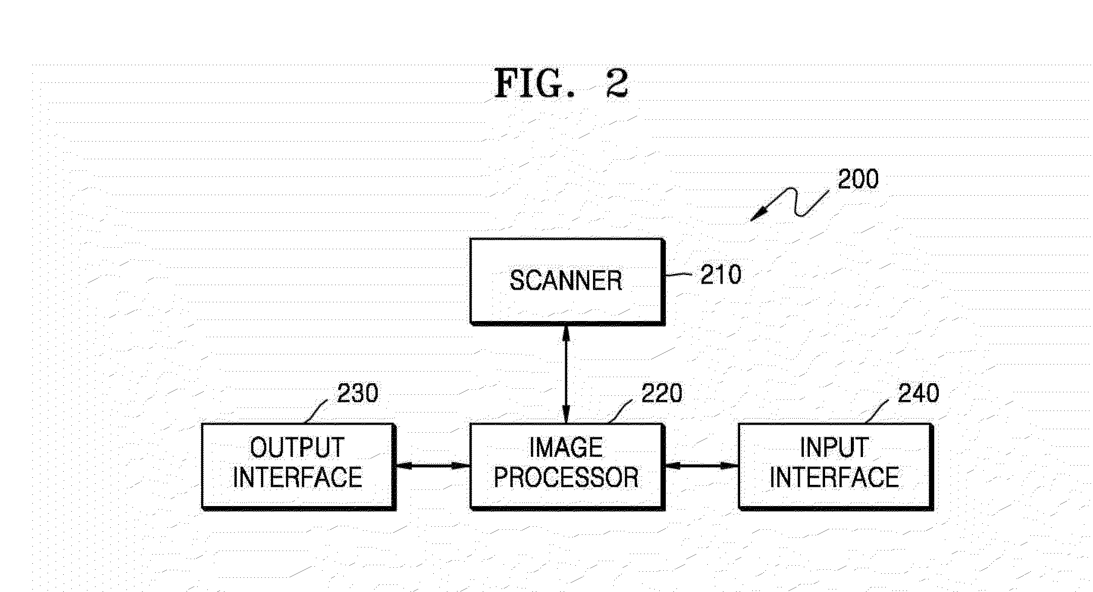
A magnetic resonance imaging (MRI) apparatus and a method of processing an MR image are provided. The MRI apparatus includes a scanner configured to acquire a first image that is a T1-weighted image and a second image that is a fluid attenuated inversion recovery (FLAIR) image by performing an MRI scan on a brain. The MRI apparatus further includes an image processor configured to determine a white matter region in the second image based on the first image and the second image, and detect a white matter hyperintensity (WMH) region in the determined white matter region. The MRI apparatus further includes an output interface configured to display the detected WMH region and a change in the WMH region over time.

Description

CROSS-REFERENCE TO RELATED APPLICATION[0001]This application claims priority from Korean Patent Application No. 10-2014-0142782, filed on Oct. 21, 2014, in the Korean Intellectual Property Office, the disclosure of which is incorporated herein in its entirety by reference.BACKGROUND[0002]1. Field[0003]Apparatuses and methods consistent with exemplary embodiments relate to a magnetic resonance imaging (MRI) apparatus and a method of processing an MR image.[0004]2. Description of the Related Art[0005]An MRI apparatus uses a magnetic field to capture an image of a subject, and is widely used in the accurate diagnosis of diseases because it shows stereoscopic images of bones, lumbar discs, joints, nerve ligaments, the heart, etc., at angles.[0006]To detect a white matter hyperintensity (WMH) region located in a white matter area of the brain, the white matter area may be defined first in an image of the brain. Then, the WMH region may be determined in the defined white matter area.[0007...

Claims


Application Information

Patent Timeline
IPC IPC(8): G06T11/60A61B5/055A61B5/00G06T7/00
CPCG06T11/60G06T7/0012A61B5/055G06T2207/10016A61B5/7425G06T2207/30016G06T2207/10088A61B5/7475A61B5/0042A61B5/4076G06T7/11G06T7/174G01R33/5602G01R33/5608
Inventor JEON, SEUNPARK, JUN-SUNGLEE, JONG-MINLEE, JUN-KI
Owner SAMSUNG ELECTRONICS CO LTD
Learn More