
Method for quantifying the level of minimal residual disease in a subject

a minimal residual disease and method technology, applied in the field of medicine, can solve the problems of not being useful, not specifically revealing a method which achieves improvement through the alignment strategy, and not being able to solve the problem of complete recovery of subjects after treatmen

Inactive Publication Date: 2016-05-05
FUNDACION DE INVESTIGACION HOSPITAL 12 DE OCTUBRE
View PDF0 Cites 6 Cited by
  • Summary
  • Abstract
  • Description
  • Claims
  • Application Information

AI Technical Summary

Benefits of technology

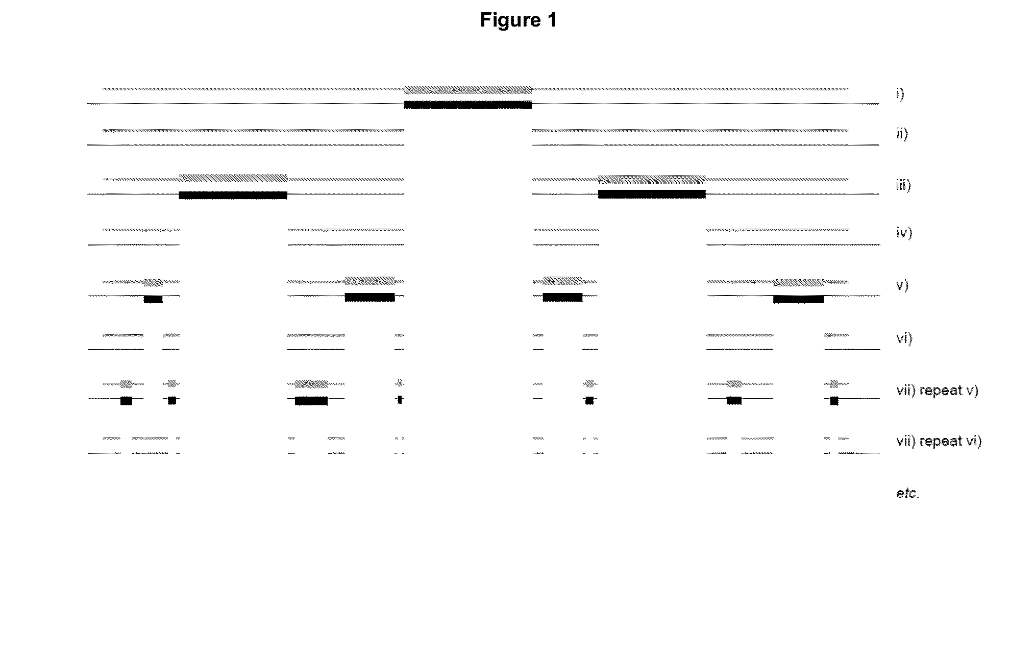
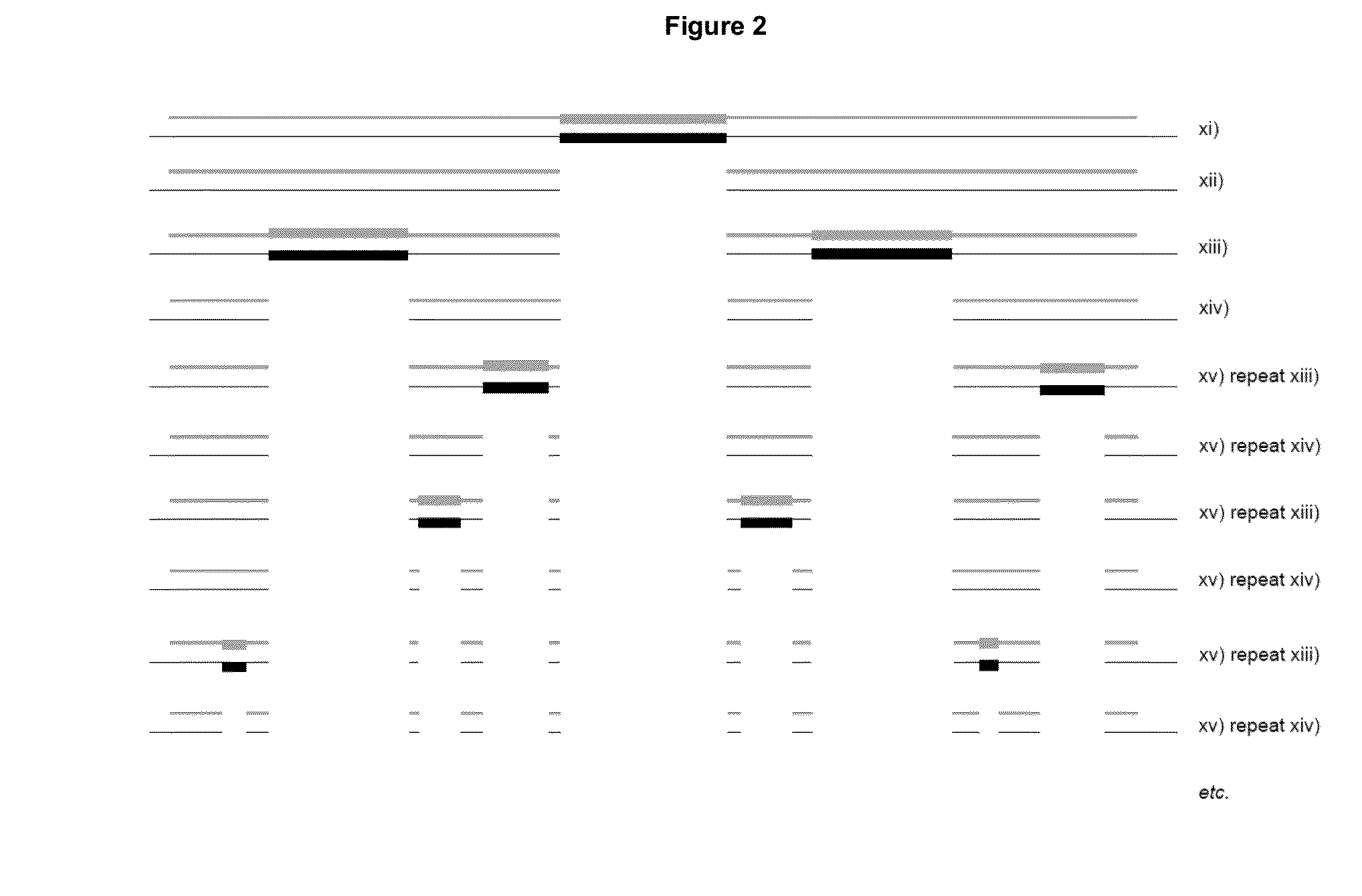
The present invention provides a method for quantifying the level of minimal residual disease (MRD) in a subject who has been treated for the disease. The method involves amplifying and sequencing nucleotide sequences from a biological sample obtained from the subject after treatment. The sequences are compared with nucleotide sequences from a biological sample obtained before treatment. The degree of similarity between the two sets of sequences is determined, and a total number of characters that are the same in both lists is obtained. This method allows for the accurate measurement of MRD in a subject and can help to guide treatment decisions.

Problems solved by technology

Nevertheless, it may be that some diseases are not detected or that the subject's recovery is not complete after treatment.
Although the aforementioned documents disclose several different approaches for detecting MRD wherein improvements in are achieved through different methods of sequencing, none of these documents specifically discloses a method which achieves improvement through the alignment strategy used.
The method of alignment used is important to the accuracy of any method which is based on comparison of nucleotide sequences because the rate of failures of sequencers using a classical binary logic—in which sequences can only be equal or different—is so high that it is not useful.
However, despite generally disclosing that alignment may be achieved using references sequences such as primer binding sequences or non-reference sequences, this document does not disclose a specific method for alignment that is capable of determining the level of disease in a subject irrespective of the genetic characteristics of the nucleotide or the disease.

Method used


Image

  • Method for quantifying the level of minimal residual disease in a subject
  • Method for quantifying the level of minimal residual disease in a subject
  • Method for quantifying the level of minimal residual disease in a subject

Examples


example 1

Quantification of MRD in Multiple Myeloma Using Massively Parallel Sequencing of Genes of Immunoglobulins

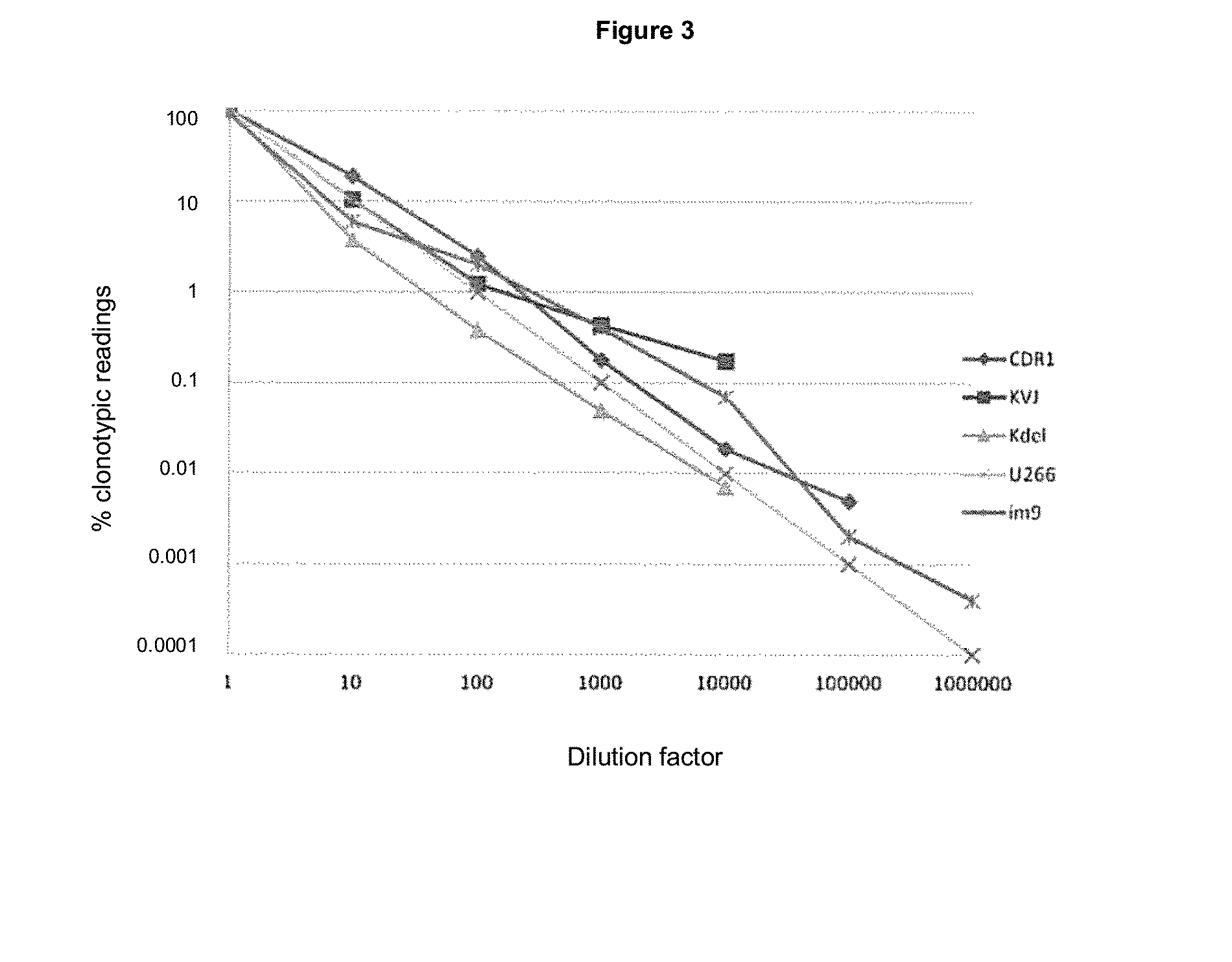
[0503]The following presents a method for quantification of tumor clonotypic sequences within the polyclonal background rearrangements of genes of immunoglobulins (Ig) via massively parallel sequencing (MPS). The detection of clonal rearrangement in B and T cell neoplasms allows the evolution of these pathologies to be monitored. To quantify these rearrangements in B cells, primers disclosed in Tables 1 and 2 for CDR3, VDJ, IgH, IgK, KVJ, KDEL, and IgL were used, because these fragments cover more than 90% of cases (Van Dongen, Leukemia 2003). The selection of these particular rearrangements is due to the design of primers which only amplify short (less than 200 bp) sequences; allowing to sequence these fragments in the PROTON platform, capable of 10 Gb.

[0504]Patients negative for VDJ, IgH, CDR3, KVJ, KDEL diagnoses may be sequenced with the rest of the BIOMED primers like IgH, V...

example 2

Quantification of SNV, MNV and Indels

[0510]The method described in the foregoing is applicable to the detection of any type of mutation, given some limitations, as follows. The average error based on massive sequencing platforms is 0.5% or, in other words, one erroneous reading in 200 for each position in the genome. The probability that an error happens reading the variant sought is 0.5% / 4 bases, or about 0.1%. This theoretical limitation has been verified experimentally for point mutations (SNV: DNM3A and IDH2) in cases of AML (acute myeloid leukemia, FIG. 5), wherein this error is 0.1% for each position. In those mutations that include more than two positions for reading, such as a multiple mutation (MNV) or an indel, the error will be (0.1×n) %, where n is the number of clonal variants present in the reading.

example 3

Quantification of Long Insertions and Translocations

[0511]In this case the sensitivity limit is not reached due to the fact that there is no background against which to compare readings, because primers amplify only those DNA fragments that have a translocation or inversion. To alleviate this problem, a control DNA was used, this being one of the genes involved in the translocation, in its wild-type form. Thus the ratio of clonotypic sequences / total sequences takes into account the number of readings of both.


PUM

PropertyMeasurementUnit
weightaaaaaaaaaa
lengthaaaaaaaaaa
Ctaaaaaaaaaa

Abstract

The present invention belongs to the field of diagnosis of disease. Thus the present invention is focused on a method and kit for quantifying the level of minimal residual disease (MRD) in a subject who has been treated for said disease, which comprises:(a) identifying, amplifying and sequencing a nucleotide sequence in a biological sample obtained from said subject after treatment for said disease, wherein the gDNA of said biological sample has an average weight, k, per cell, and wherein said nucleotide sequence is identified using primers and is amplified using an amount, D, to afford a first list of characters;(b) identifying, amplifying and sequencing a nucleotide sequence in a biological sample obtained from a subject with said disease using the same primers as in step (a) to afford a second list of characters;(c) determining, for each first list of characters obtained in step (a), the degree of similarity, DS, with each second list of characters obtained in step (b);(d) selecting, for each first list of characters obtained in step (a), the DS of highest value, DSHV;(e) adding up the number of first lists of characters obtained in step (a) which have a DSHV that is greater than a threshold value, T, to obtain Lc;(f) adding up the total number of lists of characters, Lt, in the first list of characters; and(g) calculating the level of minimal residual disease (MRD) according to either of the following formulae:MRD=(Lc×k) / (Lt×D)orMRD=Lc×(D / k) / Lt2.

Description

CROSS-REFERENCE TO RELATED APPLICATIONS[0001]This application is filed under the provisions of 35 U.S.C. §119(a) and claims the priority of European Application No. 14191967.0 filed on 5 Nov. 2014, which is incorporated by reference herein in its entirety.FIELD OF THE INVENTION[0002]The present invention may be included in the field of medicine in general, more particularly in the field of diagnosis of disease.[0003]In particular, the present invention is focused on a method and kit for quantifying the level of minimal residual disease in a subject. In addition, the present invention is focussed on use of the method and / or kit for quantifying the level of minimal residual disease in a subject.BACKGROUND TO THE INVENTION[0004]Current methods for the detection and treatment of disease mean that it is possible to control many diseases at a clinical level, thereby obliterating all traces of the disease. Nevertheless, it may be that some diseases are not detected or that the subject's re...

Claims


Application Information

Patent Timeline
Patent Type & Authority Applications(United States)
IPC IPC(8): C12Q1/68G16B30/10
CPCC12Q1/6874G06F19/22C12Q1/6806C12Q2600/118G16B30/00C12Q1/6886C12Q1/6858C12Q2527/113C12Q1/6851G16B30/10C12Q2535/122C12Q2537/165C12Q1/6883
Inventor BARRIO GARC A, SANTIAGOMART NEZ LOPEZ, JOAQUINMAR N SEBASTI N, CARLOSRAPADO MART NEZ, MARIA INMACULADAAYALA D AZ, ROSA MARIAS NCHEZ VEGA CARRION, BEATRIZ
Owner FUNDACION DE INVESTIGACION HOSPITAL 12 DE OCTUBRE
Learn More