
System and method for acoustic radiation force creep-recovery and shear wave propagation elasticity imaging

a technology of elasticity imaging and acoustic radiation, applied in the field of systems and methods for medical imaging using vibratory energy, can solve the problems of rheological models not describing the material behavior at all frequencies, the difficulty of measuring shear wave attenuation in the field of elasticity imaging, and the undesirable amount of computational burden on the quantification process

Active Publication Date: 2016-05-19
MAYO FOUND FOR MEDICAL EDUCATION & RES
View PDF3 Cites 5 Cited by
  • Summary
  • Abstract
  • Description
  • Claims
  • Application Information

AI Technical Summary

Benefits of technology

The present invention provides a way to measure the viscoelastic properties of tissue using an ultrasound system. This is done by applying a mechanical stress to the tissue and measuring the resulting displacement during a recovery period. A complex modulus value is extracted from the displacement, and a loss tangent is calculated from this value. This enables the calculation of a viscoelastic tissue property metric. This method can provide a non-destructive and non-invasive way to measure tissue viscoelastic properties, which can have various applications in medicine and biotechnology.

Problems solved by technology

Quantitative mechanical properties can be measured in a model independent manner if both shear wave speed and attenuation are known; however, measuring shear wave attenuation is challenging in the field of elasticity imaging.
Current elasticity imaging techniques are useful to indentify tissue mechanical properties; however, to quantify these properties a rheological model must be used, which introduces an undesirable amount of computational burden to the quantification process.
In additional, rheological models may not describe the material behavior at all frequencies, may not be appropriate for the physical test being performed, and are less general than a model-free approach.
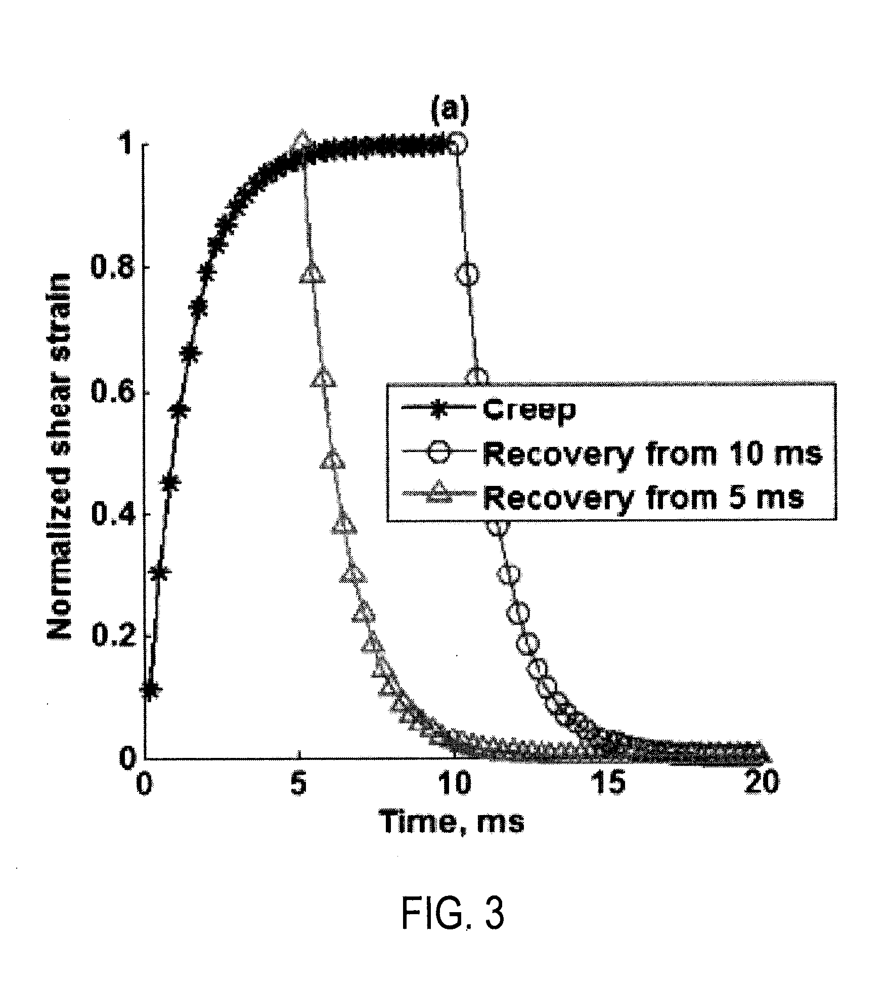
A limitation of this current method, is that to study viscoelastic parameters in a wider range of frequencies, the creep period needs to be maintained for long times, for instance up to 30 ms, which means that the total exposure time to acoustic radiation force will increase.

Method used


Image

  • System and method for acoustic radiation force creep-recovery and shear wave propagation elasticity imaging
  • System and method for acoustic radiation force creep-recovery and shear wave propagation elasticity imaging
  • System and method for acoustic radiation force creep-recovery and shear wave propagation elasticity imaging

Examples


Embodiment Construction

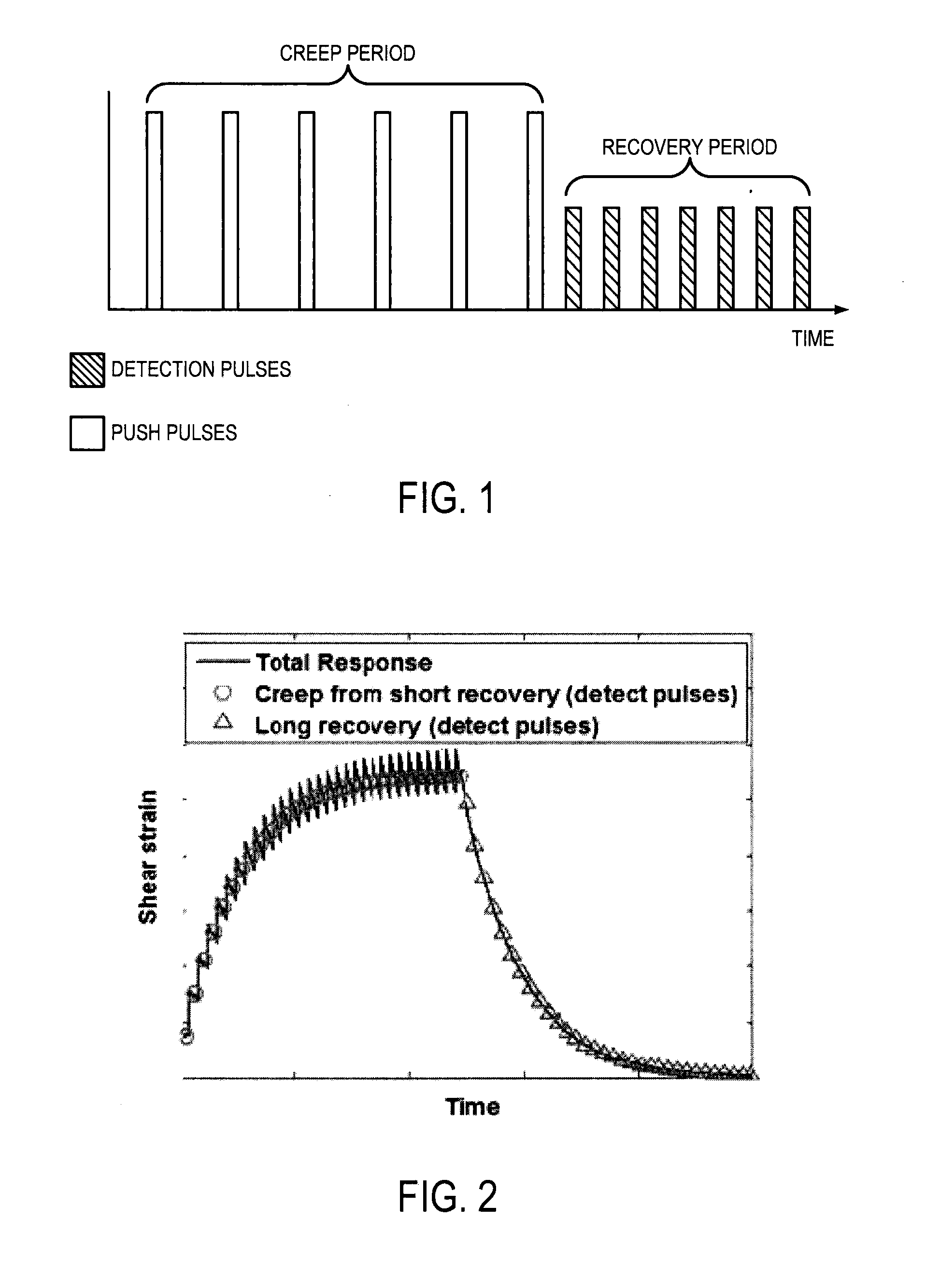
[0022]Described here are systems and methods for model-independent quantification of tissue viscoelastic properties by estimating a complex shear modulus from a time-dependent creep-recovery response induced by acoustic radiation force. A creep response is generated and data is acquired during a recovery period following the creep response, as illustrated in the example pulse sequence in FIG. 1. In this approach, the creep response is generated using either multiple push pulses each having a short duration, or a continuous push having a long duration. Generating the creep response using these techniques overcomes the problem of exceeding FDA limits of mechanical index (“MI”) during creep period. The push pulses generate a temporal step-force, and following the cessation of the push pulses there is a recovery period, as illustrated in FIG. 2, during which data is acquired using detection pulses. In addition to the creep response, shear waves are also generated outside the focal area....


PUM


Abstract

A model-independent method for producing a viscoelastic tissue property metric using ultrasound is provided. A mechanical stress, such as an acoustic force, is applied to a tissue during a creep period using an ultrasound system to generate a creep response. Tissue displacement resulting from the applied acoustic force is measured during a recovery period following the creep period. From the tissue displacement measurements, a relative complex modulus is extracted, and a loss tangent is calculated based on extracted complex modulus. Using the calculated loss tangent, viscoelastic tissue property metrics may be calculated.

Description

CROSS-REFERENCE TO RELATED APPLICATIONS[0001]This application claims the benefit of U.S. Provisional Patent Application Ser. No. 61 / 833,215, filed on Jun. 10, 2013.STATEMENT REGARDING FEDERALLY SPONSORED RESEARCH[0002]This invention was made with government support under EB002640, EB002167, and DK082408 awarded by the National Institutes of Health. The government has certain rights in the invention.BACKGROUND OF THE INVENTION[0003]The field of the invention is systems and methods for medical imaging using vibratory energy, such as ultrasound imaging. More particularly, the field of the invention is systems and methods for shear wave dispersion ultrasound vibrometry (“SDUV”).[0004]Tissue mechanical properties are linked to tissue pathology state. Shear wave propagation methods have been proposed to quantify tissue mechanical properties. In these methods, shear waves that result from a transient (impulsive or short tone burst) excitation of tissue propagate only a few millimeters, as ...

Claims


Application Information

Patent Timeline
Patent Type & Authority Applications(United States)
IPC IPC(8): A61B8/08
CPCA61B8/5223A61B8/485A61B8/085G01S7/52042G16H50/30
Inventor GREENLEAF, JAMES F.CARRASCAL, CAROLINA AMADOR
Owner MAYO FOUND FOR MEDICAL EDUCATION & RES
Learn More