
Automated 3D Reconstruction of the Cardiac Chambers from MRI and Ultrasound

a 3d reconstruction and cardiac chamber technology, applied in the field of automatic 3d reconstruction of cardiac chambers from mri and ultrasound, can solve the problems of significant methodological variability, time-consuming manual segmentation, and loss of important details of objects

Active Publication Date: 2016-05-19
RGT UNIV OF CALIFORNIA
View PDF5 Cites 23 Cited by
  • Summary
  • Abstract
  • Description
  • Claims
  • Application Information

AI Technical Summary

Benefits of technology

The patent describes a method for creating a 3D segmentation of the heart chamber from MRI or ultrasound data. The method involves minimizing an energy function to create a smooth and accurate boundary of the heart chamber. This can help with the diagnosis and treatment of various heart conditions.

Problems solved by technology

Currently many of the commercially available software platforms for CMR post-processing either provide suboptimal automated segmentation or require a substantial amount of manual segmentation support from the user, resulting in significant methodological variability (Janik M, Cham M D, Ross M I, et al.
Additionally, manual segmentation is time consuming, and requires dedicated operator training that makes it inefficient due to the extent of information in CMR images.
Therefore, some important details of the object would be lost during the procedure; thus the resultant objects usually possess rough surfaces.
Although there are some methods to automate consecutive “2D segmentation” and “3D multiplanar reconstruction” steps, this approach fails to exploit the benefit of a true, 3D volumizing technique.
Additionally, most segmentation approaches in 2D cannot readily handle cases where an object of interest (e.g., papillary muscles) appears to be separated into several cross-sections (i.e., non-convex object).
This separation and discontinuity commonly can be seen in CMR images, which incur further challenges in 2D segmentation.
However, none of these singular approaches has resulted in an accurate and fast segmentation algorithm that requires no prior statistical model.

Method used


Image

  • Automated 3D Reconstruction of the Cardiac Chambers from MRI and Ultrasound
  • Automated 3D Reconstruction of the Cardiac Chambers from MRI and Ultrasound
  • Automated 3D Reconstruction of the Cardiac Chambers from MRI and Ultrasound

Examples


Embodiment Construction

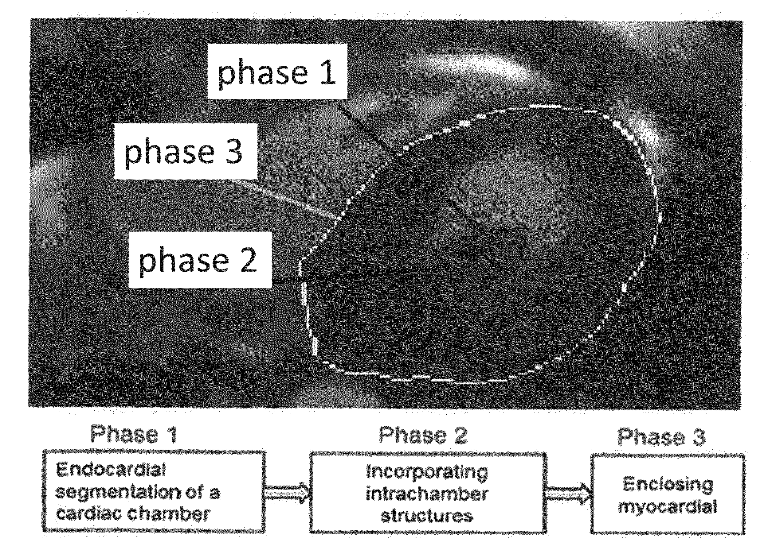
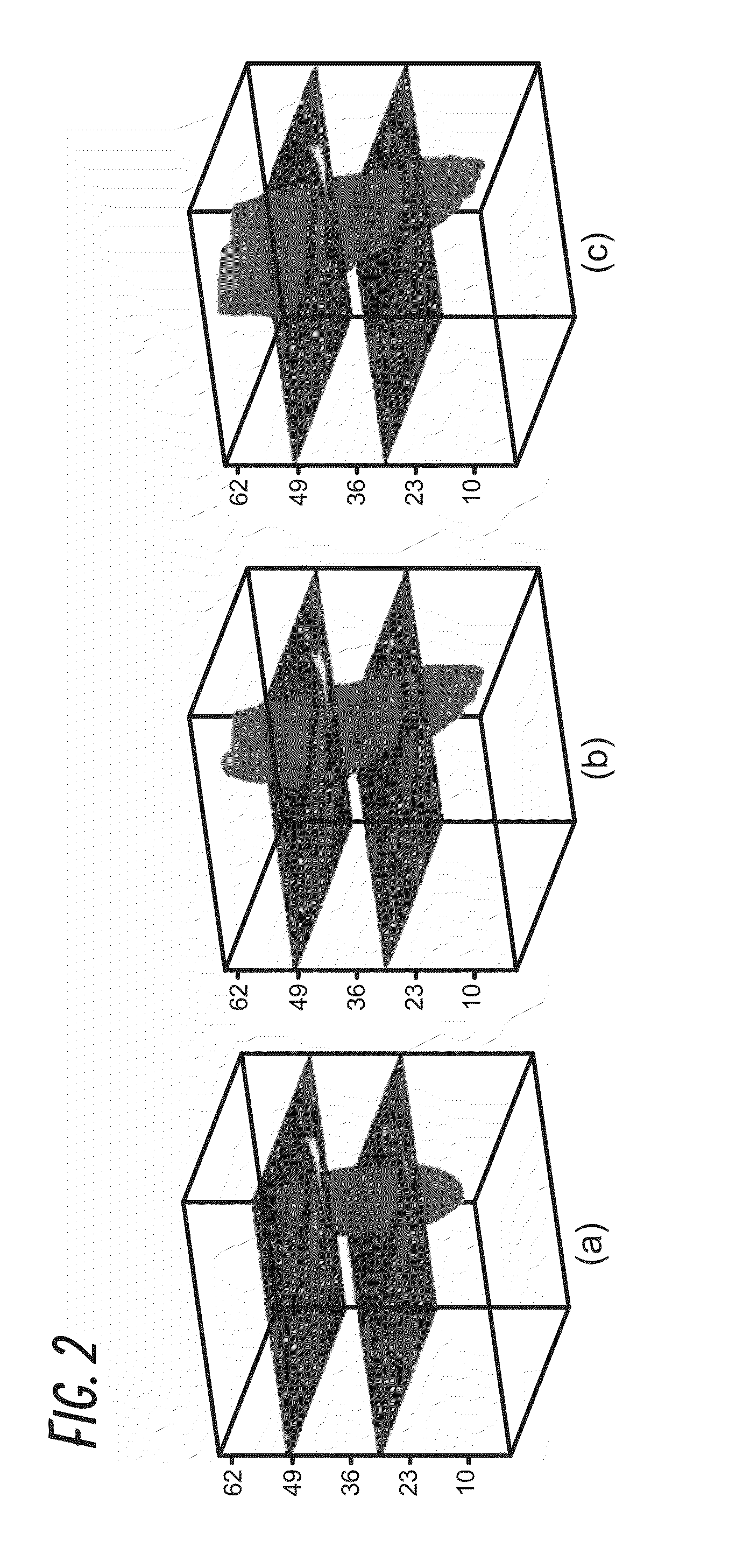
[0044]We have developed and tested a fast, automated 3D segmentation tool for cardiac Magnetic Resonance Imaging (MRI) or cardiac Ultrasound imaging. The segmentation algorithm automatically reconstructs raw cardiac MRI or Ultrasound data to a 3D model (i.e., direct volumetric segmentation), without relying on any prior statistical knowledge, making it widely applicable and useful for many clinical applications.

[0045]To overcome limitations of previous methodologies, the current invention utilizes emerging principles in image processing to develop a true 3D reconstruction technique without the need for training datasets or any user-driven segmentation. This was accomplished by developing an automatic segmentation framework that exploits the benefit of full volumetric imaging in an anatomically natural way. Because the current method does not rely on prior statistical knowledge, it offers dramatically more malleability than current algorithms by being broadly applicable across differ...


PUM


Abstract

The disclosure relates to a method of automatically producing a three-dimensional (3D) segmentation of a heart chamber, the method comprising: obtaining data sets from cardiac magnetic resonance imaging (MRI) or ultrasound, generating a 3D segmentation of the heart chamber from the data sets using an active contour method, modifying the 3D segmentation by adding a plurality of intra-chamber structures; and identifying an enclosing myocardium using the 3D segmentation generated by the method.

Description

INCORPORATION BY REFERENCE TO ANY PRIORITY APPLICATIONS[0001]This application claims priority U.S. Provisional Application No. 62 / 073,688 filed on Oct. 31, 2014. Any and all applications for which a foreign or domestic priority claim is identified in the Application Data Sheet as filed with the present application are hereby incorporated by reference under 37 CFR 1.57.BACKGROUND[0002]Cardiac magnetic resonance imaging (CMR) is a valuable tool that provides important information for diagnosis and evaluation of cardiac anatomic abnormalities, and cardiovascular disease (Frangi A F, Niessen W J, Viergever M A. Three-dimensional modeling for functional analysis of cardiac images, a review. Medical Imaging, IEEE Transactions on 2001; 20(1):2-5). CMR is a safe modality that does not require ionizing radiation or iodinated contrast but delivers images with high spatial and temporal resolution (Yuan C, Kerwin W S, Ferguson M S, et al. Contrast-enhanced high resolution MRI for atheroscleroti...

Claims


Application Information

Patent Timeline
IPC IPC(8): G06T15/08G06T19/20A61B8/08G06T7/00A61B5/055A61B5/00
CPCG06T15/08A61B5/055A61B5/0044A61B8/0883G06T7/0081G06T2207/30048G06T19/20G06T2207/10136G06T2207/10088G06T2207/20116G06T7/0089A61B8/466A61B8/483A61B8/5223A61B2576/02G06T7/12G06T7/149G06T2207/10132G06T2219/2021G16H30/40
Inventor JAFARKHANI, HAMIDHAJIAGHAYI, MAHDIGROVES, ELLIOTTKHERADVAR, ARASH
Owner RGT UNIV OF CALIFORNIA
Learn More